Geographic polymorphism of
P
element in populations of
Drosophila sturtevanti
Luciane M. de Almeida
1, Francisco Langeani
2and Claudia M.A. Carareto
11
Departamento de Biologia, Universidade Estadual Paulista (UNESP), São José do Rio Preto, SP, Brazil.
2Departamento de Zoologia e Botânica, Universidade Estadual Paulista (UNESP), São José do Rio Preto,
SP, Brazil.
Abstract
The aim of this report was to detect full-sized P element sequences in eight strains of Drosophila sturtevanti populations from distant geographic regions and to assess the structural geographic variation amongP element sequences. PCR analysis confirmed the presence of a putative completeP element in all strains. Southern blot analysis indicated bands shared by all strains, and bands restricted to geographically related strains. Parsimony analysis corroborated the hybridization pattern that reflected the geographic relationships.
Key Words:canonicalPelements,Drosophila, Pelement polymorphism, saltansgroup, transposable elements.
Received: August 9, 2002; accepted: December 4, 2003.
Introduction
ThePelement, one of the best studied transposable elements in eukaryotes, was first discovered inDrosophila melanogaster(Kidwellet al., 1977; Binghamet al., 1982) in which multiple copies per genome are typically present but only a few are autonomous. The complete canonicalP element is 2.9 kb long and has 31 bp inverted terminal re-peats, 11 bp inverted subterminal repeats and four ORFs that encode the transposase (O’Hare and Rubin, 1983). The Pelement structure, distribution of insertions, and transpo-sition and regulatory mechanisms have been extensively studied inD.melanogaster, but only a few reports have ad-dressed these characteristics in thesaltansspecies group (Daniels and Strausbaugh, 1986; Danielset al., 1990; Clark et al., 1995; Clark and Kidwell, 1997; Clarket al.,1998; Silva and Kidwell, 2000). These studies have shown thatP elements from the saltans and willistoni species groups form four subfamilies. Of these, the most prevalent is the canonicalPelement, which form a compact subfamily with a maximum sequence divergence of 10% (Clark and Kidwell, 1997). ThePelement subfamilies in thesaltans andwillistoni species groups have been described based mainly on partial sequences (Clarket al., 1995; Clark and Kidwell, 1997; Haring et al., 2000; Silva and Kidwell, 2000). There has been no assessment of whether the
se-quences from the different subfamilies are complete or de-fective, or whether strains of the same species are polymorphic in theirPelement structure.
Since D. sturtevanti (sturtevanti subgroup) is the most widespread species of thesaltansgroup, it is ideal for investigating the existence of full-sized sequences and structural geographic variation in the P elements of this group. In this work, we analyzed eight strains of D. sturtevantifrom different geographic regions (from Mex-ico to the extreme south of Brazil) in order to assess the geographic polymorphism in the transposable element of this species.
Materials and Methods
Fly stocks
TheD.sturtevantistrains used in this study were from (1) Apazapan, Veracruz (APA; 19°11’ N 96°10’ W), and Matlapa, San Luis Potosi, Veracruz (MAT; 22°10’ N 101° W) in Mexico; both strains were collected in 1998 by J.C. Silva, University of Arizona, Tucson, USA; (2) Villavicencio (COL; 4°09’ N 73°38’ W) in Colombia, (H 193.3, The Genetics Foundation, University of Texas, Aus-tin, Texas, USA); (3) Santana do Riacho, MG (I27, 19° S
44° W), Mirassol, SP (BRA; 20°47’ S 49°28’ W), São José do Rio Preto, SP (RP1and RP2; 20°50’ S 49°20’ W and
20°60’ S 49°18’ W, respectively) and Novo Horizonte, SP (NHO; 21°29’ S 49°18’ W), in Brazil. The BRA strain was established with flies collected in 1971 by W.J. Tadei
Send correspondence to Cláudia Márcia A. Carareto. Departa-mento de Biologia, Rua Cristovão Colombo, 2265, Jardim Naza-reth, 15054-000, São José do Rio Preto, SP, Brazil. Phone: +55-17-2212382. E-mail: [email protected].
(UNESP, São José do Rio Preto, SP), I27was established in
1995 by C.R. Vilela (USP, São Paulo, SP), RP1in 1997 by
L.M. Almeida and RP2and NHO in 1998 by F.R. Torres
(UNESP, São José do Rio Preto, SP). The Harwich-w strain of D. melanogaster, a strong P strain isolated by M.G. Kidwell (University of Arizona, Tucson, AZ, USA), was used as a positive control.
PCR amplification
Genomic DNA from each strain was amplified by PCR using the primer M-IR which anneals to the terminal repeats (nucleotides 14 to 31 and 2894 to 2877) of full-sized, and internally deleted copies of P elements (Haringet al., 1995). The reaction mixture consisted of 200 ng of genomic DNA, 2 mM MgCl2, 0.16 mM of each
dNTP, 0.25 pmol of pM-IR/µL, 0.1 unit of Taq DNA poly-merase and 1x buffer. Temperature cycling involved heat-ing the solutions to 94 °C for 7 min, followed by 30 cycles of 94 °C for 45 s, 57 °C for 45 s, 72 °C for 1.5 min, and a fi-nal extension at 72 °C for 5 min.
Southern blot analysis
ThePelement insertion sites and the variation in the restriction fragment sizes were investigated using Southern blot analysis (Sambrooket al., 1989). For both methods, a chemioluminescent hybridization system (ECLTM direct nucleic acid labelling and detection systems, Amersham Life Science) was used according to the manufacturer’s in-structions.
Total genomic DNA was isolated from pools of about 50 individuals, as described by Jowett (1986). Approxi-mately 10µg of DNA from each strain were digested with the appropriate restriction endonucleases (Figure 1). The DNA fragments were then electrophoresed, transferred to a nylon membrane, and fixed. The blot was hybridized with
ECL hybridization buffer and the appropriate probe. The membrane was washed for 40 min in primary buffer (6 M urea, 0.4% SDS and 0.5 M xSSC) at 42 °C, and for 10 min in secondary buffer (2xSSC) at room temperature.
Restriction fragment lenght polymorphism (RFLP) and probes
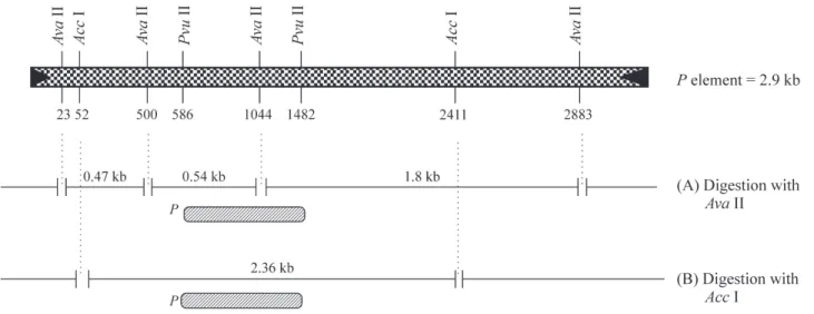
To assess P element restriction fragment polymor-phism, genomic DNA was digested with the endonucleases AvaII andAccI and probed with the 896 bpPvuII fragment of thePelement extracted from pπ25.1 by digesting theD. melanogaster Pelement sequence withPvuII. Digestion of the canonicalPelement byAvaII generated three internal fragments of 478 bp, 544 bp and 1838 bp in length; whereas AccI generated a single 2360 kb internal fragment that em-braced 81% of the complete sequence. Figure 1 depicts the restriction sites for the enzymes used and the positions where the probes hybridized.
Evolutionary analysis
Relationships among thePelement sequences of each strain based on the absence (0) or presence (1) of restriction fragments were inferred using the maximum parsimony method, as implemented in PAUP v.4.0b10 (Swofford, 1997). Maximum parsimony searches were done using the branch-and-bound algorithm. Bootstrap analyses were done using parsimony and consisted of 500 replicates with the branch-and-bound algorithm.
Results and Discussion
Identification of putative full-sizedPelements
We analyzed strains of D. sturtevanti collected in Mexico (19°11’ N 96°10’ W), Colombia (4°09’ N 73°38’ W) and southeastern Brazil (19° S 44° W to 21°29’ S
49°18’ W) in order to assess the existence of full-sizedP el-ement sequences and to determine whether these sequences showed geographical variation based on their endonuclease restriction sites. The occurrence of an approximately 2.9 kb fragment amplified in all strains (apparently the same as amplified in thePelement of theD. melanogasterpositive control) revealed the presence of at least one potentially full-length element (Figure 2) and several smaller defective sequences. This complete element may belong to the ca-nonical subfamily but could also be a more divergent se-quence since our amplification conditions were not stringent. In addition to the 2.9 kb fragment, a slightly smaller fragment was also observed only inD. sturtevanti. This second sequence could belong to a P element subfamily different from the canonical one since pM-IR can amplify the completePelement sequence belonging to subfamily M or others such as subfamily T in species of the obscuragroup (Hagemannet al., 1996).
Pelement RFLP analysis
Two RFLP maps were used to evaluate the geo-graphic variation ofPelement. The restriction enzymeAva II cleaves a full-sizedPelement at four sites (positions 23, 500, 1044 and 2883) to generate internal fragments of 478 bp, 544 bp and 1838 bp. The absence of one of these frag-ments, or variation in their sizes, indicates internal dele-tions or the absence of restriction sites as a result of point mutations. Figure 3A shows theAvaII digests hybridized with the 0.9 kbPvuII fragment carried by pπ25.1. Since this probe hybridized with the 0.54 kb and 1.8 kbAvaII fragments, we expected to find these bands, or smaller ones, as a result of deletions. Both fragments were present in all D. sturtevanti strains and also in D. melanogaster-Harwich. All of the strains had fragments smaller than 1.8 kb, indicating internal deletions. Several of these bands were shared by all of theD. sturtevantistrains (black arrows), but some of them were restricted to Colom-bian and Mexican strains (dotted arrows), and another was restricted to Brazilian strains (hatched arrows). The bands
larger than 1.8 kb represented polymorphisms caused by mutations in the first twoAvaII restriction sites. Southern blotting also showed that all of theD.sturtevantistrains, but not D. melanogaster, had bands with a molecular weight > 1.8 kb.
Figure 3B shows theAccI digest hybridized with the Pvu II fragment. Acc I cleaves the canonical P element close to its ends (positions 53 and 2412) to generate a single 2360 kb internal fragment that embraces 81% of the com-plete sequence. This blot allowed us to include in the analy-sis of polymorphism the initial sequence the ofPelement that did not hybridized in the experiments withAvaII (53 bp up to 500 bp). TheD.melanogaster-Harwich positive control showed the expected 2.4 kb band (column 9) and smaller ones derived from defective elements. The
Brazil-Figure 2- PCR amplification of the 2.9 kb full-size sequence of theP ele-ment inD. sturtevanti. 1.λHindIII DNA marker, 2. COL, 3. APA, 4. MAT, 5. I27, 6. BRA, 7. RP1, 8. NHO, 9. RP2and 10. pπ25.1 (positive
con-trol), (ï) 2.9 kb band.
Figure 3- Southern blot ofD.sturtevantigenomic DNA digested with re-striction endonucleases:A. Digested withAvaII and probed with thePvuII fragment of thePelement from pπ25.1.B. Digested withAccI and probed with thePvuII fragment of thePelement from pπ25.1 (samples: 1- COL, 2- APA, 3- MAT, 4- I27, 5- BRA, 6- RP1, 7- NHO, 8- RP2and 9-D.
ian strains ofD. sturtevantishowed bands corresponding to 2.4 kb (white double arrow); whereas, the Mexican and Co-lombian strains had a slightly larger band (black double ar-row). In addition to the similarity between the Mexican-Colombian strains on the one hand and the Brazil-ian strains on the other, there were other fragments com-mon to these groups as shown by the dotted arrows for the Mexican-Colombian group and the hatched arrows for the Brazilian group. As in theAvaII analysis, bands larger than 2.4 kb were present in allD.sturtevantistrains but not inD. melanogaster.
The PCR analysis and theAvaII andPuvII blots to-gether indicate the presence of apparently full-sizedP ele-ments in D. sturtevanti, although divergent P element sequences were seen when the Brazilian strains were com-pared with the other three. RFLP analysis revealed a group of sequences inD. sturtevantithat were common to most of the strains and another group restricted to the most geo-graphically related strains. The fragments common to all strains (black arrows in Figure 3A and B) may represent se-quences that have been inactive since the dispersion ofD. sturtevantiin the Americas and have not undergone muta-tion at the restricmuta-tion sites for the enzymes used in this study. The sequences common to the Colombian and Mexi-can strains (dotted arrows) and to the Brazilian (hatched ar-rows) were different and probably originated more recently in the ancestral populations of each group. The existence of different groups of sequences coexisting in the same ge-nome reflects the presence of multiples P element subfamilies in thesaltansspecies group and may have re-sulted from different horizontal transfer events (Silva and Kidwell, 2000).
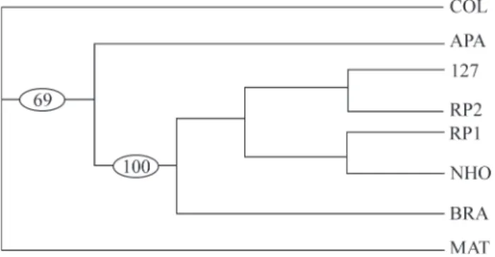
Figure 4 summarizes the results of the parsimony analysis used to determine the relationships among theP el-ement restriction fragments of each strain. Of 73 charac-ters, 9 were constant, 14 were uninformative, and 50 were parsimony informative. All of the six equally parsimonious trees corroborated the hybridization pattern that reflected geographic relationships;i.e., the presence of a group of se-quences clearly associated with the Brazilian strains (100% of 500 bootstrap replicates). Although the tree shows dif-ferent relationships among the Brazilian strains, bootstrap analysis indicated that the branches would collapse to cre-ate a polytomie if the maximum branch length were zero.
The lack of significant variation noted above was confirmed by the similar banding patterns of strains BRA, RP1and RP2. Together with the bi-directionality of the low
hybrid dysgenesis indices previously reported for this spe-cies (Almeida, 2000; Almeida and Carareto, 2002), this sta-tistical homogeneity allows us to reinforce the suggestion of P element inactivity in D. sturtevanti. These findings also contribute to our knowledge of the structural differen-tiation and geographical variation in P elements of D. sturtevanti.
Acknowledgments
The authors thank M.G. Kidwell, J.C. Silva, C.R. Vilela, W.J. Tadei and F.R. Torres for providing us with strains ofD. sturtevanti. M.G. Kidwell, A.J. Holyoake and J.P. Castro provided helpful comments on an early version of the manuscript and S. Aranha edited the manuscript. This work was supported by FAPESP (Grant nos. 95/7192-2 and 98/08734-1 to C.M.A.C and 97/14646-5 to L.M.A) and CNPq.
References
Almeida LM (2000) Genomic distribution of P element in
Drosophila sturtevanti populations. Genet Mol Biol 23:503-504.
Almeida LM and Carareto CMA (2002) Gonadal hybrid dysgenesis in Drosophila sturtevanti (Diptera, Drosophilidae). Iheringia, Série Zoologia 92:71-79. Bingham PM, Kidwell MG and Rubin GM (1982) The molecular
basis of the P-M hybrid dysgenesis: the role ofPelement, a
Pstrain transposon family. Cell 29:995-1004.
Clark JB, Altheide TK, Schlosser MJ and Kidwell MG (1995) Molecular evolution of P transposable elements in genus
DrosophilaI. Thesaltansandwillistonispecies groups. Mol Biol Evol 12:902-913.
Clark JB and Kidwell MG (1997) A phylogenetic perspective on
Ptransposable element evolution inDrosophila.Proc Natl Acad Sci USA 94:11428-11433.
Clark JB, Kim P and Kidwell MG (1998) Molecular evolution of
P transposable elements in genus Drosophila III. The
melanogasterspecies group. Mol Biol Evol 15:746-755. Daniels SB and Strausbaugh LD (1986) The distribution ofP
ele-ment sequences in Drosophila: thewillistoni and saltans
species groups. J Mol Evol 23:138-148.
Daniels SB, Peterson KR, Straubaugh LD, Kidwell MG and Chovnick A (1990) Evidence for horizontal transmission of Figure 4- Relationships estimated by parsimony analysis using the branch-and-bound method based on restriction maps ofPelements in eight strains ofD. sturtevanti. All characters were unordered and gaps were treated as missing data. This tree is one of the six most parsimonious reconstructions and required 99 steps. The consistency index is 0.6465 and the retention index is 0.6067. The numbers in the ovals indicate the percent of 500 bootstrap replications that contain the indicated clade. The strains are denoted by codes (APA - Apazapan and MAT - Matlapa, both from Mexico; COL - Villavicencio, Colombia, and I27- Santana do Riacho,
BRA- Mirassol, RP1and RP2, São José do Rio Preto, and NHO - Novo
thePtransposable element betweenDrosophilaspecies. Ge-netics 124:339-355.
Hagemann S, Haring E and Pinsker W (1996) Repeated horizontal transfer ofPtransposons betweenScaptomyza pallidaand
Drosophila bifasciata.Genetica 98:43-51.
Haring E, Hagemann S and Pinsker W (1995) Different evolution-ary behaviour ofPelement subfamilies: M-type and O-type elements in Drosophila bifasciata and D. imaii. Gene 163:197-202.
Haring E, Hagemann S and Pinsker W (2000) Ancient and recent horizontal invasions of drosophilids byPelements. J Mol Evol 51:577-586.
Jowett T (1986) Preparation of nucleic acids. In: Roberts DB (ed)
Drosophila: a Pratical Approach. edited by D.B. Roberts, IRL Press, Oxford, pp 275-286.
Kidwell MG, Kidwell JF and Sved J (1977) Hybrid dysgenesis in
Drosophila melanogaster: a syndrome of aberrant traits in-cluding mutation, sterility and male recombination. Genet-ics 86:813-833.
O’Hare K and Rubin GM (1983) Structure ofPtransposable ele-ment and their sites of insertion and excision in the
Drosophila melanogastergenome. Cell 34:25-35.
Sambrook J, Fritsch EF and Maniatis T (1989) Molecular Clon-ing: a Laboratory Manual 2 ed, v.1, Cold Spring Harbor Lab-oratory Press, New York, 9.31-9.57 pp.