www.cbpv.org.br/rbpv ISSN 0103-846X (Print) / ISSN 1984-2961 (Electronic)
Doi: http://dx.doi.org/10.1590/S1984-29612017010
New morphological data and molecular diagnostic of
Henneguya friderici
(Myxozoa: Myxobolidae), a parasite
of
Leporinus friderici
(Osteichthyes: Anostomidae) from
southeastern Brazil
Novos dados morfológicos e diagnóstico molecular de
Henneguya friderici
(Myxozoa: Myxobolidae), parasito
de
Leporinus friderici
(Osteichthyes: Anostomidae) do sudeste do Brasil
Letícia Poblete Vidal
1; José Luis Luque
2*
1 Programa de Pós-graduação em Ciências Veterinárias, Universidade Federal Rural do Rio de Janeiro – UFRRJ, Seropédica, RJ, Brasil 2 Departamento de Parasitologia Animal, Universidade Federal Rural do Rio de Janeiro – UFRRJ, Seropédica, RJ, Brasil
Received December 20, 2016 Accepted February 16, 2017
Abstract
The myxozoan
Henneguya friderici
is a parasite of the gills, intestine, kidney and liver of
Leporinus friderici
, a characiform
fish belonging to the family Anostomidae. Forty-two specimens of
L. friderici
that had been caught in the Mogi Guaçú
River, state of São Paulo, were studied. Elongated white plasmodia were found in the gill filaments of 10 host specimens
(24%). The mature spores had an ellipsoidal body with polar capsules of equal size and caudal length greater than body
length. This study also described 18S rDNA sequencing of
H. friderici
infecting the gill filaments. This produced a
sequence of 1050 bp that demonstrated significant genetic differences with previously described species of
Henneguya
.
Similarity analysis using sequences from species that clustered closest to those produced by this study showed that the
species with greatest genetic similarity to
H. friderici
was
H. leporinicola
, with 94% similarity.
Keywords:
Myxosporea, Characiformes, 18S rDNA, phylogeny.
Resumo
O myxozoa
Henneguya friderici
é um parasito encontrado nas brânquias, fígado, intestino e rins de
Leporinus friderici,
(Characiformes: Anastomidae). Foram capturados e examinados quarenta e dois espécimes de
L. friderici
oriundos do
Rio Mogi Guaçú, estado de São Paulo. Cistos alongados e brancos foram encontrados nos filamentos branquiais de
10 (24%) hospedeiros. Os esporos maduros apresentaram o corpo alongado com as cápsulas polares em tamanhos iguais e
o comprimento caudal maior do que o comprimento corporal. Com isso, o presente trabalho, descreve o sequenciamento
de 1050 pb do gene 18S rDNA de
H. friderici
infectando os filamentos branquiais, o que demonstrou diferenças genéticas
significativas em comparação com espécies previamente descritas de
Henneguya
. A análise de similaridade utilizando as
sequencias de espécies que se agruparam mais próximas às produzidas por este estudo mostrou que a espécie com maior
semelhança genética com
H. friderici
foi
H. leporinicola
, com 94% de similaridade.
Paravras-chave:
Myxosporea, Characiformes,18S rDNA, filogenia.
*Corresponding author: José Luis Luque. Departamento de Parasitologia Animal, Universidade Federal Rural do Rio de Janeiro – UFRRJ, CP 74540, CEP 23851-970, Seropédica, RJ, Brasil. e-mail: luqueufrrj@gmail.com
Introduction
The diversity of known myxozoans has grown greatly since
the early work of Kudo (1919). Around 2.200 species have now
been described (LOM & DYKOVÁ, 2006) and these represent
around 18% of cnidarian species diversity, as far as is currently
known (OKAMURA et al., 2015).
Henneguya
Thélohan, 1892,
is one of the most diverse genera of Myxosporea and currently
includes more than 200 known and described species (LOM
& DYKOVÁ, 2006). This widespread genus includes typical
coelozoic and histozoic species and predominantly infects marine
and freshwater fish (EIRAS & ADRIANO, 2012).
Vidal, L.P.; Luque, J.L. Braz. J. Vet. Parasitol. 82
Of these, around 28 have been found to infect fish species of the
order Characiformes.
Identification of the species in this genus, like those in other
genera of myxozoans, is based almost exclusively on spore morphology.
In the class Myxosporea, morphology has been the main criterion
for classification of species (KUDO, 1933; MOLNÁR, 1994).
In fact, this method has always failed to identify highly similar
species that are found in the same infection site and host and which
only have subtle differences in spore structures (YE et al., 2012).
Fortunately, this problem has been solved through molecular
approaches (SMOTHERS et al., 1994; ANDREE et al., 1999;
HOLZER et al., 2004). 18S rDNA is the molecular marker that
has most commonly been used for detection, identification and
phylogenetic analysis on myxozoans (HOLZER et al., 2006).
The difficulties of relying on spore morphology for species
identification have led authors to recommend that SSU rDNA
sequencing should be included when new species are described
(ANDREE et al., 1999; KENT et al., 2001; LOM & DYKOVÁ,
2006).
Leporinus friderici
(Bloch, 1794) is a characiform fish belonging
to the family Anostomidae that is, popularly known in Brazil
as “piau”. It is widely distributed in the Amazon and Paraguay
river basins (FROESE & PAULY, 2016). Among the species of
Henneguya
, only
Henneguya friderici
(CASAL et al., 2003) has
been reported from
L. friderici.
Henneguya friderici
was found infecting the gills, intestine,
kidney and liver of “piau” from an estuarine region of the Amazon
River, in the state of Pará, Brazil. Relative organelle preservation
occurred in the liver tissue and, in some cases, development of
the parasite caused gradual and generalized degeneration in the
intestine, gills and kidney (CASAL et al., 2003)
The present paper supplements the original description
of
H. friderici
with new data on morphology and 18S rDNA
sequencing on samples from gill filaments of
L. friderici
from
the Mogi Guaçú River, state of São Paulo, Brazil. The new data
support the original diagnosis by Casal et al. (2003).
Materials and Methods
Forty-two specimens of
L. friderici
were caught by local fishermen
with nets and hooks in the Mogi Guaçú River near Pirassununga,
state of São Paulo, Brazil (21°55’36” S; 47°22’6” W), between
January 2014 and January 2016. Gills extracted from the fish
were placed in Petri dishes with tap water and were examined for
myxozoans using a dissecting microscope. Infected gill filaments
were preserved using two different methods: frozen (for spore
measurements) and in 95% ethanol (for DNA analysis).
Parasitological examinations were conducted using standard
methods with the aid of an optical microscope (Olympus BX51)
with differential interference contrast (DIC). Images were captured
using a 3.2 mp UC30 digital camera and were analyzed by means
of photomicrography software (Cell
D3.4, Olympus Soft Imaging
Solutions GmbH, Germany). At least 30 measurements were
made for each relevant spore dimension, following the guidelines
of Lom & Arthur (1989).
Gill filaments from three hosts were used for DNA extraction by
means of the DNeasy Blood & Tissue Kit, following the manufacturer’s
instructions (QIAGEN Inc., California, USA). The polymerase
chain reaction (PCR) was performed as described by Whipps et al.
(2015) in 50 µl reaction volumes of the Quick-Load Taq 23 Master
Mix (New England Biolabs, Ipswich, Massachusetts, USA), with
0.5 µM of each primer and 3 µl of template DNA. A first round
of amplification targeting the small subunit (SUU) rDNA was
performed using the primers 18E and 18R (WHIPPS et al., 2003),
followed by a second round of PCR with 18E and Myxgen2R
(KENT et al., 2000) or with 18R and Myxgen3F (KENT et al.,
2000). The amplifications were performed in a C1000
TMthermal
cycler (BioRad Laboratories, Hercules, California, USA) with
initial denaturation at 95 °C for 3 min, followed by 35 cycles
of 94 °C for 30 s, 56 °C for 45 s and 68 °C for 90 s, and a final
extension at 72 °C for 7 min. Product amplification was evaluated
by observation on 1% agarose gel, and the remainder of the
sample was purified using the E.Z.N.A. Cycle Pure Kit (Omega
Bio-Tek, Norcross, Georgia, USA). DNA was quantified using a
DNA spectrophotometer (NanoDrop Technologies, Wilmington,
Delaware, USA). Sequencing reactions were carried out by means
of the ABI BigDye Terminator Cycle Sequencing Ready Reaction
Kit version 3.1, using the ABI3730xl Genetic Analyzer (Applied
Biosystems, Foster City, California, USA). Contiguous sequences
were assembled in Geneious (Geneious version 9, created by
Biomatters, available from (http://www.geneious.com/) and were
deposited in GenBank (Table 1).
Alignments were subjected to maximum likelihood (ML) and
Bayesian inference (BI) (rates = invgamma) analyses; additionally,
Tamura & Nei (TRN) distance values were performed using Geneious.
ML and BI trees were calculated under the TRN + I + G model
for the sequences of the rDNA 18S, using PHYML (GUINDON
& GASCUEL, 2003) and MrBayes (HUELSENBECK &
RONQUIST, 2001) Geneious plug-ins for ML and BI, respectively.
These models were selected using jModelTest2 (DARRIBA et al.,
2012). Nucleotide frequencies were estimated from the data
(A = 0.2824, C = 0.1634, G = 0.2826, T = 0.2715). Six rates
of nucleotide substitution were (AC) = 1.0000, (AG) = 3.2212,
(AT) = 1.0000, (CG) = 1.0000, (CT) = 6.0419, (GT) = 1.0000;
proportion of invariable sites = 0.1460; gamma distribution = 0.3960
estimated with 4 rate categories. ML nodal support was estimated
by 1000 nonparametric bootstrap replications. Bayesian posterior
probability were determined running the Markov chains (two runs
and four chains) for 4 × 10
6generations, discarding the initial 1/4
of sampled trees (trees sample every 4 × 10
3generations) as burn
in fraction. Phylogenetic trees were rooted using
Ceratonova shasta
(Noble, 1950) as outgroup based upon previous Myxobolidae
phylogenies (ADRIANO et al., 2009; CAPODIFOGLIO et al.,
2015; NALDONI et al., 2015).
Results
and convex in lateral view. The polar capsules were elongated and
equal in size and occupied a little less than half of the spore body
(Figure 1). Spores (N = 30) were 12.8 ± 2.1 (7.4-14.8) µm in
length, 4.4 ± 0.4 (3.4-5.2) µm in width and 32.8 ± 2.6 (2.49-40)
µm in total length. The bifurcated caudal processes were cylindrical,
equal in size, 19.6 ± 2.2 (16.1-24.4) µm in length, and extended
behind the spore. Two equal capsules were pyriform, tapering
toward their anterior end and occupying nearly half of the spore,
and they measured 5.1 ± 0.5 (3.7-5.9
)
µm in length and 1.5 ± 0.1
(1.2-1.8) µm in width (Figure 1). Table 2 provides a comparison
between the data on the spore dimensions, infection sites and
host of
H. friderici
obtained in this study and the data from the
original descriptions.
The 18S rDNA sequencing on
H. friderici
spores resulted in a
sequence containing 1050 bp, which was deposited in the GenBank
database under accession number KY315824. This sequence was
used for phylogenetic analysis. A BLAST comparison between the
sequence obtained and other myxosporean sequences available in
GenBank revealed that the 18S rDNA sequence of
H. friderici
had 92% similarity to that of
Henneguya leporinicola
Martins,
Table 1. List of myxozoan whose sequences were used for analyses and the obtained in the present study.Parasite GenBank
accession No. Host Country Reference
Ceratomyxa shasta AF001579 Oncorhynchus mykiss USA Bartholomew et al. (1997)
Henneguya adiposa EU492929 Ictalurus punctatus USA Griffin et al. (2009)
Henneguya bulbosus KM000055 Ictalurus punctatus USA Rosser et al. (2014)
Henneguya. cerebralis JX131380 Thymallus nigrescens Mongolia Batueva et al. (2013)
Henneguya. corruscans JQ654971 Pseudoplatystoma corruscans Brazil Adriano et al. (2012)
Henneguya creplini EU732597 Zingel zingel Hungary Eszterbauer et al. (2006)
Henneguya cuniculator KF732840 Pseudoplatystoma corruscans Brazil Naldoni et al. (2014)
Henneguya cutanea AY676460 Abramis brama Hungary Kallert et al. (2005)
Henneguya dogieli KJ725078 Siniperca chuatsi China Unpublished
Henneguya doneci LC011456 Carassius gibelio China Li et al. (2015)
Henneguya doneci EU344898 Carassius auratus China Unpublished
Henneguya doneci HM146129 Carassius gibelio China Ye et al. (2012)
Henneguya doori HDU37549 Perca fluviatilis Canada Siddall et al. (1995)
Henneguya exilis AF021881 Ictalurus punctatus USA Lin et al. (1999)
Henneguya friderici KY315824 Leporinus friderici Brazil Present study
Henneguya gurlei DQ673465 Ameiurus nebulosus USA Iwanowicz et al. (2008)
Henneguya ictaluri AF195510 Ictalurus punctatus USA Pote et al. (2000)
Henneguya jocu KF264964 Lutjanus jocu Portugal Azevedo et al. (2014)
Henneguya leporinicola KP980550 Leporinus macrocephalus Brazil Capodifoglio et al. (2015)
Henneguya lobosa EU732600 Esox lucius Germany Eszterbauer et al. (2006)
Henneguya maculosus KF296344 Pseudoplatystoma corruscans Brazil Carriero et al. (2013)
Henneguya mississippiensis KP404438 Ictalurus punctatus USA Rosser et al. (2015)
Henneguya multiplasmodialis JQ654969 Pseudoplatystoma corruscans Brazil Adriano et al. (2012)
Henneguya pellis FJ468488 Ictalurus punctatus USA Griffin et al. (2009)
Henneguya pellucida KF296352 Piaractus mesopotamicus Brazil Carriero et al. (2013)
Henneguya piaractus KF597016 Piaractus mesopotamicus Brazil Müller et al. (2013)
Henneguya pseudoplatystoma KP981638 Pseudoplatystoma corruscans Brazil Milanin et al. (2015)
Henneguya pseudorhinogobii AB447994 Rhinogobius sp. Japan Kageyama et al. (2009)
Henneguya psorospermica EU732602 Esox lucius Germany Eszterbauer et al. (2006)
Henneguya rhinogobii AB447992 Rhinogobius sp. Japan Kageyama et al. (2009)
Henneguya rotunda KJ416130 Salminus brasiliensis Brazil Moreira et al. (2014a)
Henneguya salminicola AF031411 Oncorhynchus kisutch Canada Hervio et al. (1997)
Henneguya sp. JQ411297 Oncorhynchus masou masou Japan Yokoyama et al. (2012)
Henneguya sp. KR704889 Cirrhinus mrigala India Unpublished
Henneguya sp. EU732601 Esox lucius Hungary Eszterbauer et al. (2006)
Henneguya sp. EU732599 Perca fluviatilis Hungary Eszterbauer et al. (2006)
Henneguya sp. JQ690355 Carassius auratus China Unpublished
Henneguya sutherlandi EF191200 Ictalurus punctatus USA Griffin et al. (2008)
Henneguya visibilis KC771143 Leporinus obtusidens Brazil Moreira et al. (2014b)
Henneguya zikaweiensis KR020026 Carassius auratus China Zhang et al. (2015)
Henneguya zschokkei HZU13827 Prosopium williamsonii USA Smothers et al. (1994)
Vidal, L.P.; Luque, J.L. Braz. J. Vet. Parasitol. 84
Souza, Moraes & Moraes, 1999 (KP980550) and 89% similarity
to that of
H. bulbosus
Rosser, Griffin, Quiniu, Khoo & Pote,
2014 (KM000055).
Similarity analysis using sequences from species that clustered
closest to those produced by the present study showed that
the species with greatest genetic similarity to
H. friderici
was
H. leporinicola
, with 94% similarity. The ML and BI phylogenetic
tree (Figure 2) showed that
H. friderici
appears as a sister species
of
H. leporinicola
in a subclade composed mainly of myxosporean
parasites of Characiformes and Esociformes.
Discussion
Henneguya friderici
was described by Casal et al. (2003)
infecting the gills, intestine, kidney and liver of
L. friderici
in the
Amazon River, near Belém, state of Pará, Brazil. Its description was
based on morphological and ultrastructural data. This was, in the
past, the main method for characterization and identification of
myxosporeans (MOLNÁR, 2002). However, Kent et al. (2001) and
Lom & Dyková (2006) suggested that amplification of 18S rDNA
is fundamental for describing new species of myxosporeans, because
of the difficulties of characterizing the spores morphologically.
The present study provided 18S rDNA sequencing on
H. friderici
that was found infecting the gill filaments of host caught in the Mogi
Guaçú River in the state of São Paulo. This enabled phylogenetic
Figure 1.(a) Mature spore of Henneguya friderici parasite of gillfilaments of Leporinus friderici in frontal view with Nomarski interference contrast; (b) schematic of Henneguya friderici myxospore demonstrating the polar capsule, spore capsule, and caudal processes. Scale bar 10 µm.
Figure 2. Maximum Likelihood from phylogenetic analysis of the sequences of 18S rDNA gene of Henneguya friderici associated with the closest species indicated by the analysis of Max Score by BLAST of the NCBI platform. First number of nodal support is from maximum likelihood bootstrap (1000 replications), the second number shows Bayesian posterior probability (for 4 × 10⁶ generations; burn-in = 4 × 10³).
. 2017
85
N
ew morphological data and molecular diagnostic of
H. friderici
Henneguya leporinicola 7.6(5.5-8.7) 4.2 (3.6-4.9)
21.8 (12.9-32.2)
- 3.0 (2.0-3.6) 1.6 (1.2-2.0) Leporinus macrocephalus
Gills Martins et al. (1999)
Henneguya azevedoi 12.0(11-13) 3.2 (3-4) 39.4 (37-40) 56.4 (52-58) 6.3 (6-7) 2.1 (2-3) Leporinus obtusidens Gill lamellae Barassa et al. (2012)
Henneguya caudicula 11.3
(11-12)
5.4 (5-6) 3.4 (3-4) 14.7 (14-16) 3.7 (3-4) 1.5 Leporinus lacustris Gill filament Eiras et al. (2008)
Henneguya friderici 10.4 (9.6-11.8)
5.7 (4.8-6.6)
23.3 (19.1-28.7)
33.8 (28.7-39.3)
4.9 (4.2-5.9) 2.1 (1.5-2.6)
Leporinus friderici Gills Casal et al. (2003)
Henneguya schizodon 13.1
(12-14)
3.3 (3.4) 16.3 (15-17) 28.9 (27-30) 5.4 (5-6) 1.3 (1-1.5) Schizodon fasciatus Kidney Eiras et al. (2004)
Henneguya visibilis 10.8 ± 0.6 3.9 ± 0.2 18 ± 1.2 26.8 ± 1.1 4.9 ± 0.3 1.4 ± 0.1 Leporinus obtusidens Connective tissue
Moreira et al. (2014b)
Henneguya friderici 12.8 ± 2.1 (7.4-14.8)
4.4 ± 0.4 (3.4-5.2)
19.6 ± 2.2 (16.1-24.4)
32.8 ± 2.6 (2.49-40)
5.1 ± 0.5 (3.7-5.9)
1.5 ± 0.1
(1.2-1.8) Leporinus friderici
Gill filament Present study
Vidal, L.P.; Luque, J.L. Braz. J. Vet. Parasitol. 86
analysis on this parasite. The 18S rDNA gene is used in molecular
systematics for determining relationships among myxozoans
because it is highly variable between very closely related species
(KENT et al., 2001). The morphometric and morphological data
obtained in the present study clearly confirmed the identification
of the species as
H. friderici
, which was originally described by
Casal et al. (2003) (Table 2).
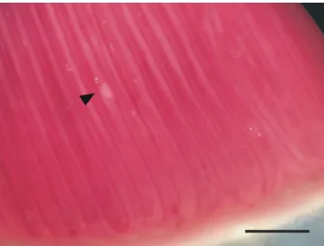
Molnár (2002) divided the formation of gill-located myxosporean
plasmodia into three types: (1) lamellar; (2) filamental; and
(3) gill arch. Among these, the filamental type is subdivided into
four types: (1) vascular; (2) epithelial; (3) intrachondral; and
(4) basifilamental. In the present study, the
H. friderici
plasmodia
developed on the filamental epithelium of the gills and deformed
the gill filaments (Figure 3).
The prevalence of
H. friderici
in piau was 24%. This was close
to the 30% reported by Casal et al. (2003), considering all the
infected organs of
L
.
friderici.
However, in fish from the Mogi
Guaçú River, infection was only observed in the gill filaments.
Furthermore, these results corroborated data from other studies
conducted in South America in which species of
Henneguya
were
found at the same infection site (NALDONI et al., 2009, 2014).
These supplementary data on the morphology, 18S rDNA
sequencing and phylogeny of
H. friderici
may facilitate accurate
diagnoses and better understanding of the phylogenetic relationships
of this parasite. Fiala (2006) indicated that host preference is very
important and that myxosporean species could group together
according to fish host species. Although host geographical
origin is particularly important, tissue tropism in myxosporean
evolution has also been revealed in numerous phylogenetic studies
(ANDREE et al., 1999; KENT et al., 2001; ESZTERBAUER,
2004; FIALA, 2006).
Acknowledgements
To M.Sc. Júlio Cesar C. de Aguiar (CEPTA/IBAMA) for
assistance in the development of the work. Letícia G. P. Vidal was
supported by a Doctoral fellowship from CNPq (Conselho Nacional
de Desenvolvimento Científico e Tecnológico do Brasil), and José
L. Luque was supported by a Researcher fellowship from CNPq.
References
Adriano EA, Arana S, Alves AL, Silva MRM, Ceccarelli PS, Henrique-Silva F, et al. Myxobolus cordeiroi n. sp., a parasite of Zungaro jahu (Siluriformes: Pimelodiade) from Brazilian Pantanal: morphology, phylogeny and histopathology. Vet Parasitol 2009; 162(3-4): 221-229. PMid:19372007. http://dx.doi.org/10.1016/j.vetpar.2009.03.030.
Adriano EA, Carriero MM, Maia AAM, Silva MRM, Naldoni J, Ceccarelli PS, et al. Phylogenetic and host-parasite relationship analysis
of Henneguya multiplasmodialis n. sp. infecting Pseudoplatystoma spp.
in Brazilian Pantanal wetland. Vet Parasitol 2012; 185(2-4): 110-120. PMid:22051071. http://dx.doi.org/10.1016/j.vetpar.2011.10.008. Andree KB, Székely C, Molnár K, Gresoviac SJ, Hedrick RP. Relationships among members of the Genus Myxobolus (Myxozoa: Bivalvidea) based on small subunit ribosomal DNA sequence. J Parasitol 1999; 85(1): 68-74. PMid:10207366. http://dx.doi.org/10.2307/3285702.
Azevedo C, Rocha S, Matos P, Matos E, Oliveira E, Al-Quraishy S, et al. Morphology and phylogeny of Henneguya jocu n. sp. (Myxosporea, Myxobolidae), infecting the gills of the marine fish Lutjanus jocu.
Eur J Protistol 2014; 50(2): 185-193. PMid:24457131. http://dx.doi.
org/10.1016/j.ejop.2013.12.002.
Barassa B, Adriano EA, Cordeiro NS, Arana S, Ceccarelli PS. Morphology and host – parasite interaction of Henneguya azevedoi n. sp., parasite of gills of Leporinus obtusidens from Mogi Guaçu River, Brazil. Parasitol Res 2012; 110(2): 887-894. PMid:21842391. http://dx.doi.org/10.1007/ s00436-011-2571-5.
Bartholomew JL, Whipple MJ, Stevens DG, Fryer JL. The life cycle
of Ceratomyxa shasta, a Myxosporean parasite of salmonids, requires a
freshwater polychaete as an alternate host. J Parasitol 1997; 83(5): 859-868. PMid:9379291. http://dx.doi.org/10.2307/3284281.
Batueva MD, Katokhin AV, Pronina SV, Pronin NM. Supplementary studies and molecular data on Henneguya cerebralis Pronin, 1972 (Myxozoa: Myxosporea), a parasite from Kosogol grayling Thymallus arcticus nigrescens in Mongolia. Parasitol Int 2013; 62(6): 530-534. PMid:23933262. http:// dx.doi.org/10.1016/j.parint.2013.07.014.
Capodifoglio KRH, Adriano EA, Silva MRM, Maia AAM. Supplementary data of Henneguya leporinicola (Myxozoa, Myxosporea) a parasite of
Leporinus macrocephalus from fish farms in the state of São Paulo, Brazil.
Acta Parasitol 2015; 60(3): 451-458. PMid:26204182. http://dx.doi.
org/10.1515/ap-2015-0062.
Carriero MM, Adriano EA, Silva MR, Ceccarelli PS, Maia AA. Molecular phylogeny of the Myxobolus and Henneguya genera with several new South American species. PLoS One 2013; 8(9): e73713. PMid:24040037. http:// dx.doi.org/10.1371/journal.pone.0073713.
Casal G, Matos E, Azevedo C. Light and electron microscopic study of the myxosporean, Henneguya friderici n. sp. from the Amazonian teleostean fish, Leporinus friderici.Parasitol 2003; 126(4): 313-319. PMid:12741510. http://dx.doi.org/10.1017/S0031182003002944.
Darriba D, Taboada GL, Ramón D, Posada D. jModelTest 2: more models, new heuristics and high-performance computing. Nat Methods 2012; 9(8): 772. PMid:22847109. http://dx.doi.org/10.1038/nmeth.2109. Figure 3. Plasmodia of Henneguya friderici infecting the gill filaments
Eiras JC. Synopsis of the species of the genus Henneguya Thélohan, 1892 (Myxozoa: Myxosporea: Myxobolidae). Syst Parasitol 2002; 52(1): 43-54. PMid:12023561. http://dx.doi.org/10.1023/A:1015016312195. Eiras JC, Adriano EA. A checklist of new species of Henneguya Thélohan, 1892 (Myxozoa: Myxosporea, Myxobolidae) described between 2002 and 2012. Syst Parasitol 2012; 83(2): 95-104. PMid:22983797. http:// dx.doi.org/10.1007/s11230-012-9374-7.
Eiras JC, Malta JC, Varela A, Pavanelli GC. Henneguya schizodon n. sp. (Myxozoa, Myxobolidae), a parasite of the Amazonian Teleost fish Schizodon
fasciatus (Characiformes, Anostomidae). Parasite 2004; 11(2): 169-173.
PMid:15224578. http://dx.doi.org/10.1051/parasite/2004112169. Eiras JC, Takemoto RM, Pavanelli GC. Henneguya caudicula n. sp. (Myxozoa, Myxobolidae) a parasite of Leporinus lacustris (Osteichthyes, Anostomidae) from the high Paraná River, Brazil, with a revision of
Henneguya spp. infecting South American fish. Acta Protozool 2008;
47(2): 149-154.
Eszterbauer E. Genetic relationship among gill-infecting Myxobolus species (Myxosporea) of cyprinids: molecular evidence of importance of tissue-specificity. Dis Aquat Organ 2004; 58(1): 35-40. PMid:15038449. http://dx.doi.org/10.3354/dao058035.
Eszterbauer E, Marton S, Rácz OZ, Letenyei M, Molnár K. Morphological and genetic differences among actinosporean stages of fish-parasitic myxosporeans (Myxozoa): difficulties of species identification. Syst Parasitol 2006; 65(2): 97-114. PMid:16676228. http://dx.doi.org/10.1007/ s11230-006-9041-y.
Fiala I. The phylogeny of Myxosporea (Myxozoa) based on small subunit ribosomal RNA gene analysis. Int J Parasitol 2006; 36(14): 1521-1534. PMid:16904677. http://dx.doi.org/10.1016/j.ijpara.2006.06.016. Froese R, Pauly D, editors. FishBase [online]. 2016. [cited 2016 Nov 20]. Available from: www.fishbase.org
Griffin MJ, Khoo LH, Torrans L, Bosworth BG, Quiniou SM, Gaunt PS, et al. New data on Henneguya pellis (Myxozoa: Myxobolidae), a parasite of blue catfish Ictalurus furcatus.J Parasitol 2009; 95(6): 1455-1467. PMid:19575542. http://dx.doi.org/10.1645/GE-2106.1.
Griffin MJ, Pote LM, Wise DJ, Greenway TE, Mauel MJ, Camus AC. A novel Henneguya species from channel catfish described by morphological, histological, and molecular characterization. J Aquat Anim Health 2008; 20(3): 127-135. PMid:18942589. http://dx.doi.org/10.1577/H07-001.1. Guindon S, Gascuel O. A simple, fast, and accurate algorithm to estimate large phylogenies by maximum likelihood. Syst Biol 2003; 52(5): 696-704. PMid:14530136. http://dx.doi.org/10.1080/10635150390235520. Hervio DML, Kent ML, Khattra J, Sakanari J, Yokoyama H, Devlim RH. Taxonomy of Kudoa species (Myxosporea), using a small-subunit ribosomal DNA sequence. Can J Zool 1997; 75(12): 2112-2119. http:// dx.doi.org/10.1139/z97-846.
Holzer ASC, Sommerville RW, Wootten R. Molecular relationships and phylogeny in a community of myxosporeans and actinosporeans based on their 18S rDNA sequences. Int J Parasitol 2004; 34(10): 1099-1111. PMid:15380681. http://dx.doi.org/10.1016/j.ijpara.2004.06.002. Holzer AS, Sommerville C, Wootten R. Molecular studies on the seasonal occurrence and development of five myxozoans in farmed Salmo trutta
L. Parasitology 2006; 132(2): 193-205. PMid:16216135. http://dx.doi.
org/10.1017/S0031182005008917.
Huelsenbeck JP, Ronquist F. MrBayes: Bayesian inference of phylogenetic trees. Bioinformatics 2001; 17(8): 754-755. PMid:11524383. http:// dx.doi.org/10.1093/bioinformatics/17.8.754.
Iwanowicz LR, Iwanowicz DD, Pote LM, Blazer VS, Schill WB. Morphology and 18S rDNA of Henneguya gurlei (Myxosporea) from
Ameiurus nebulosus (Siluriformes) in North Carolina. J Parasitol 2008;
94(1): 46-57. PMid:18372621. http://dx.doi.org/10.1645/GE-1092.1. Kageyama T, Yanagida T, Ohara K, Yokoyama H. Henneguya pseudorhinogobii n. sp. (Myxozoa: Myxosporea) parasitizing the gills of the freshwater goby
Rhinogobius sp. or from the Nagara River and redescription of Henneguya
rhinogobii.Fish Sci 2009; 75(3): 657-663. http://dx.doi.org/10.1007/
s12562-009-0096-y.
Kallert DM, Eszterbauer E, El-Matbouli M, Erséus C, Haas W. The life cycle of Henneguya nuesslini Schuberg & Schro 1905 (Myxozoa) involves a triactinomyxon-type actinospore. J Fish Dis 2005; 28(2): 71-79. PMid:15705152. http://dx.doi.org/10.1111/j.1365-2761.2004.00599.x. Kent ML, Andree KB, Bartholomew JL, El-Matbouli M, Desser SS, Devlin RH, et al. Recent advances in our knowledge of the Myxozoa.
J Eukaryot Microbiol 2001; 48(4): 395-413. PMid:11456316. http://
dx.doi.org/10.1111/j.1550-7408.2001.tb00173.x.
Kent ML, Khattra J, Hedrick RP, Devlin RH. Tetracapsula renicola n. sp. (Myxozoa: Saccosporidae); the pkx myxozoan: the cause of proliferative kidney disease of salmonid fishes. J Parasitol 2000; 86(1): 103-111. http://dx.doi.org/10.1645/0022-3395(2000)086[0103. PMid:10701572. Kudo R. Studies on Myxosporidia: a synopsis of genera and species of Myxosporidia. Illinois Biol Monogr 1919; 5(3): 1-265.
Kudo R. A taxonomic consideration of Myxosporidia. Trans Am Microsc Soc 1933; 52(3): 195-216. http://dx.doi.org/10.2307/3222254. Li Y, Zhang Y, Siriguleng, Sato H. Siriguleng, Hiroshi S. Henneguya doneci (Myxosporea: Bivalvulida) in the gill filaments of Prussian carp Carassius
gibelio (Bloch) from the upper Yellow River running through Inner
Mongolia, China. J Vet Med Sci 2015; 77(8): 1001-1005. PMid:25843612. http://dx.doi.org/10.1292/jvms.14-0666.
Lin D, Hanson LA, Pote LM. Small subunit ribosomal RNA sequence
of Henneguya exilis (Class Myxosporea) identifies the actinosporean
stage from an oligochaete host. J Eukaryot Microbiol 1999; 46(1): 66-68. PMid:10188262. http://dx.doi.org/10.1111/j.1550-7408.1999.tb04585.x. Lom J, Arthur JR. A guideline for the preparation of species descriptions in Myxosporea. J Fish Dis 1989; 12(2): 151-156. http://dx.doi. org/10.1111/j.1365-2761.1989.tb00287.x.
Lom J, Dyková I. Myxozoan genera: definition and notes on taxonomy, life-cycle terminology and pathogenic species. Folia Parasitol (Praha) 2006; 53(1): 1-36. PMid:16696428. http://dx.doi.org/10.14411/fp.2006.001. Martins ML, Souza VN, Moraes JRE, Moraes FR. Gill infection of Leporinus
macrocephalus Garavello & Britski, 1988 (Osteichthyes: Anostomidae)
by Henneguya leporinicola n. sp. (Myxozoa: Myxobolidae). Description,
histopathology and treatment. Rev Bras Biol 1999; 59(3): 527-534. PMid:10765464. http://dx.doi.org/10.1590/S0034-71081999000300018. Milanin T, Maia AAM, Silva MR, Carriero MM, Adriano EA. Molecular phylogeny and ultrastructure of Myxobolus cf. cuneus, a parasite of patinga hybrid and Henneguya pseudoplatystoma, a parasite of pintado hybrid.
Acta Parasitol 2015; 60(3): 442-450. PMid:26204181. http://dx.doi.
org/10.1515/ap-2015-0061.
Molnár K. Comment on the host, organ and tissue specificity of fish myxosporeans and on the types of their intrapiscine development. Parasitol Hung 1994; 27: 5-20.
Molnár K. Site preference of fish myxosporeans in the gill. Dis Aquat
Organ 2002; 48(3): 197-207. PMid:12033706. http://dx.doi.org/10.3354/
Vidal, L.P.; Luque, J.L. Braz. J. Vet. Parasitol. 88
Moreira GSA, Adriano EA, Silva MRM, Ceccarelli PS, Maia AAM. The morphological and molecular characterization of Henneguya rotunda n. sp., a parasite of the gill arch and fins of Salminus brasiliensis from the Mogi Guaçú River, Brazil. Parasitol Res 2014a; 113(5): 1703-1711. PMid:24535737. http://dx.doi.org/10.1007/s00436-014-3815-y. Moreira GSA, Adriano EA, Silva MRM, Ceccarelli OS, Maia AAM. Morphology and 18S rDNA sequencing identifies Henneguya visibilis n. sp., a parasite of Leporinus obtusidens from Mogi Guaçu River, Brazil.
Parasitol Res 2014b; 113(1): 81-90. PMid:24100607. http://dx.doi.
org/10.1007/s00436-013-3629-3.
Müller MI, Adriano EA, Ceccarelli PS, Silva MRM, Maia AAM, Ueta MT. Prevalence, intensity, and phylogenetic analysis of Henneguya piaractus
and Myxobolus cf. colossomatis from farmed Piaractus mesopotamicus in
Brazil. Dis Aquat Organ 2013; 107(2): 129-139. PMid:24334355. http:// dx.doi.org/10.3354/dao02668.
Naldoni J, Arana S, Maia AAM, Ceccarelli PS, Tavares LER, Borges FA, et al.
Henneguya pseudoplatystoma n. sp. causing reduction in epithelial area of
gills in the farmed pintado, a South American catfish: Histopathology and ultrastructure. Vet Parasitol 2009; 166(1-2): 52-59. PMid:19695782. http://dx.doi.org/10.1016/j.vetpar.2009.07.034.
Naldoni J, Maia AAM, Silva MRM, Adriano EA. Henneguya cuniculator sp. nov., a parasite of spotted sorubim Pseudoplatystoma corruscans in the São Francisco Basin, Brazil. Dis Aquat Organ 2014; 107(3): 211-221. PMid:24429472. http://dx.doi.org/10.3354/dao02685.
Naldoni J, Zatti SA, Capodifoglio KRH, Milanin T, Maia AAM, Silva MRM, et al. Host-parasite and phylogenetic relationships of Myxobolus
filamentum sp. n. (Myxozoa: Myxosporea), a parasite of Brycon orthotaenia
(Characiformes: Bryconidae) in Brazil. Folia Parasitol (Praha) 2015; 62(1): 4-11. http://dx.doi.org/10.14411/fp.2015.014. PMid:25960558. Okamura B, Gruhl A, Bartholomew JL. An introduction to Myxozoan evolution, ecology and development. In: Okamura B, Gruhl A, Bartholomew
JL. Myxozoan evolution, ecology and development. Heidelberg: Springer;
2015. p. 1-20. http://doi.org/10.1007/978-3-319-14753-6
Pote LM, Hanson LA, Shivaji R. Small subunit ribosomal RNA sequences link the cause of proliferative gill disease in channel catfish to Henneguya n. sp. (Myxozoa: Myxosporea). J Aquat Anim Health 2000; 12(3): 26-34. http://dx.doi.org/10.1577/1548-8667(2000)012<0230:SSRRSL>2.0.CO;2. Rosser TG, Griffin MJ, Quiniou SMA, Khoo LH, Pote LM. 18S rRNA gene sequencing identifies a novel species of Henneguya parasitizing the
gills of the channel catfish (Ictaluridae). Parasitol Res 2014; 113(12): 4651-4658. PMid:25270236. http://dx.doi.org/10.1007/s00436-014-4156-6. Rosser TG, Griffin MJ, Quiniou SMA, Khoo LH, Greenway TE, Wise DJ, et al. Small subunit ribosomal RNA sequence links the myxospore stage of Henneguya mississippiensis n. sp. from channel catfish Ictalurus punctatus to an actinospore released by the benthic oligochaete Dero
digitata.Parasitol Res 2015; 114(4): 1595-1602. PMid:25716821. http://
dx.doi.org/10.1007/s00436-015-4345-y.
Siddall ME, Martin DS, Bridge D, Desser SS, Cone DK. The demise of a Phylum of Protists: Phylogeny of Myxozoa and other parasitic Cnidaria. J Parasitol 1995; 81(6): 961-967. PMid:8544072. http:// dx.doi.org/10.2307/3284049.
Smothers JF, von Dohlen CD, Smith LH Jr, Spall RD. Molecular evidence that the myxozoan Protists are Metazoans. Science 1994; 265(5179): 1719-1721. PMid:8085160. http://dx.doi.org/10.1126/science.8085160. Whipps CM, Adlard RD, Bryant MS, Lester RGJ, Findlav V, Kent ML. First report of three Kudoa species from eastern Australia: Kudoa
thyrsites from mahi mahi (Coryphaena hippurus), Kudoa amamiensis
and Kudoa minithyrsites n. sp. from sweeper (Pempheris ypsilychnus).
J Eukaryot Microbiol 2003; 50(3): 215-219. PMid:12836879. http://
dx.doi.org/10.1111/j.1550-7408.2003.tb00120.x.
Whipps CM, Murray KN, Kent ML. Occurrence of a Myxozoan parasite Myxidium streisingeri n. sp. in Laboratory Zebrafish Danio
rerio.J Parasitol 2015; 101(1): 86-90. PMid:25277837. http://dx.doi.
org/10.1645/14-613.1.
Ye LT, Li WX, Wu SG, Wang GT. Supplementary studies on Henneguya
doneci Schulman, 1962 (Myxozoa: Myxosporea) infecting the gill filaments
of Carassius auratus gibelio (Bloch) in China: histologic, ultrastructural, and
molecular data. Parasitol Res 2012; 110(4): 1509-1516. PMid:21989578. http://dx.doi.org/10.1007/s00436-011-2655-2.
Yokoyama H, Urawa S, Grabner D, Shirakashi S. Henneguya cartilaginis n. sp. (Myxozoa: Myxosporea) in the head cartilage of masu salmon
Oncorhynchus masou masou. Parasitol Int 2012; 61(4): 594-598.
PMid:22664475. http://dx.doi.org/10.1016/j.parint.2012.05.013. Zhang J, Mo X, Li N, Chen W, Yang C. Supplementary description of Henneguya zikawiensis Sikama, 1938 and its molecular phylogeny. J
Neijiang Norm Univ 2015; 35(4): 42-46. http://dx.doi.org/10.13603/j.