GROWTH INHIBITION AND MORPHOLOGICAL ALTERATIONS OF TRICHOPHYTON RUBRUM INDUCED BY
ESSENTIAL OIL FROM CYMBOPOGON WINTERIANUS JOWITT EX BOR
Fillipe de Oliveira Pereira1*, Paulo Alves Wanderley2, Fernando Antônio Cavalcanti Viana3, Rita Baltazar de Lima4, Frederico Barbosa de Sousa5, Edeltrudes de Oliveira Lima1.
1
Laboratório de Micologia, Departamento de Ciências Farmacêuticas, Centro de Ciências da Saúde, Universidade Federal da
Paraíba, João Pessoa, PB, Brasil; 2Departamento de Agricultura, Centro de Formação de Tecnólogos, Universidade Federal da
Paraíba, Bananeiras, PB, Brasil; 3Horto de Plantas Medicinais, Laboratório de Tecnologia Farmacêutica, Centro de Ciências da
Saúde, Universidade Federal da Paraíba, João Pessoa, PB, Brasil; 4Laboratório de Botânica, Departamento de Sistemática e
Ecologia, Centro de Ciências Exatas e da Natureza, Universidade Federal da Paraíba, João Pessoa, PB, Brasil; 5Laboratório de
Microscopia e Imagem Biológica, Centro de Ciências da Saúde, Universidade Federal da Paraíba, João Pessoa, PB, Brasil;
Submitted: September 17, 2009; Returned to authors for corrections: March 05, 2010; Approved: November 04, 2010.
ABSTRACT
Trichophyton rubrum is one of the most common fungi causer of dermatophytosis, mycosis that affect
humans and animals around the world. Researches aiming new products with antifungal activity become
necessary to overcome difficulties on treatment of these infections. Accordingly, this study aimed to
investigate the antifungal activity of essential oil from Cymbopogon winterianus against the dermatophyte T.
rubrum. The antifungal screening was performed by solid medium diffusion method with 16 T. rubrum
strains, minimum inhibitory concentration (MIC) and minimum fungicide concentration (MFC) were
determined using the microdilution method. The effects on mycelial dry weight and morphology were also
observed. Screening showed essential oil in natura inhibited all the tested strains, with inhibition zones
between 24-28 mm diameter. MIC50 and MIC90 values of the essential oil were 312 µg/mL for nearly all the
essayed strains (93.75 %) while the MFC50 and MFC90 values were about eight times higher than MIC for all
tested strains. All tested essential oil concentrations managed to inhibit strongly the mycelium development.
Main morphological changes on the fungal strains observed under light microscopy, which were provided by
the essential oil include loss of conidiation, alterations concerning form and pigmentation of hyphae. In the
oil presence, colonies showed folds, cream color and slightly darker than the control, pigment production was
absent on the reverse and with evident folds. It is concluded that C. winterianus essential oil showed activity
against T. rubrum. Therefore, it could be known as potential antifungal compound especially for protection
against dermatophytosis.
Key words:Trichophyton, essential oil, Cymbopogon.
INTRODUCTION
Dermatophytes are a fungi group with capacity to invade
keratinized tissues (skin, hair and nails) of humans and other
animals producing dermatophytosis. The incidence of this
infection has increased over recent years, particularly in
immunocompromissed patients and it is considered the most
common and widespread infectious disease worldwide (39,
41).
Trichophyton rubrum is a worldwide pathogen causing
various superficial infections, accounting for at least 60 % of
dermatophytosis, such as tinea capitis, tinea corporis, tinea
inguinalis, tinea manuum, tinea unguium and tinea pedis (15).
Researchers from South and North America and Europe
mention this microorganism as one of the most commonly
isolated in cases of dermatophytosis in these regions and with
recognized local resistance to therapy (25, 30, 31, 40). In
Brazil, it also remains the most frequently isolated (7, 20).
Dermatophytosis treatment has been cause of great
concern among researchers around the world. This fact is
justified by increasing prevalence of these diseases in the
world, extensive use of antifungal agents and consequent
emergence of fungal strains resistant to the main drugs
commonly used in clinical therapy (24). For overcome this
problem, the development of new antifungal products is
necessary. In this situation, interest in plants with antifungal
properties has increased as a consequence of current problems
associated with the antifungal therapy. Since antiquity,
medicinal plants have been used to treat common infectious
diseases and the essential oils of these plants have been widely
used in the treatment of infectious pathologies in many body
parts including skin (23, 29).
Cymbopogon winterianus Jowitt ex Bor (Poaceae)
popularly known as “citronella” or “java citronella”, is a
perennial herb which cultivated in India and Brazil. This
medicinal plant is used by population as repellent, antimycotic
and acaricide activities (26).
This study was carried out aiming to investigate the
antifungal activity of the essential oil of C. winterianus from
Bananeiras city in Paraiba state-Brazil, analyzing its effects on
growth and morphology of some T. rubrum strains known as
one of the most important species causing dermatophytosis.
MATERIAL AND METHODS
Plant material
Leaves of C. winterianus were collected from the
Formation Center for Technicians in Federal University of
Paraiba (campus IV), Bananeiras city, Paraiba state-Brazil, in
February 2007. Botanical identification of the plant was
obtained and a voucher sample (JPB 41387) has been deposited
at the Herbarium Professor Lauro Pires Xavier, in the Federal
University of Paraiba.
Essential oil
Fresh leaves of C. winterianus were cut into pieces and
subjected to water-distillation using a Clevenger apparatus. The
essential oil obtained (density = 0.8790 g/mL) was kept in
amber bottle flask and maintained at temperature lower than
4°C. The oil emulsion used in antifungal essays were obtained
according to following procedure: 34 µL of essential oil, 10 µL
of tween 80 and q.s.f. 3 mL of sterile distilled water were
added in a sterile tube and shaken for 3 minutes using a vortex
instrument, thus obtaining a stock emulsion with 10000 µg/mL
final concentration. Seriate dilutions were performed in
proportion of two in order to obtain emulsions from 5000 – 5
µg/mL.
Test fungal strains
Dermatophytes used in antifungal essays were obtained
from the collection of Mycology Laboratory (LM),
Pharmaceutical Science Department, Health Science Center,
Federal University of Paraiba. They included these following
LM333, LM422, LM600, LM 629, LM640, LM710, LM713,
LM720, LM722 and LM730. Quality control T. rubrum strain
(ATCC 1683) was also included. Fungi were maintained in
potato dextrose agar (PDA) (Difco®) at 28°C and 4°C until
tests procedures.
Inoculum preparation
Stock inoculums suspensions of T. rubrum strains were
prepared from 10-day culture in PDA at 28°C to induce
sporulation. Fungal colonies were covered with 5 mL of sterile
saline solution (NaCl 0,85 % w/v), the surface gently scraped
with a sterile loop and this resultant mixture of fungal units
was transferred to a sterile tube. The turbidity of the final
inoculum was standardized according to McFarland scale 0.5
tube and adjusted for presenting the fungal population of 106
colony former units (CFU). The confirmation of the inoculum
quantification was made by plating 0.01 ml of inoculum
suspension in Sabouraud dextrose agar (SDA). The plates were
incubated at 28°C and were examined daily for the presence of
fungal colonies which were counted as soon as growth became
visible (3, 18, 33).
Antifungal activity screening
Solid medium diffusion method using filter paper discs
was used for antifungal activity screening as preliminary test
for evaluating potential activity of the essential oil (1, 18).
Sterile Sabouraud dextrose agar (SDA) (Difco®) was prepared
and distributed uniformly into sterile petri plates, where 1 mL
of the fungal suspension was previously inoculated.
Afterwards, filter paper discs (diameter 6 mm) were soaked
with 20 µl of essential oil and placed on the surface of the
inoculated agar. Plates have been incubated at 28°C for 8 days.
At the end of the incubation period, fungal growth inhibition
zone diameter was measured and expressed in millimeters. It
was considered positive that antifungal activity when the
geometric mean values of growth inhibition zone in two
independent essays were equal or higher than 10 mm diameter
(19, 38).
Determination of minimum inhibitory concentration (MIC)
The MIC values were determined for the fungi strains
which were sensitive to the essential oil in solid medium
diffusion assay. Broth microdilution bioassay was used to
determine the MIC of C. winterianus essential oil (22, 32). For
this, it was used 96-well plates (flatted bottom) and cap. The
96-well plates were prepared by dispensing 100 µL of doubled
concentrated Sabouraud dextrose broth (SDB) (Difco®) into
each well. 100 µL from the stock emulsion of essential oil was
added into the first wells. Then, 100 µL from their serial
dilutions were transferred into consecutives wells, excluding
the last ones. The last well contained 100 µL of broth
inoculated with fungal inoculum to confirm the cell viability
(viability control). At the same way positive control was
carried out with standard antifungal using Ketoconazole
(Sigma-Aldrich®). In both cases, the highest concentration
(5000 µg/mL) was added into the first wells and the lowest
concentration (5 µg/mL) was added into the penultimate wells.
Sensitivity control of the essayed strains to the tween 80
without essential oil was carried out by microdilution. A 100
µL of 5% tween 80 in broth was added into wells and 10 µL of
fungal suspensions were inoculated into each respective well.
Also, a sterility control was performed to verify whether the
broth used in antifungal essay was contaminated before test
procedures. For this, 100 µL of broth was dispensed into a
well, without both essential oil and inoculum.
All plates were aseptically sealed followed by mixing on
plate shaker (300 rpm) for 30 seconds, incubated at 28°C being
read after 5 days incubation. The MIC values were determined
by visual inspection of the growth inhibition of each well
compared with that of the control (without drugs) well. MIC
was defined as the lowest essential oil concentration able to
inhibit 100 % the fungal growth. The test was performed in
duplicate and the geometric mean values were calculated.
isolates were inhibited and MIC90 is the MIC at which 90 % of
the isolates were inhibited (34).
Determination of the minimum fungicide concentration
(MFC)
MFC was determined the microdilution method to verify
if the inhibition was reversible or permanent (12, 28). Aliquot
of 20 µL from the wells that did not show growth in MIC
procedure was transferred to 96-well plates previously
prepared with 100 µL of SDB. The plates were aseptically
sealed followed by mixing on plate shaker (300 rpm) for 30
seconds, incubated at 28°C and being read 5 days of
incubation. The test was performed in duplicate and the
geometric mean values were calculated. MFC was defined as
the lowest essential oil concentration in which no visible
growth occurred when subcultured on the 96-well plates
contained broth without antifungical products.
Effects on mycelial growth
Analysis of the interference of the essential oil of C.
winterianus on mycelial growth was performed by determining
the dry mycelial weight of T. rubrum ATCC 1683 (28, 35). For
determination of the oil effects on the dry weight, flasks
containing 2500, 625, 312 and 156 µg/mL of essential oil in
SDB medium were inoculated with suspension of test T.
rubrum strains. In the correspondent control, the same amount
of essential oil was replaced by distilled water. The system was
incubated at 28°C for 15 days. From the sixth day of
incubation, micelia dry weight was determined every 3 days.
Flasks containing mycelia were filtered through Whatman filter
no. 1 (particle retention: 11 µm) and then washed with distilled
water. The mycelia were dried at 60°C for 6 h and then at 40°C
over night. The filter paper containing dry mycelia from two
independent essays were weighed and the mean values
obtained. Percent growth inhibition based on the dry weight, at
each time of analysis, was calculated as:
[(control weight – sample weight) x 100] / Control weight.
Microscopic study of fungal morphology
The evaluation on the micromorphological alterations
caused by the essential oil of C. winterianus in T. rubrum
ATCC 1683 was performed in duplicate by the slide culture
technique. At first, a SDA block containing the essential oil
(78, 156, 312 L/mL) was transferred to the center of a glass
slide in a Petri dish with a piece of filter paper. Subsequently, a
mycelium sample was taken from the periphery of a
10-days-old fungal colony grown on PDA and inoculated onto the
center of the sides of the agar medium block. About 1.5 mL of
sterile water was placed in the Petri dish bottom and incubated
at 28ºC for 5 days. After incubation, slides with reproductive
structures were fixed in lacto-phenol–cotton blue stain and
observed under the optical microscope (Olympus® model
CH-30) at 400x to examine morphological abnormalities. Structural
changes observed in optical microscopy in test essays were
recorded and compared with the normal growth found in the
control experiment. Control essay without essential oil was
tested in the same way (14, 16).
Macroscopic study of fungal morphology
The evaluation on the macromorphological alterations
caused by the essential oil of C. winterianus was performed by
macroscopic examination of cultures of T. rubrum ATCC 1683
in duplicate. Petri dishes were initially prepared with 15 mL of
medium SDA added of 78, 156 and 312 µg/mL the essential oil
and the whole system was homogenized. After solidification of
the culture medium, fungi were inoculated onto each agar
surface. Control essay without essential oil was tested in the
same way. After assembling the dishes, the whole system was
incubated at 28 °C for 10 days. Afterwards, analysis were
finally performed based on the visual observation of colonies
of control and test experiments characterizing them by
appearance, texture and surface color, in addition to aspects of
colonies’ color reverse (1, 9).
Statistical analysis
oil on mycelial weight, was performed to determine
statistically significant differences (P <0.05) employing
analysis of variance (one-way ANOVA) using Kruskal-Wallis
test followed by Dunn post-test. For this, the implementation of
statistical analysis was performed using GraphPad Prism
version 4.03 for Windows, San Diego, California, USA.
RESULTS AND DISCUSSION
Results of antifungal activity of C. winterianus essential
oil on T. rubrum strains are shown in Table 1. The essential oil
in natura had inhibitory effect on all strains tested with
inhibition zones diameters between 24-28 mm. The most
sensitive strain was T. rubrum LM63, due to the essential oil
has produced the largest inhibition zones with 28 mm diameter.
The smallest inhibition zones (24 mm) were found to T.
rubrum LM333. MIC50 and MIC90 values are also summarized
in Table 1. As it can be seen, MIC50 and MIC90 for the essential
oil was 312 µg/mL for the essayed strains. The lowest MIC
value was 156 µg/mL for T. rubrum LM63, confirming the
results found in the assessment of the fungal activity of the
essential oil, thus these strains were the most sensitive at
screening. Ketoconazole showed MIC50 and MIC90 lower than
essential oil, the values found were 19.5 µg/mL for
ketoconazole. Control results showed absence of fungal growth
inhibition by Tween 80, fungal growth in broth without
addition of essential oil and absence of fungal growth in broth
without suspensions. The essential oil fungicide effect was
stronger on T. rubrum LM63 which is shown by their lowest
MFC values (1250 µg/mL). MFC50 and MFC90 values of the oil
were ever about eight times higher than MIC values for all
strains tested (2500 µg/mL). However MFC50 and MFC90 for
ketoconazole were about thirty-two times than MIC values
(625 µg/mL).
Table 1. Antifungal activity of C. winterianus essential oil and ketoconazole against T. rubrum strains.
MIC (µg/mL) MFC50 (µg/mL)
Products inhibition
zones*
MIC50 MIC90 MFC50 MFC90
Essential oil 24-28 312 312 2500 2500
Ketoconazole - 19.5 19.5 625 625
*Range of antifungal activity screening results are showed in fungal growth inhibition zones diameters (mm); ¯ Not assessed.
It is known that medicinal plants have been source of
many drugs applied in clinical procedures. In nature, essential
oils are involved in many important actions showing an
important role in plants protection as antimicrobials and
insecticides. It has long been recognized that some essential
oils have antimicrobial properties and these characteristics are
possibly related to the function of these compounds in plants
(2, 5).
Earlier, this essential oil has demonstrated the
antimicrobial power against various microorganisms with
emphasis in protozoa, bacteria and fungi pathogenic to humans
and plants, being useful for controlling mycotic diseases in
large plantations, in addition to its activity as repellent and
insecticide (11, 13, 27, 37). However, little information about
its effect against classical important fungi involved in human
infections, including dermatophytes species such as T. rubrum
still exists. This fact support the importance of this study;
allowing to add the antifungal action against major causative
agents of dermatophytosis to the properties inherent to this
essential oil.
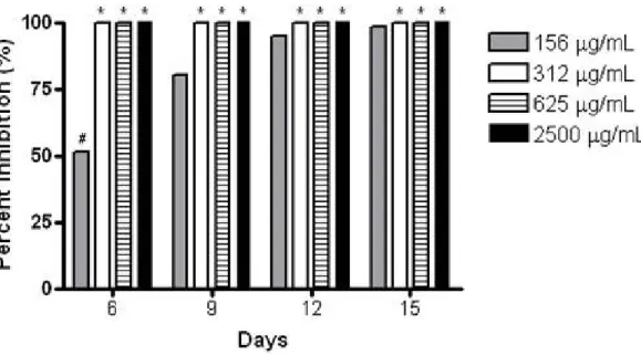
The effects of C. winterianus essential oil on mycelial
mycelial weight of T. rubrum ATCC 1683 and showed in
Figure 1. This study revealed that all tested essential oil
concentrations managed to inhibit the mycelium development.
When analyzing each concentration separately, it is confirmed
a significant statistical difference (P<0.05) between the results
obtained only by essential oil at 156 µg/mL, at the times of 6,
9, 12 and 15 days. This fact confirms that in sub-inhibitory
concentration, the impediment of mycelial growth of T. rubrum
ATCC 1683 is getting worse with increasing interaction time
between drug and fungal cells. The same information was not
observed with the other essential oil concentrations, once
fungus did not develop mycelium until the 15 days culture with
the oil, that means 100 % inhibition at all times analyzed.
Comparing the results of all concentrations tested, it was
found a significant difference between 156 and 312, 625, 2500
µg/mL values, at each time separately. The analysis on the
essential oils effects on fungal growth in function of time used
in this work has stood out among researchers around the world.
Good fungal growth of Trichophyton species, similarly to other
filamentous fungi, produce hyphae which can penetrate the
innermost skin layer and aggravate the damage in the host (17,
43). Therefore, some researchers are investigating the essential
oils potential in inhibiting mycelial growth of pathogenic fungi
due to their importance in the mycosis development.
* P<0.05 when compared with values of 156 µg/mL. # P<0.05 among values of 156 µg/mL.
Figure 1. Percentage of inhibition on dry mycelial weight of T. rubrum ATCC 1683 in the presence of several concentrations of
C. winterianus essential oil.
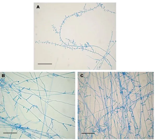
Observations on T. rubrum ATCC 1683, examined under
light microscope at 400x magnification after exposure to C.
winterianus essential oil, showed some morphological
abnormalities. Essential oil induced similar alterations in all
concentrations, however they increased in large quantities as
the oil concentration increased (Figure 2). Although the form
of T. rubrum conidia have not changed, their production was
severely impaired in all essential oil concentrations making it
rarely found from the 313 µg/mL of oil in culture medium. The
normal hyphae, with losses in pigmentation and presence of
vacuoles inside them. From the 156 µg/mL concentration, the
presence of chlamydospores was detected having production
increased in proportion to the increase of essential oil
concentration.
Microscopic examination of the control mycelium
(untreated cell) showed a regular cell structure with
homogenous cytoplasm, chlamydospores absent, abundant
conidiation, long clear septate hyphaes with lateral tear-shaped
microconidia and macroconidia absent. Chlamydospores are
usually round, increased volume and thick walled. They are
formed from hyphae under unfavorable environmental
conditions to fungal development. Maybe the excessive
production of chlamydospores was induced by the increased oil
concentration. Such phenomenon might be necessary for fungi
take their growth in the presence of this adverse component.
These modifications in the cytological structure may be related
to the interference of the essential oil with the enzymes
responsible for synthesis or maintenance of fungal cell wall as
previously cited by other researchers, impairing normal growth
and cell morphogenesis (6, 10, 16, 42).
(A) Control experiment showing typical forms of species, bar 100 m. (B) Modifications on hyphae development induced by essential oil (156 mg/mL) and presence of chlamydospores (C), bar 100 m.
Figure 2. Light microphotographs of T. rubrum ATCC 1683 mycelium growing on ASD without or with C. winterianus essential
The morphological evaluation of T. rubrum ATCC 1683
cultures was performed in SDA added of essential oil at 78,
156 and 312 µg/mL concentrations. At 78 µg/mL of essential
oil, no visible alteration was observed in the culture. However,
in presence of 156 µg/mL oil concentration, the colonies
showed folds and a small projection in the center, cream color,
slightly darker than the control, pigment production was absent
on the reverse and with evident folds. In the presence of 312
µg/mL essential oil, there was less colonies development and
same characteristics present in concentrations above were
observed. In contrast, the control experiment showed colonies
with classic features of T. rubrum, with smooth velvety white
colonies. The colony reverse presented no pigmentation. The
results in this report direct affect on disease pathogenesis, once
dermatophytosis depend on morphogenesis normal capacity of
fungi and their growth in infection locus.
Essential oils of C. winterianus leaves have more than 80
components, but citronellal, citronellol and geraniol are
terpenes cited as the mainly constituents of the oil (4, 21).
Earlier studies reported citronellal and geraniol with antifungal
activity against Aspergillus niger, Fusarium oxysporum e
Penicillium digitatum, with 100 g/ml MIC (20).
According to the scientific literature, the antifungal
property of phytochemicals found in C. winterianus essential
oil (e.g. mono-terpenes) involves inhibition of extracellular
enzymes synthesis and the disruption of cell wall structure
resulting in lack of cytoplasm, damage of integrity and
ultimately the mycelial death. Damage to microorganism’s
membrane, collapse the proton pump, cytoplasm granulation
and break down of the electron transport chain are some events
possibly related to the antifungal property of essential oils (8,
36). Several records in the scientific literature worldwide
reported that macromolecules found in fungi of which
functionality is related to growth, survival, cell morphogenesis
and virulence, are identified as promising targets for new
antifungal agents (24).
Therefore, the results found in this report may be
considered relevant and promising. In conclusion, this report
indicates that essential oils can have a practical and rational use
in the inhibition of dermatophyte growth. Even though, it is
important to develop studies with the essential oil of C.
winterianus and their isolates compounds on toxicological and
pharmacological analyses for developing new antifungal agents
for treatment of serious mycosis, especially the
dermatophytosis.
ACKNOWLEDGEMENTS
The authors are thankful to CNPq and FAPESQ-PB for
grants and fellowships.
REFERENCES
1. Adam, K.; Sivropouou, A.; Kokkini, S.; Lanaras, T.; Arsenakis, M. (1998). Antifungal activities of Origanum vulgare subsp. hirtum, Mentha spicata, Lavandula angustifólia and Slavia frticosa essential oils against
human pathogenic fungi. J. Agr. Food Chem. 46 (5), 1739-1745. 2. Bakkali, F.; Averbeck, S.; Averbeck, D.; Idioomar, M. (2008). Biological
effects of essential oils – A review. Food. Chem. Toxicol. 46 (2), 446-475.
3. Barros, M.E.S.; Santos, D.A.; Hamdan, J.S. (2006). In vitro methods for antifungal susceptibility testing of Trichophyton spp. Mycol. Res. 110 (11), 1355-1360.
4. Blank, A.F.; Costa, A.G.; Arrigoni-Blank, M.F.; Cavalcanti, S.C.H.; Alves, P.; Innecco, R.; Ehlert, P.A.D.; Sousa, I.F. (2007). Influence of season, harvest time and drying on Java citronella (Cymbopogon winterianus Jowitt) volatile oil. Braz. J. Pharmacogn. 17 (4), 557-564.
5. Burt, S. (2004). Essential oils: their antibacterial properties and potential applications in food – a review. Int. J. Food Microbiol. 94 (3), 223-253. 6. Carmo, E.S.; Lima, E.O.; Souza, E.L.; Sousa, F.B. (2008). Effect of
Cinnamomum zeylanicum Blume essential oil on the growth and
morphogenesis of some potentially pathogenic Aspergillus species. Braz. J. Microbiol. 39 (1), 91-97.
7. Costa, M.; Passos, X.S.; Souza, L.K.H.; Miranda, A.T.B.; Lemos, J.A.; Oliveira Júnior, J.G.; Silva, M.R.R. (2002). Epidemiology and etiology of dermatophytosis in Goiania, GO, Brazil. Rev. Soc. Bras. Med. Trop. 35 (1), 19-22.
of essential oil of Melaleuca alternifolia (tea tree oil). J. Appl. Microbiol. 88 (1), 170-175.
9. Daferera, D.J.; Ziogas, B.N.; Polissiou, M.G. (2003). The effectiveness of plant essentiol oils on the grouth of Botrytis cinerca, Fusarium sp and Clavibacter michiganessis. Crop Protection. 22 (1), 39-44.
10. Debilberck, V.G.; De Roques, C.G.; Bessiere, J.M.; Fonvielle, J.L.; Dargent, R. (2001). Effect of Cymbopogon nardus (L) W. Watson essential oil on the growth and morphogenesis of Aspergillus niger. C. J. Microbiol. 47 (1), 17-19.
11. De-Blasi, V.; Debrot, S.; Memoud, P.A.; Gendre, L.; Schowing, J. (1990). Amoebicidal effect of essential oils in vitro. J. Toxicol. Clin. Exp. 10 (6), 361-361.
12. Denning, D.W.; Hanson, L.H.; Perlman, A.M.; Stevens, D.A. (1992). In vitro susceptibility and synergy studies of Aspergillus species to conventional and new agents. Diag. Microbiol. Infect. Dis. 15 (1), 21-34. 13. Duarte, M.C.T.; Figueira, G.M.; Sartoratto, A.; Rehder, V.L.G.; Delarmelina, C. (2005). Anti-Candida activity of Brazilian medicinal plants. J. Ethnopharmacol. 97 (2), 305-311.
14. Frost, D.J.; Brandt, K.D.; Cugier, D.; Goldman, R.A. (1995). A whole-cell Candida albicans assay for the detection of inhibitors towards fungal cell wall synthesis and assembly. J. Antibiot. 48 (4), 306-309.
15. Gräser, Y.; Kuijpers, A.F.A.; Prersber, W.; de HOOG, G.S. (2000). Molecular Taxonomy of the Trichophyton rubrum Complex. J. Clin. Microbiol. 38 (9), 3329–3336.
16. Gunji, S.; Arima, K.; Beppu, T. (1983). Screening of antifungal antibiotics according to activities inducing morphological abnormalities. Agric. Biol. Chem. 47 (9), 2061-2069.
17. Gupta, A.K.; Ahmadm I.; Porretta, M.; Summerbell, R.C. (2003). Arthroconidial formation in Trichophyton raubitschekii. Mycoses. 46 (8), 332-338.
18. Hadacek, F.; Greger, H. (2000). Testing of antifungical natural products: methodologies, comparability of results and assay choise. Phytochem. Anal. 11 (9),137-147.
19. Lima, E.O.; Gompertz, O.F.; Giesbrecht, A.M.; Paulo, M.Q. (1993). In vitro antifungal activity of essential oil obtained from officinal plants against dermatophytes. Mycoses. 36 (9-10), 333-336.
20. Lima, E. O.; Pontes, Z.B.V.S.; Oliveira, N.M.C.; Carvalho, M.F.F.P.; Guerra, M.F.L.; Santos, J.P. (1999). Freqüência de dermatofitoses em João Pessoa-Paraíba-Brasil. An. Bras Dermatol. 74 (2), 127-132. 21. Marco, C.A.; Inneccom R.; Mattos, S.H.; Borges, S.S.; Nagao, E.O.
(2007). Características do óleo essencial de capim-citronela em função de espaçamento, altura e época de corte. Hort. Bras. 25 (3), 429-432. 22. Moreira, A.C.P.; Lima, E.O; Wanderley, P.A.; Carmo, E.S.; Souza, E.S.
(2010). Chemical composition and antifungal activity of Hyptis suaveolens (L. ) poit leaves essential oil against Aspergillus species.
Braz. J. Microbiol. 41 (1), 28-33.
23. Nunes, X.P.; Maia, G.L.A.; Almeida, J.R.G.S.; Pereira, F.O.; Lima, E.O. (2006). Antimicrobial activity of the essential oil of Sida cordifolia L. Braz. J. Pharmacogn. 16 (1), 642-644.
24. Odds, F.C.; Alistair, J.P.; Gow, B.; Gow, A.R. (2003). Antifungal agents: mechanisms of action. Trends in Microbiol. 11 (6), 272-279.
25. Padilla, A.; Sampedro, A.; Sampedro, P.; Delgado, D. (2002). Clinical and epidemiological survey of dermatophytoses in Jaen (Spain). Rev. Iberoam. Micol. 19 (1), 36-39.
26. Pandey, A.K.; Rai, M. (2003). Antimycotic potential in some naturally occurring essential oils. In: Donatella, M., Raim M.(eds). Plant-derived antimycotics: current trends and future prospects. Haworth Press,
London, England, p. 344-345.
27. Pattnaik, S.; Subramanyan, V.R.; Kole, C.R. (1996). Antibacterial and antifungal activity of ten essential oils in vitro. Microbios. 86 (349), 237-246.
28. Rasooli, I.; Abyaneh, M.R. (2004). Inhibitory effects of thyme oils on growth and aflatoxin production by Aspergillus parasiticus. Food Control. 15 (6), 479-483.
29. Rios, J.L.; Recio, M.C. (2005). Medicinal plants and antimicrobial activity. J. Ethnopharmacol. (perspective paper), 100 (1), 80-84. 30. Rubio, M.C.; Rezusta, A.; Tomás, J.G.; Ruesca, R.B. (1999). Perspectiva
micológica de los dermatofitos en el ser humano. Rev. Iberoam. Micol. 16 (1), 16-22.
31. Ruiz, L.R.B.; Zaitz, C. (2001). Dermatófitos e dermatofitoses na cidade de São Paulo no período de agosto de 1996 a julho de 1998. An. Bras. Dermatol. 76 (4), 391-401.
32. Sahin, F.; Gulluce, M.; Daferera, D.; Sokmen, A.; Polissiou, M.; Agar, G.; Ozer, H. (2004). Biological activities of the essential oil and methanol extract of Origanum vulgare ssp vulgare in the Eastern Anatolia region of Turkey. Food Control. 15 (7), 549-557.
33. Santos, D.A.; Barros, M.E.S.; Hamdan, J.S. (2006). Establishing a method of inoculum preparation for susceptibility testing of Trichophyton rubrum and Trichophyton mentagrophytes. J. Clin. Microbiol. 44 (1), 98-101.
34. Santos, D.A.; Hamdan, J.S. (2005). Evaluation of Broth Microdilution Antifungal Susceptibility Testing Conditions for Trichophyton rubrum. J. Clin. Microbiol. 43 (4), 1917-1920.
35. Sharma, N.; Tripathi, A. (2006). Effects of Citrus sinensis (L.) Osbeck epicarp essential oil on growth and morphogenesis of Aspergillus niger (L.) Van Tieghem. Microbiol. Res. 163 (3), 337-344.
36. Sikkema, J.; De Bont, J.A.M.; Poolan, B. (1995). Mechanisms of membrane toxicity of hydrocarbons. Microbiol. Rev. 59 (2), 201-222. 37. Simic, A.; Ran ic, A.; Sokovic, M.D.; Ristic, M.; Grujic-Jovanovic, S.;
Vukojevic, J.; Marin, P.D. (2008). Essential oil composition of Cymbopogon winterianus. and Carum carvi. and their antimicrobial
38. Souza, E.L.; Stamford, T.L.M.; Lima, E.O.; Trajano, V.N. (2007). Effectiveness of Origanum vulgare L. essential oil to inhibit the growth of food spoiling yeasts. Food Cont. 18 (5), 409-413.
39. Squeo, R.F.; Beer, R.; Silvers, D.; Weitzman, I.; Grossman, M. (1998). Invasive Trichophyton rubrum resembling blastomycosis infection in the immunocompromised host. J. Am. Acad. Dermatol. 39 (2.2), 379–380. 40. Valdigen, G.L.; Pereira, T.; Macedo, C.; Duarte, M.L.; Oliveira, P.;
Ludovico, P.; Souza-basto, A.; Leão, C.; Rodrigues, F. (2006). A twenty year survey of dermathophytoses in Braga, Portugal. Int. J. Dermatol. 45
(7), 822-827.
41. Weitzman, I.; Summerbell, R.C. (1995). The dermatophytes. Clin. Microbiol. Rev. 8 (2), 571-578.
42. Zambonelli, A.; Zechini D’Aurelio, A.; Bianchi, A.; Albasini, A. (1996). Effects of essential oil on phytopathogenic fungi. Phytopathol. 144 (9-10), 491-494.