ISSN: 2067-533X
INTERNATIONAL JOURNAL
OF
CONSERVATION SCIENCE
Volume 4, Issue 2, April-June 2013: 133-144 www.ijcs.uaic.ro
PRELIMINARY STUDY ON CONTROLLING
BLACK FUNGI DWELLING ON STONE MONUMENTS
BY USING A MICROWAVE HEATING SYSTEM
Oana-Adriana CUZMAN1*, Roberto OLMI2, Cristiano RIMINESI1, PieroTIANO1
1 CNR-ICVBC Institute for the Conservation and Valorization of Cultural Heritage,
Via Madonna del Piano 10, 50019, Sesto Fiorentino, Italy
2 CNR-IFAC Institute of Applied Physics “N. Carrara” – CNR,
Via Madonna del Piano 10, 50019, Sesto Fiorentino, Italy
Abstract
Microcolonial black fungi have their natural ecological niche on rocks and walls of hypogean environments, playing an important role in the deterioration of materials and aesthetical alteration of monumental stones and mortars. Three black fungi (Sarcinomyces sp., Pithomyces sp. and Scolecobasidium sp.) have been isolated from cultural assets of historical interest. These fungal strains have been used to test the microwave heating method as a new control methodology for eradicating the fungal biological growth on cultural stone artifacts. This methodology is based on a 2.45 GHz microwave electromagnetic radiation, generated by a new apparatus with an appropriate applicator. The first results showed the best dose of 65°C for three minutes, for all the investigated fungal strains. This methodology is very promising because is safety for the operator and the environment, and can be easily applied on site. The use of this method to kill biodeteriogens can avoid the application of chemicals formulates potentially dangerous for substrates and environment.
Keywords: black fungi; marble; hypogean monument; biodeterioration; microwave heating
Introduction
Blackening of artistic marbles and mortars in hypogean structures is often due to the colonization by darkly pigmented fungi containing melanin [1], the so-called black-fungi, meristematic fungi or microcolonial fungi [2]. They are slow-growing fungi that are forming cauliflower-like microcolonies [3], being real rock inhabitants [1]. They were commonly isolated from stone monuments and in rocks in the Mediterranean Basin [4, 5]. These fungi share common growth characteristics and morphological features, numerous showing a meristematic growth [6], many others having highly pleomorphic habitus and some morphologically very similar fungi can be phylogenetically distant [7]. Dematiaceous fungi are found to be polyphyletic within the Ascomycota and occur in at least three orders, namely Chaetothyriales, Dothideales and Pleosporales [2, 8].
In terms of geomicrobiology black and yeast-like fungi have their natural ecological niche on rocks [4], stones [9] and even caves [10], where the occurrence of melanised fungi was discrete and Scolecobasidium sp. has been isolated from black stains. This fungus synthesizes a characteristic melanin and it could be responsible for the black stain formation and
*
Physical control methods, such as ionizing radiation, gamma radiation and UV radiation, were applied on several work of arts [14-17], as an alternative for the conservation of stone colonized by biodeteriogens. Within these methods, the efficacy of the one based on ionizing radiations was challenged by several authors [18-20].
Another new physical controlling method of great interest is the use of the microwave radiation (non-ionizing radiation). Many authors investigated the bioeffects produced by the microwave both on organs and organ systems [21] and on different micro- [22-24] and macro-organisms [25]. The effects of microwave were attributed especially to thermal and less to the non-thermal processes. The thermal energy conversion is due to the interaction of the dipolar moment of the several molecules in living organisms with the electromagnetic waves, generating therefore enthalpic phenomena (heating), essential in destroying microorganisms. The athermal effects are related with the alteration of macromolecules conformation of the organisms, as a result of the interaction between their vibrational modes and the electromagnetic field [24, 26, 27].
The microwave are principally used in food industries, in commercial operations for sterilization and pasteurization [28-30] and in medical field [31, 32]. Only few studies are focused on the inactivation of microorganisms by microwave irradiation on building surfaces [27] and aerosols [33, 34]. These studies are especially conducted using bacteria and yeasts [22-24, 30-32] and only few of them were carried out by using fungi [27, 34-36].
The microwave heating was also tested in the cultural heritage field, as a physical method for controlling the biological growth on or within the artifacts. It has proved to be an effective means to kill woodworms and beetle in wooden objects [37-39], by increasing the temperature of the insect, in different evolution stages, to a lethal level, with low risks for the wood damage. Other authors [40] investigated the microwave action on a common wood decay fungus, Serpula lacrymans. The microwave heating method was tested also on lichens, by using a microwave system with an applicator able to concentrate the power in a thin thickness (few millimeters) keeping it at safety levels for the operators [41, 42].
This paper is a preliminary study focused on the application of microwave treatment system proposed by Olmi et al. [41], for controlling the black fungi growing on stone monuments. The aim of this research was to detect the lethal dose (time/heating treatment temperature) for different strains of black fungi. The original data obtained from laboratory experiments made on three black fungal strains isolated from artistic stones are reported in this work.
Materials and methods
Microwave heating methodology
been always maintained about 3mm. All tested fungal strains (Sarcinomyces sp, Pithomyces sp. and Scolecobasidium sp.) were cultured on malt extract agar (MEA) and the microwave radiation was performed on 10 days old colonies.
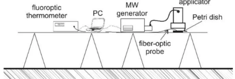
Fig 1. Set-up scheme of the microwave treatment system used for the laboratory tests.
During the microwave treatment, the temperature into MEA was controlled by a fluoroptic thermometer (LUXTRON 1000A/A) which is not sensitive to microwave radiation. The fiber-optic probe has firstly been positioned in the centre of the Petri dish where the microwave radiation is focalised, and then, it was placed near the edge of the Petri dish. There were no substantial differences between the temperatures registered in different places of the Petri dish during the same treatment, therefore, on the MEA substrate, so the fungal colony was uniformly heated in each Petri dish.
Different lethal doses (exposure time per exposure temperature for a fixed value of microwave power) were considered in order to evaluate the effectiveness of the method. The temperature exposure was maintained almost constant (standard deviation about 1.0°C) by feedback control system that adjusts the radiation power switching ON/OFF the magnetron. The temperature, measured using a fluoroptic thermometer, was recorded over time by a control unit (PC). The temperature behaviour for doses of 55°C/3min and 65°C/3min is plotted in figure 2. The treatment interval was considered to start when the selected treatment temperature was achieved, therefore immediately after the arising time interval. The increasing temperature rate depends on the characteristic of the materials (in particular, on the moisture content), and on the power level of the microwave radiation chosen for the application. The temperature distribution on one colony of black fungal strain (Fig. 3a) is shown in figure 3b. The IR image has been acquired by the IR thermo-camera (FLIR mod. B335) several seconds after the microwave applicator removal. Obviously the edges of the Petri dish are cooled faster than the center.
Fig 3. Pure colony of a black fungus (Phytomyces sp.) on MEA before the microwave heating treatment (a) and its IR image 15 seconds after the microwave treatment at 65°C for 3 minutes (b).
Several combinations (temperature and exposure time) were investigated, in order to obtain the best dose, for irreversible biological damages and lower risk of thermal stress into the support. Each Petri dish containing a pure colony (in triplicate) was submitted to microwave applications at the chosen operational conditions.
Black fungal strains from monumental assets
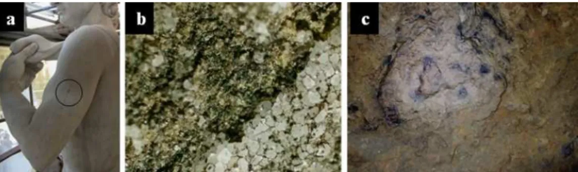
Three fungal strains Sarcinomyces sp. (Fig. 4a), Pithomyces sp. (Fig. 4b) and
Scolecobasidium sp. (Fig. 4c) were used in this study. They were isolated from three different black biological colonization (Fig. 5) developed on the following cultural assets:
Fig 4. Black fungal strains isolated from monumental assets: (a) Sarcinomyces sp. (630 x) isolated from the left arm of the copy of David of Michelangelo; (b) Pithomyces sp. (400 x) isolated from a roman marble capital belonging to Marble Museum of Carrara Municipality; (c) Scolecobasidium sp. (400 x) strain isolated from Lascaux cave and kindly
supplied by Saiz-Jimenez.
Fig 5. Biocenosis developed on the three cultural assets: (a) black spot on the left arm of the copy of David marble statue; (b) microscope observation of the sample collected from the marble capital (40x); and (c) black spots developed
A) The duplicate of the David of Michelangelo statue, located in front of the “Palazzo Vecchio” in Florence, Italy. This marble statue was sculptured by the Florentine artist Luigi Arrighetti in the first years of 1900, and placed on site in 1910. Dark lines and spots often associate with green colour are present in several places on the surface. These biocenosis (Fig. 5a) were mainly composed by cyanobacteria and microfungal stains with dominance of
Sarcinomyces sp., a black yeast like fungus, that have been isolated.
B) A fragment of a roughly sculptured roman marble capital (200 BC) coming from the Gioia marble cave, Carrara, Italy, and kept since its discovery in 2006, in the outdoor premises of Marble Museum of Carrara Municipality. A black intergranular patina (Fig. 5b) covered this marble block in few years after its outdoor exposure. The biological growth was very similar with the one observed on the copy of David statue. Pithomyces sp. was the strain isolated from this roman capitalError! Reference source not found..
C) The Lascaux cave is setting in south-western France and is famous for its Palaeolithic paintings. It has suffered, in a recent past, two main biological crises: the “green” and the “white” sickness, being the former due mainly to a proliferation of the green algae (Bracteacoccus minor), and the latter to the growth of a hyphomycete, Fusarium solani, characterised by white filaments, which colonized the cave’s sediments and walls [10]. More recently, the progressive presence of black spots has been observed in few zones of the cave. They are olivaceus to black colonies of dematiaceous hyphomycete, belonging to the genera
Scolecobasidium, isolated and kindly supplied by Saiz-Jimenez.
Morphological characterization of isolated fungi was carried out by optical microscopic examinations with a Nikon Eclipse 600 microscope.
Assessment of the microwave treatment effectiveness
The evaluation of the fungal strains vitality, after the microwave treatment, was performed both by traditional method (sowing small parts of treated colony on MEA) and by the use of a mixture of two fluorescent dyes (Plant Cell Viability Kit, Sigma Co.) according to provided protocol. Propidium iodide stains red the dead mycelium, while fluorescent stains in green the live one. The fluorescence of the samples was observed using the Nikon Eclipse 600 microscope equipped with filter cubes UV-2A (Ex. 330-380 nm, DM 400 nm, BA 420 nm) and FITC (Ex. 465-495 nm, DM 505 nm, BA 515-535 nm) for red and green fluorescence, respectively, and the images were collected with a Nikon DXM1200F digital CCD camera. At least five different parts of each fungal colony were used for each type of assessment.
Results and discussions
The microwave heating showed a clear influence on the fungal vitality, both on mycelium and fruiting bodies, being related with the applied dose (time/temperature).
Fig 6. Optical microscopical examination (by TL, FTIC and UV filters) of
Sarcinomyces sp. treated at 55°C for 3, 6 and 9 minutes.
Fig 7. Ten days old colonies of Pithomyces sp. developed on MEA starting from a small fragment of mycelia treated with the microwave heating system at 55°C for 6 minutes (a) and 9 minutes (b),
and at 65°C for 1 minutes (c) and 3 minutes (d).
for all three black fungi. All black fungi treated at 65°C for 3 minutes did not re-grown on MEA after 10 days of incubation. The combination of 3 minutes at 65°C resulted to be the best lethal dose for all tested black fungi, with respect to the case of 9 minutes at 55°C, equally effective but inducing a higher thermal stress.
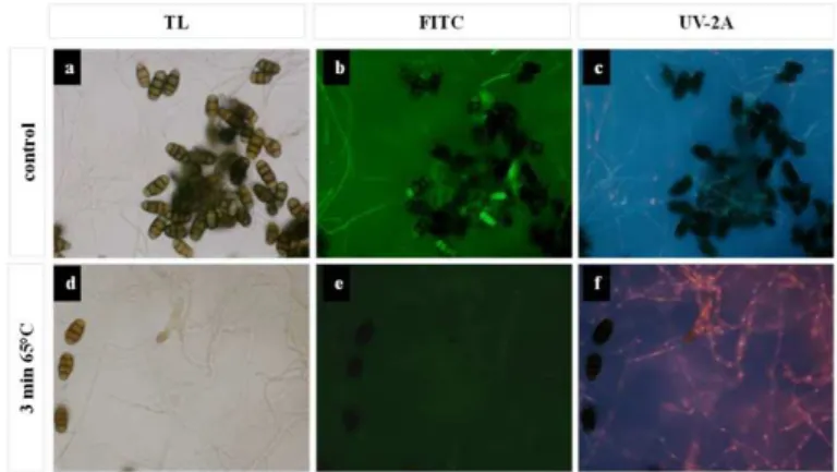
The vitality staining of each fungal strain, treated by microwave heating for 3 min at 65°C, revealed that all fungi were dead. A clearly red colour was observed for the hyphae that have been damaged by the microwave treatment, as shown in Fig. 8f (Sarcynomyces sp.), Fig. 9f (Pithomyces sp.) and Fig. 10f (Scolecobasidium sp.). Green colour of the mycelia and/or conidia can be observed for all untreated fungi (Fig.8b, Fig. 9b, Fig. 10b). The selected dose (3 minutes for 65°C) was enough for killing the mycelia of all three investigated dematiaceous fungi as well as the conidia of Pithomyces sp. were clearly harmed (Fig. 9f), allowing the penetration of the big molecule of propidium iodide dye, which is membrane impermeable for the viable cells. In fact, the living young conidia of this black fungus were fully coloured in green (Fig.9b) due to the exclusive penetration of the fluorescein dye.
Fig 8. Vitality assessment by microscopical examination of the mycelium of
Sarcinomyces sp. after the microwaves treatment at 65°C, and the untreated control observed in transmitted light (TL) and in epifluorescence, under green and ultraviolet (UV-2A) light.
Fig 9. Vitality assessment by microscopical examination of the mycelium of Pithomyces sp. after the microwaves treatment at 65°C and the untreated control observed in transmitted light (TL) and in epifluorescence, under green and ultraviolet (UV-2A) light. Dead hyphae and conidia (arrow)
Fig 10. Vitality assessment by microscopical examination of the mycelium of Scolecobasidium sp. grown on MEA and the untreated control observed in transmitted light (TL) and in epifluorescence,
under green and ultraviolet (UV-2A) light.
Generally speaking, no serious damaging effects were observed when fungal colonies were treated for 3 or 6 minutes at 55°C and for 1 minutes at 65°C. While the treatments for 9 minutes at 55°C and 3 minutes at 65°C, using microwave radiation at 2.45 GHz, have had a lethal consequence for all three types of fungal strains.
On the basis of the results achieved, the microwave technology applied in laboratory on three different black fungal strains isolated from cultural assets, seems very promising for a further in situ application on stone supports. It must be considered that the laboratory treatment conditions can slightly differ for a real in situ application, because the stone or mortar material have different properties and variable moisture content with respect to the MEA culture media. Thus the most suitable dose has to be tailored case by case, verifying the effective lethal action. The microwave system for treating microorganisms growing on monumental surfaces made on stone must have some peculiar characteristics, related both to the specific properties of the biotic agent, to the thickness of biological patina and to the type of stone material. Compared to microwave systems for disinfesting wood, which need a "deep-volume" treatment, the microwave system for treating biological patinas must be able of inducing an effective temperature increase in a controlled small thickness (1-5 mm), while the heating stress should be the lower possible for a real application, in order to limit any over-heating of the substrate, usually stone material or mortar.
Moreover, the treatment of an extended and homogeneous area, at a fixed treatment temperature, depends on the thermal characteristics of the support (moisture content, porosity) as well. This is due to the thermal diffusion which starts from the center of the microwave spot area (about 5cm in diameter). Larger colonized areas can be treated with successive contiguous treatments.
Anyway, in order to have an optimal employing of this method for controlling the biodeteriogens on stone monuments, several key points must be asserted, such as:
- the influence of microwave heating on water diffusion mechanism into the substrate, which may cause changes of the surface pattern (color variations, whitening due to salts efflorescence);
- the possible side effects on other organisms, such as the changes in the electronic properties of fungal melanin or the stimulation of the heterotrophic growth due to the presence of dead biomass that could be used as a carbon source for these secondary colonizers.
Conclusions
The laboratory experiments performed in this study showed that the proposed microwave heating system is able to uniformly heat an area of about 30 cm2 of culture medium with black fungus on it, having a total height (fungus layer + MEA layer) of maximum 5 mm. The lethal dose for 3 minutes at 65°C resulted the most suitable for the required targets. In fact, the heating at 65°C induced by the microwave radiation can affect not only the mycelium but also the fruiting bodies which are usually more resistant to any treatments, and the 3 minutes period is the shorter effective time in order to have the maximum lethal effect and to reduce the thermal stress of the substrate. The lethal dose for 9 minutes at 55°C induces higher thermal stress in the support and seems too long for its application on cultural heritage assets made on stone. Eventually the temperature can be increased when the time of application is reduced.
The microwave heating methodology proposed in this paper is fully portable, being developed for further in situ applications.
Acknowledgments
This work has been co-founded by the Regione Toscana projects TDT-BioArt (Innovative Techniques for the Diagnostics and Treatment of Biodeteriogens for Artistic/Archaeological assets) and TeCon@BC (Innovative Techniques for Conservation and Promotion of Cultural Heritage). The authors wish to thank Dr. Tulio Turchetti of the Institute for Plant Protection of CNR (IPP-CNR) for assistance and support on the analysis of fungal strains.
References
[1] E. Diakumaku, A.A. Gorbushina, W.E. Krumbein, L. Panina, S. Soukharjevski, Black fungi in marble and limestones - an aesthetical, chemical and physical problem for the conservation of monuments, Science of the Total Environment, 167(1-3), 1995, pp. 295-04.
[2] K. Sterflinger, Biodiversity and ecophysiology of yeast, Rosa CA., Péter G., Springer-Verlag, Berlin, 2006, p. 501-514.
[3] H.B. Sert, H. Sümbül, K. Sterflinger, Microcolonial fungi from antique marbles in
Perge/Side/Termessos (Antalya/Turkey), Antonie van Leeuwenhoek Journal of
Microbiology, 91(3), 2007, pp. 217–227.
communities in an urban environment (Vienna, Austria), Antonie van Leeuwenhoek Journal of Microbiology, 80, 3, 2001, pp. 275–286.
[9] E.V. Bogomolova, D.W. Minter, A new microcolonial rock-inhabiting fungus from marble in Chersonesos (Crimea, Ucraine), Mycotaxon, 86, 2003, pp. 195-204.
[10]F. Bastian, V. Jurado, A. Nováková, C. Alabouvette, C. Saiz-Jimenez, The Microbiology of Lascaux Cave. Microbiology, 156, 2010, pp. 644–652.
[11]C. Saiz-Jimenez, J.J. Ortega-Calvo, J.W. De Leeuw, The chemical structure of fungal melanins and their possible contribution to black stains in stone monuments, Science of the Total Environment, 167, 1-3, 1995, pp. 305–314.
[12]K. Sterflinger, Fungi as geological agents, Geomicrobiology Journal, 17, 2, 2000, pp. 97-124.
[13]K. Sterflinger, W.E. Krumbein, Dematiaceous fungi as a major agent of biopitting on Mediterranean marbles and limestones, Geomicrobiology Journal, 14, 3, 1997, pp. 219-230.
[14]L. Van Der Molen, J. Garty, B.W. Aardema, W. Krumbein, Growth control of algae and cyanobacteria on historical monuments by a mobile UV unit (MUVU), Studies in Conservation, 25, 2, 1980, pp. 71-77.
[15]J. Teply, C. Franek, R. Kraus, V. Cervanka, Mobile irradiator and its application in the preservation of the objects of art. International Journal of Radiation, Applications and Instrumentations, Part C. Radiation Physics and Chemistry, 28, 5-6, 1986, pp. 585-588.
[16]R. Ramière, Les contaminants biologiques des biens culturels, Muséum national d’histoire naturelle et Editions scientifiques et médicales, Elsevier SAS, Paris, 2002, p. 291-302.
[17]M. Guiomar Carneiro Tomazello, F. Wiendl , The Applicability of Gamma Radiation to the Control of Fungi in Naturally Contaminated Paper, Restaurator, 16, 2, 2009, pp. 93–99. [18]A.A. Gorbushina, K. Whitehead, T. Dornieden, A. Niesse, A. Schulte, J.I. Hedges, Black
fungal colonies as units of survival: hyphal mycosporines synthesized by rock-dwelling microcolonial fungi, Canadian Journal of Botany, 81, 2, 2003, pp. 131-138.
[19]E. Dadachova, R.A. Bryan, X. Huang, T. Moadel, A.D. Schweitzer, P. Aisen, J.D. Nosanchuk, A. Casadevall, Ionizing radiation changes the electronic properties of melanin
and enhances the growth of melanized fungi. PLoS ONE, 2, 5, 2007,
doi:10.1371/journal.pone.0000457.
[21]S.M. Michaelson, Microwave biological effect: an overview, Proceedings of the IEEE, 68, 1, 1980, pp. 40-49.
[22]G.R. Vela, J.F. Wu, Mechanism of lethal action of 2,450MHz radiation on microorganisms, Applied and Environmental Microbiology, 37, 3, 1979, pp. 550-553. [23]S. Banik, S. Bandyopadhyay, S. Ganguly, Bioeffects of microwave – a brief review,
Bioresource Technology, 87, 2, 2003, pp. 155-159.
[24]O.A. Valsechi, J. Horii, D. de F. De Angelis, The effect of microwaves on microorganisms.
Arquivos Instituto Biologico São Paolo, 71, 2004, pp. 399-404.
[25]M. Urech, B. Elcher, J. Siegenthaler, Effects of microwave and radio frequency electromagnetic fields on lichens, Bioelectromagnetics, 17, 1966, pp. 327-334.
[26]A.W.J. Dawkins, N.R.V. Nightingale, G.P. South, R.J. Sheppard, E.H. Grant, The role of water in microwave absorption by biological material with particular reference to microwave hazards, Physics in Medicine and Biology, 24, 6, 1979, pp. 1168-1176.
[27]R.L. Górny, G. Mainelis, A. Wlazło, A. Niesler, D.O. Lis, S. Marzec Viability of fungal and actinomycetal spores after microwave radiation of building materials, Annals of Agriculturals and Environmental Medicine, 14, 2, 2007, pp. 313-324.
[28]H. Schubert, M. Regier, Advances in microwave and radio frequency processing: Report from the 8th International Conference on Microwave and High Frequency Heating, Bayreuth, Germany, Sept 3-7, 2001, Willert-Porada M., Springer; 2006, p. 259-270.
[29]R.S. Chavan, S.R. Chavan, Microwave baking in food industry: a review, International Journal of Diary Science, 5, 3, 2010, pp. 113-127.
[30]A. Dumuţa-Codre, O. Rotaru, L. Giurgiulescu, F. Boltea, L. Crisan, B. Neghelea,
Preliminary researches regarding the microwaves influence on the milk microflora,
Analele Universitatii din Oradea, Fascicula Biologie, XVII, 1, 2010, pp. 103-107. [31]D.L. Dixon, L.C. Breeding, T.A. Faler, Microwave disinfection of denture base materials
colonized with Candida albicans, Journal of Prosthetic Dentistry, 81, 1, 1999, pp. 207-214.
[32]N.H. Campanha, A.C. Pavarina, I.L. Brunetti, C.E. Vergani, A.L. Machado, D.M. Spolidorio, Candida albicans inactivation and cell membrane integrity damage by microwave irradiation. Mycoses, 50, 2, 2007, pp. 140-147.
[33]J.H. Jung, J.E. Lee, C.H. Lee, S.S. Kim, B.U. Lee, Treatment of fungal bioaerosols by a high-temperature, short time process in a continuous-flow system, Applied and Environmental Microbiology, 75, 9, (2009), pp. 2742-2749.
[34]Y. Wu, M. Yao, Inactivation of bacteria and fungus aerosols using microwave irradiation, Journal of Aerosol Sci 2010; 41:682-93.
[35]T. Mężykovski, J. Bal, H. Dębiec, K. Kwarecki, Response of Aspergillus nidulans and Physarium polycephalum to microwave irradiation, Journal of Microwave Power, 15, 2, 1980, pp. 75-80.
[36]T. Ishitani, T. Kojo, S. Yanai, Effects of microwave irradiation of mold spores,
Representative Natural Food Research Institute, 38, 1981, pp. 102-106.
2008, Braga, Portugal, Gril J, University Press, Firenze, 2010, p. 148.
[41]R. Olmi, M. Bini, A. Ignesti, S. Priori, C. Riminesi, Evanescent-Field Microwave Applicators for the Treatment of Biodeteriogens on Stone Materials of Archaelogical Interest, Proceedings of XVIII RiNEm, 6th-10th September, 2010, Benevento, Italy, p. 474.
[42]R. Olmi, M. Bini, O.A. Cuzman, A. Ignesti, P. Frediani, S. Priori, C. Riminesi, P. Tiano,
Investigation of the microwave heating method for the control of biodeteriogens on cultural heritage assets, Proceedings of 10th International conference on non-destructive investigations and microanalysis for the diagnostics and conservation of cultural and environmental heritage, 13th-15th April 2011, Florence, Italy (on CD proceedings, code E2), available at http://www.tdt-bioart.eu/index.php?lang=en.