The Proper Splicing of RNAi Factors Is Critical for
Pericentric Heterochromatin Assembly in Fission Yeast
Scott P. Kallgren1, Stuart Andrews2., Xavier Tadeo1., Haitong Hou1., James J. Moresco3
, Patricia G. Tu3, John R. Yates III3, Peter L. Nagy2, Songtao Jia1*
1Department of Biological Sciences, Columbia University, New York, New York, United States of America,2Department of Pathology and Cell Biology, Columbia University College of Physicians and Surgeons, New York, New York, United States of America,3Department of Chemical Physiology, The Scripps Research Institute, La Jolla, California, United States of America
Abstract
Heterochromatin preferentially assembles at repetitive DNA elements, playing roles in transcriptional silencing, recombination suppression, and chromosome segregation. The RNAi machinery is required for heterochromatin assembly in a diverse range of organisms. In fission yeast, RNA splicing factors are also required for pericentric heterochromatin assembly, and a prevailing model is that splicing factors provide a platform for siRNA generation independently of their splicing activity. Here, by screening the fission yeast deletion library, we discovered four novel splicing factors that are required for pericentric heterochromatin assembly. Sequencing total cellular RNAs from the strongest of these mutants,
cwf14D, showed intron retention in mRNAs of several RNAi factors. Moreover, introducing cDNA versions of RNAi factors significantly restored pericentric heterochromatin in splicing mutants. We also found that mutations of splicing factors resulted in defective telomeric heterochromatin assembly and mis-splicing the mRNA of shelterin component Tpz1, and that replacement oftpz1+with its cDNA partially rescued heterochromatin defects at telomeres in splicing mutants. Thus, proper splicing of RNAi and shelterin factors contributes to heterochromatin assembly at pericentric regions and telomeres.
Citation:Kallgren SP, Andrews S, Tadeo X, Hou H, Moresco JJ, et al. (2014) The Proper Splicing of RNAi Factors Is Critical for Pericentric Heterochromatin Assembly in Fission Yeast. PLoS Genet 10(5): e1004334. doi:10.1371/journal.pgen.1004334
Editor:Hiten D. Madhani, University of California San Francisco, United States of America
ReceivedOctober 11, 2013;AcceptedMarch 6, 2014;PublishedMay 29, 2014
Copyright:ß2014 Kallgren et al. This is an open-access article distributed under the terms of the Creative Commons Attribution License, which permits unrestricted use, distribution, and reproduction in any medium, provided the original author and source are credited.
Funding:This work was supported by National Institutes of Health grants R01-GM085145 to SJ, R01-NS064253 to PLN, the National Center for Research Resources (P41-RR011823), and National Institute of General Medical Sciences (P41-GM103533). SPK was supported by NIH training grant T32-GM008798. XT is a Fulbright Scholar. The funders had no role in study design, data collection and analysis, decision to publish, or preparation of the manuscript.
Competing Interests:The authors have declared that no competing interests exist.
* E-mail: jia@biology.columbia.edu
.These authors contributed equally to this work.
Introduction
Eukaryotic genomic DNA associates with histone and non-histone proteins to form chromatin, which is necessary for the spatial and temporal organization of chromosomes. A distinction is commonly drawn between two types of chromatin: euchromatin and heterochromatin. Euchromatin is less condensed and often associated with genes that are actively transcribed. Heterochroma-tin is highly condensed and often forms over repetitive DNA elements such as transposons. The formation of heterochromatin prevents expression of transposons, improper recombination of repetitive genomic loci, and missegregation of chromosomes during mitosis and meiosis, thus maintaining genome stability [1,2].
Histones within heterochromatin regions are usually hypo-acetylated and methylated at histone H3 lysine 9, which serves as a binding site for heterochromatin protein 1 (HP1) [3–5]. HP1 subsequently recruits diverse proteins to regulate cellular processes such as transcriptional silencing, recombination suppression, and chromosome segregation [1]. The mechanism that attracts histone methyltransferases and deacetylases to repetitive DNA elements is under intensive study. In certain cases, sequence-specific DNA binding proteins are directly involved in recruitment of these enzymes [6–9]. Alternatively, the repetitive nature may itself be sufficient to trigger heterochromatin assembly [10,11]. Recent
work has shown that DNA repeats are transiently transcribed, and the transcripts are processed by the RNA interference (RNAi) machinery into small interfering RNAs (siRNAs), which help target histone-modifying enzymes to repeat regions. However, the mechanistic details of RNAi-mediated heterochromatin assembly is not yet well understood [12–14].
between siRNAs and the nascent transcripts and recruits the H3K9 methyltransferase complex CLRC, which contains SET domain protein Clr4 as its catalytic subunit. H3K9 methylation recruits HP1 family proteins Swi6 and Chp2, which in turn recruit histone deacetylases (HDACs) such as SHREC to further compact chromatin (see review [12–14]).
Surprisingly, in addition to these complexes, splicing factors are required for heterochromatin assembly at pericentric regions [15– 17]. In fission yeast, 43% of genes contain introns [18], indicating the prevalence of splicing in this organism. The spliceosome and the splicing reactions of fission yeast are also highly conserved with those of higher eukaryotes, which utilize snRNAs U1, U2, U4, U5, and U6, as well as over one hundred protein components (see review [19]). The process of splicing starts when U1 and U2 snRNPs bind to the 59 splice site and branch point on a pre-mRNA, respectively. The U4/U6.U5 snRNP and the Prp19 complex (also known as NineTeen Complex, or NTC) are then recruited to form the precatalytic spliceosome. After the release of U1 and U4, the spliceosome is activated and the 59 splice site is cleaved and fused to the branch point to form a lariat structure. Next, 39 intron cleavage is coupled to exon ligation in a post-spliceosomal complex of U2, U5, and U6. The mature mRNA is then released and the snRNPs recycled. Most notably, tempera-ture-sensitive mutants prp10-1 (component of U2) and cwf10-1 (component of U5) exhibit silencing defects at pericentric regions [15]. Like dcr1D, these mutants lose most siRNAs derived from pericentric repeats [15]. Additionally, the spliceosome associates with RDRC component Cid12 [15,20], indicating a possible direct role of splicing factors in connecting the nascent transcripts and the RNAi machinery during heterochromatin formation. It was hypothesized that splicing factors act in the RNAi pathway independently of RNA splicing because silencing defects are obvious when the splicing of a controltbp1mRNA is intact and introducing cDNAs of ago1+
or hrr1+
was unable to rescue pericentric heterochromatin silencing defects of prp10-1 cells [15,17]. However, the possibility that splicing factors regulate the proper processing of RNAi factors has not been rigorously tested. Notably, a number of recently identified factors involved in
RNAi (arb1+ , arb2+
, ers1+
, anddsh1+
) contain introns and might require splicing factors for their proper expression [21–24].
In this study, we performed a screen of theS. pombenonessential gene deletion strain library and discovered four new putative splicing factors involved in pericentric heterochromatin assembly. We demonstrated that the phenotype of the strongest of these, cwf14D, is similar to those of RNAi mutants in regulating pericentric heterochromatin assembly. RNA-seq analyses further found thatcwf14Dresulted in mis-splicing of a subgroup of genes, including a number of RNAi factors. Moreover, we showed that introducing the cDNAs of three RNAi factors,ago1+
, arb2+ , and ers1+
, significantly alleviated silencing defects associated with cwf14D. Furthermore, we found that the mRNA of telomere shelterin protein Tpz1, which is involved in telomeric silencing, was also mis-spliced in splicing mutants and that introducingtpz1+ cDNA partially rescues telomeric silencing defects of splicing factor mutants. Thus splicing factors are involved in heterochro-matin assembly mainly through regulating the proper splicing of heterochromatin assembly factors.
Results
A high-throughput screen for mutants affecting pericentric heterochromatin
To comprehensively identify factors required for pericentric heterochromatin assembly, we performed a screen of the fission yeast haploid deletion library for mutants that affect silencing of a reporter inserted into pericentric heterochromatin, otr::ade6+ (Figure 1A–C) [25]. In wild-type cells,otr::ade6+is silenced, causing red colony color on low adenine medium. However, when heterochromatic silencing is lost at the pericentric region,otr::ade6+ is expressed, and colonies are white. Strains with intermediate silencing defects show variable degrees of pink/red color, allowing rough phenotypic quantification (Figure 1C). In order to eliminate strains that have inherent metabolic defects causing lighter-than-red color, we performed a control screen with anade6-M210query strain lacking theotr::ade6+reporter (Figure 1B), which allowed us to filter false positives out of the screen. Our finalized list of hits is shown in Figure 1C. Each hit colony was assigned a score between 1 and 4, with 4 indicating the strongest silencing defects.
Among the mutants identified, 25 were previously known to be required for pericentric heterochromatin assembly, validating the effectiveness of our screen. These were mutants in the complexes of CLRC, ARC, RITS, RDRC, CUL4-DDB1, HIRA, Clr6C, TRAMP, and SHREC, and in individual factors such as HP1 homolog Swi6, NAD+
histone deacetylase Sir2, CENP-B homolog Cbp1, and CHD1 remodeler Hrp1 (Figure 1C). There were also a number of previously reported heterochromatin mutants that are listed in the library but were missed in our screen, such asago1D, rdp1D, anddcr1D. We confirmed by PCR that the null mutation was missing in these strains, indicating that false negatives are more likely the result of incorrect deletions present in the library than due to methodological bias.
Most interestingly, there were eight novel mutants identified in this screen, of which four are uncharacterized genes implicated in various steps of mRNA splicing: cwf14 (SPBC24C6.11), dre4 (SPAC13C5.02),cwf12(SPBC32F12.05c), and the human SRRM1 homolog we named srm1 (SPCC825.05c). Each of these strains showed elevated levels of pericentric dh transcripts, a common phenotype of heterochromatin mutants, suggesting that these mutants indeed affected pericentric heterochromatin assembly (Figure 1D). Ascwf14Dshowed the strongest phenotype among these four splicing mutants, we chose it as the focus of our subsequent experiments.
Author Summary
Cwf14 is required for RNAi-mediated pericentric heterochromatin assembly
We first confirmed by serial dilution analysis thatcwf14Dcells containingotr::ade6+
formed white colonies similar to those ofdcr1D (Figure 2A). We also examined the effect ofcwf14Don silencing of another reporter inserted at the same location,otr::ura4+
[26]. The
silencing of this reporter gene in wild-type cells allows them to grow on counterselective medium containing 5-fluoroorotic acid (FOA). Serial dilution analyses showed that cwf14D has compa-rable silencing defects todcr1D, as indicated by attenuated growth on FOA medium (Figure 2A). Moreover, chromatin immunopre-cipitation (ChIP) analyses showed increased enrichment of RNA
Figure 1. A genetic screen for nonessential genes required for pericentric heterochromatin silencing in fission yeast.(A) Schematic diagram of theotr::ade6+-natMX6reporter. (B) Workflow to introduceotr::ade6+into the deletion library. A control screen was performed in parallel. The selection of onlyade6-M210-hphMX6progeny generates a uniform background for color development. White arrows denote a mutant that affects silencing, and yellows arrows denote a false positive that affects colony color independently of silencing. (C) List of mutants identified affecting pericentric silencing. Hit colonies were assigned color scores between 1 and 4, as indicated. (D) qRT-PCR analyses of transcripts derived from pericentricdhrepeats, normalized toact1. Wild-type was set to 1. Error bars represent standard deviation of three experiments.
Polymerase II at pericentric regions incwf14Dcells, indicating that the effect on pericentric transcript levels was at least in part due to increased transcription (Figure 2B).
Further ChIP analyses showed strong reduction in levels of heterochromatin hallmarks such as H3K9me2 and Swi6 at otr::ura4+and to a lesser extent at the endogenous dhrepeats in cwf14Dcells (Figure 2C). This pattern is similar to RNAi mutants such asdcr1D, but less pronounced than that ofclr4D, suggesting that Cwf14 might regulate RNAi-mediated heterochromatin assembly. Consistent with this idea, siRNAs derived from pericentric repeats were eliminated in cwf14D cells, similar to dcr1Dcells (Figure 2D).
It was shown that a 1.6 kb fragment of pericentric dg repeats (termed L5) induces heterochromatin assembly and silencing of adjacent genes when inserted into an ectopic site in an RNAi-dependent manner [27,28]. Silencing is lost in cwf14D cells as well, consistent with the idea that Cwf14 is involved in RNAi-mediated heterochromatin assembly (Figure 2E).
Since pericentric heterochromatin promotes proper loading of cohesin near centromeres to promote chromosome bi-orientation [29–31], most heterochromatin mutants show defects in chromo-some segregation [26] and sensitivity to the microtubule-destabi-lizing drug thiabendazole (TBZ) [32]. As expected,cwf14Dwas
Figure 2. Cwf14 is required for pericentric heterochromatin assembly through the RNAi pathway.(A, E, F, and G) Serial dilution analyses to measure the expression of reporter genes and sensitivity to TBZ. (B and C) ChIP analyses of Pol II (Rpb1), H3K9me2, and Swi6 levels at pericentricdh repeats andotr::ura4+. Pol II ChIP was normalized toact1gene, and H3K9me2 and Swi6 ChIP was normalized toact1promoter. Error bars represent standard deviation of three experiments. (D) Northern blot analyses of siRNAs derived from pericentricdhrepeats.
also sensitive to TBZ (Figure 2A). Moreover, both the silencing and TBZ-sensitivity phenotypes were rescued by complementation of cwf14D with a plasmid containing an intact copy of cwf14+ under the control of its endogenous regulatory elements (Figure 2F). Sincecwf14Dcells had some growth defects, we also performed PCR-mediated random mutagenesis and isolated a mutant (cwf14-F26L) that resulted in silencing defects without significantly affecting growth (Figure 2G).
To test whethercwf14Daffects H3K9 methyltransferase activity of CLRC, we used a system in which Clr4 is tethered via a fused Gal4 DNA binding domain (GBD) to an ectopically integrated ade6+ reporter adjacent to three copies of the Gal4 binding site (3xgbs-ade6+
) [33] (Figure 3A). This tethering induces heterochromatin formation over a 6 kb locus, silencing transcription of theade6+
reporter. RNAi factors are not required for this silencing, consistent with the idea that RNAi is required for the targeting of CLRC to DNA repeats [33]. Dilution analysis showed thatcwf14Dhad no effect on Clr4-tethered silencing of 3xgbs-ade6+
, and ChIP analyses showed that H3K9me2 was enriched at nearby loci 2 and 3 kilobases away in cwf14Dcells, similar to wild-type cells (Figure 3B). Collectively, these data suggest that Cwf14 is involved in heterochromatin formation at pericentric repeats through the RNAi pathway.
Cwf14 associates with the spliceosome
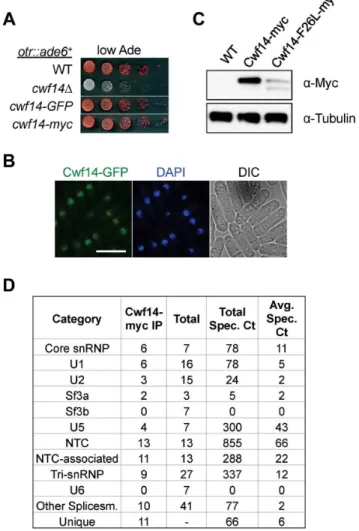
Cwf14 is highly conserved across species. Its budding yeast homolog, Bud31p, co-purifies with the spliceosome [34], and BUD31Dcauses mis-splicing of the mRNAs of ARP2and SRC1, two factors required for proper budding [35]. Cwf14 was initially identified in a purification of splicing factor Cdc5, together with a number of spliceosome components [36]. However, whether Cwf14 is a stable component of the spliceosome and involved in splicing has not been tested directly. In order to further specify the mechanism of Cwf14 action, we constructed C-terminally tagged versions ofcwf14at its endogenous locus. GFP and Cwf14-myc were fully functional, as they did not show silencing defects of otr::ade6+(Figure 4A). Imaging of Cwf14-GFP showed that Cwf14 localizes predominantly in the nucleus (Figure 4B). Western blot analysis showed that Cwf14-myc is a 40 kD protein. Moreover, the F26L mutation resulted in reduced Cwf14 protein levels, indicating that it is a partial loss-of-function allele (Figure 4C). We also performed immunoprecipitation of Cwf14-myc followed by
MudPIT mass spectrometry to identify its interacting proteins. Most factors that co-immunoprecipitated with Cwf14, but not in a control purification, were components of the spliceosome, especially from subcomplexes NTC and U5 (Figure 4D and Table S1). This result corroborates data that Cwf14 co-precipitates with Prp17, Prp19, and Cwf2, all members of U5-associated NTC [19,37], as well as a component of U5 snRNP, Cwf10 [38].
Cwf14 is involved in splicing of specific mRNAs
Previous work suggests that the spliceosome is involved in heterochromatin assembly through tethering RDRC to pericentric transcripts [15]. This is because RDRC component Cid12 associates with the spliceosome [15,20], and no splicing defects were observed in the well-characterized splicing substratetbp1in splicing factor mutants when silencing defects were apparent [15,17]. However, purifications of spliceosome components, including our purification of Cwf14, have not identified any other heterochromatin components [36,37,39] (Table S1). These results indicate either that the physical connection between the spliceo-some and RDRC is very weak, or that Cid12 is present in two separate complexes, RDRC and spliceosome. It remains a
Figure 3. Cwf14 is not required for CLRC activity.(A) Schematic diagram of the ura4::3xgbs-ade6+
reporter. (B) Left, serial dilution analysis to measure reporter gene expression. Right, ChIP analysis of H3K9me2 levels at the reporter, normalized toact1promoter. Error bars represent standard deviation of three experiments.
doi:10.1371/journal.pgen.1004334.g003
Figure 4. Cwf14 associates with the spliceosome. (A) Tagged versions of Cwf14 are functional as indicated by serial dilution analysis. (B) Imaging of cells expressing Cwf14-GFP, with DAPI to stain nucleus. Scale bar, 10mm. (C) Western blot analysis of Cwf14-myc expression. (D) Mass spectrometry analyses of purified Cwf14-myc complex. Factors were binned into annotated subcomplexes.
possibility that splicing factors regulate the correct processing of mRNAs involved in RNAi-mediated heterochromatin assembly.
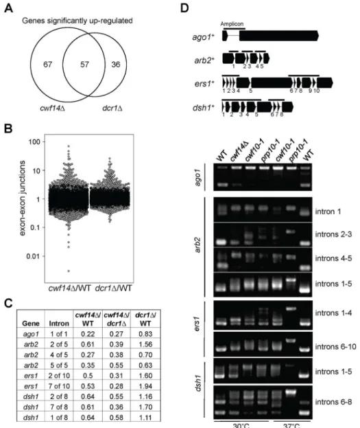
In order to test whethercwf14Dhas a general splicing defect, we performed RNA-seq analyses of total cellular RNAs from wild-type, dcr1D, and cwf14D cells. The gene expression profiles of cwf14D and dcr1D showed strong overlap of significantly up-regulated genes (Figure 5A and Tables S2 and S3), consistent with the idea that Cwf14 functions in the RNAi pathway. We found that cwf14D indeed resulted in intron retention of a portion of genes (Figure 5B and Table S4), consistent with the finding that it is associated with the spliceosome. The majority of introns were properly processed, which indicates that Cwf14 only moderately affected the activity of the spliceosome. Interestingly, many RNAi factors contain introns. Although unbiased ranking of exon-exon junction ratios between wild-type andcwf14DRNAs did not show preferential enrichment of RNAi or heterochromatin assembly
factors (Table S4), significant intron retention was observed within mRNAs fromago1,arb2,ers1, anddsh1incwf14Dcells as compared to those in wild-type cells, despite similar sequencing depths genome-wide and similar levels of each gene transcript in both samples (Figure 5C). RT-PCR analyses confirmed that these mRNAs were indeed spliced inefficiently incwf14Das well as incwf10-1andprp10-1 cells (Figure 5D). Moreover, Western blot analysis showed strong reduction of protein levels of Flag-Ago1, the RNAi factor most severely affected by cwf14D, in both cwf14D and cwf10-1 strains (Figure S1), indicating that the mis-splicing of ago1 mRNA, and possibly other RNAi factors as well, resulted in altered protein levels. Previously, it was shown that introducing cDNA versions of ago1+orhrr1+failed to rescue silencing defects ofprp10-1[15]. We also generated a cDNA version of ago1+ at its endogenous chromosomal location and under the control of its endogenous regulatory elements (ago1+::cDNA
). This cDNA construct showed
Figure 5. Cwf14 is required for the proper splicing of RNAi factors.(A) Area-proportional overlap of genes up-regulated incwf14Danddcr1D (each compared to wild type) at p,0.05. (B) Ratios of exon-exon junction counts from RNA-seq for each junction in the indicated samples. A ratio of 1 indicates no difference. The bottom tail ofcwf14D/WT, which is less pronounced indcr1D/WT, indicates splicing. (C) RNA-seq analysis shows mis-splicing of a number of RNAi factor introns incwf14D. Ratios are exon-exon junction counts from RNA-seq for each junction in the indicated samples. (D) Top, diagram of RNAi genes with intron positions indicated. Bars represent PCR fragments used to analyze intron retention. Bottom, RT-PCR analyses of RNA with primers flanking introns.
no defects in silencing ofotr::ura4+
, indicating that this replacement created a functional Ago1 protein (Figure S2). However, ago1+
::cDNAwas unable to rescue otr::ura4+
silencing defects and TBZ sensitivity of cwf14D (Figure 6A), even though it restored Flag-Ago1 protein levels (Figure S1). We reasoned that the inability ofago1+
::cDNAto rescuecwf14Ddefects is because other RNAi factors are also mis-spliced. We thus generated cDNA versions ofarb2+
anders1+
at their endogenous chromosomal loci, which were both functional (Figure S2). Neitherarb2+
::cDNAnor ers1+
::cDNA alone showed any effect on silencing ofotr::ura4+ in cwf14D cells (Figure 6A). However, when combinations of two cDNAs were introduced together into cwf14Dcells, there was a detectable rescue of silencing defects and TBZ sensitivity, and the effect was stronger when all three cDNAs were introduced (Figure 6A). Further ChIP analysis showed that both H3K9me and Swi6 protein levels were significantly increased at both otr::ura4+
and pericentricdhrepeats incwf14Dcells supplemented withago1+
,arb2+
, anders1+
cDNAs (3cDNAs) (Figure 6B). We also found that introducingago1+::cDNA was sufficient to rescue the silencing defects of otr::ade6+
associated with cwf10-1 (Figure S3A). Moreover, there is a significant increase in both
H3K9me and Swi6 levels at bothotr::ade6+
anddhrepeats in cwf10-1 agocwf10-1+
::cDNAcells (Figure S3B). Altogether, these results suggest that mutations in different splicing factors affect the splicing of diverse RNAi factors to regulate heterochromatin assembly at pericentric regions.
Thus our results clearly demonstrated that splicing factors mainly exert their effects on pericentric heterochromatin assembly by promoting the proper splicing of RNAi factors. However, we caution that the rescue of heterochromatin silencing defects of cwf14D cells was incomplete even with ago1+, arb2+, and ers1+ cDNAs. This probably reflects the requirement of Cwf14 for the proper splicing of other RNAi factors such as dsh1+, rdp1+, arb1+, hrr1+, or some unidentified factors involved in heterochromatin assembly. Such an idea is supported by the fact that the rescue ofcwf14Dsilencing defects correlated with the number of cDNA constructs that were introduced. It is also possible that splicing factors have a direct contribution in pericentric heterochromatin assembly. However, the strong rescue of pericentric silencing incwf14Dcells with cDNA constructs suggests that the regulation of RNAi factor splicing is a major role of splicing factors in this process.
Figure 6. Introducing cDNAs of RNAi factors rescues pericentric heterochromatin defects ofcwf14Dcells.(A) Serial dilution analyses to measure the expression ofotr::ura4+and sensitivity to TBZ. (B) ChIP analyses of H3K9me2 and Swi6 levels at pericentricdhrepeats andotr::ura4+, normalized to anact1promoter fragment. Error bars represent standard deviation of three experiments.
Cwf14 regulates the proper splicing oftpz1to control telomere silencing
We also found that telomere shelterin componenttpz1+ showed a strong reduction in exon-exon junction sequencing reads in cwf14D cells relative to wild-type cells, and this phenotype was confirmed by RT-PCR analyses in cwf10-1 and prp10-1 as well (Figure 7A). A C-terminally Flag-tagged version of Tpz1 [40] affected silencing of a reporter gene inserted near telomere repeats (Figure S4), suggesting that Tpz1 is required for telomere silencing. We then analyzed the effect of splicing factor mutations on silencing of a reporter inserted near telomere repeats of chromosome two (Tel2::ura4+
) [41]. Indeed, cwf14D resulted in silencing defects at this reporter gene (Figure 7B), accompanied by a reduction of H3K9me levels at tlh1, which is embedded at telomeric heterochromatin, as well as the accumulation of tlh1 transcripts (Figure 7C and 7D). Both cwf10-1 and prp10-1 cells showed increased tlh1 transcript levels, indicating that loss of telomeric silencing is a general feature of splicing mutants (Figure 7C and 7D). Because multiple DNA sequences contribute to heterochromatin assembly at telomeres, including tlh1+
, telomere associated sequences (TAS), and terminalTEL repeats [8], we further tested silencing atTEL::ade6+, which is inserted on the mini-chromosome Ch16 adjacent to telomere repeats [42]. We found that cwf14D, cwf14-F26L, and cwf10-1 resulted in loss of silencing of this reporter (Figure 7E). Interestingly, replacement of tpz1+with its cDNA partially rescued telomeric silencing phenotypes of thecwf14-F26L and cwf10-1mutants (Figure 7E and 7F). We could only marginally rescue the telomere silencing defects of cwf14Dcells withtpz1+
::cDNA(Figure 7F and data not shown). Thus it is possible that additional factors involved in telomere silencing might also contain introns and depend on the spliceosome to properly regulate their splicing. Alternatively, splicing factors might affect telomere silencing through additional mechanisms. Nonethe-less, these results demonstrate that inefficient splicing of tpz1 contributes to telomeric silencing defects in splicing mutants.
Discussion
The formation of heterochromatin requires RNAi-mediated processing of repeat-derived transcripts and the targeting of histone modifying activities to repeat regions, leading to H3K9me and the recruitment of HP1 proteins. It has been shown that splicing factors are required for RNAi-mediated heterochromatin assembly in fission yeast, although the mechanism by which they are involved is not well characterized. The previously prevailing model was that the spliceosome physically associates with RNAi factors to regulate heterochromatin assembly, rather than acting through its splicing activity [15,16]. One of the main evidences for this idea is that splicing mutants show severe silencing defects even though the splicing of tbp1 mRNA was not affected [15,17]. However, whether these splicing factor mutants selectively affect the splicing of RNAi factors has not been rigorously tested.
Our RNA-seq analyses showed prominent intron retention of a subgroup of mRNAs incwf14Dcells, even though the majority of introns (including those of tbp1) are still properly processed (Figure 5B and Table S4). Interestingly, a number of key RNAi factors were among the list of strongly mis-spliced introns, a result that is further corroborated by RT-PCR analyses of a selective set of RNAi factor mRNAs in other spliceosome mutants such as cwf10-1andprp10-1(Figure 5D). Most importantly, we found that introducing a combination of cDNAs of RNAi factors significantly alleviated pericentric heterochromatin defects associated with cwf14D and cwf10-1 (Figure 6 and S3). Thus splicing factors regulate the proper splicing of RNAi factors, which is a major, if
not sole, contributor to heterochromatin assembly defects in splicing mutants. That introducing tpz1+
::cDNA was able to partially rescue telomere silencing defects associated with splicing factors further supports the idea that mis-splicing of heterochro-matin factors is the reason splicing factor mutants show heterochromatin assembly defects (Figure 7E and 7F).
It is noteworthy thatcwf14Dhas only moderate splicing defects, with some introns show very strong sensitivity, whereas most others show little to no defects (Figure 5B and Table S4). This raises the question of whether specific intron sensitivity is a result of introns that are inherently difficult to splice. Consistent with this idea, our RT-PCR analyses showed prominent unspliced precur-sor mRNAs of RNAi factors even in wild-type cells (Figure 5D). It is also a striking pattern that heterochromatin factors in the same complexes tend to either have or not have introns. For example, many members of RNAi, such asago1+
,arb1+ ,arb2+
,ers1+ ,dsh1+
, rdp1+
, and hrr1+
, have introns, but none of CLRC (clr4+ , rik1+
, raf1+
,raf2+ ,cul4+
,stc1+
), SHREC (clr1+ ,clr2+
,clr3+ ,mit1+
), orswi6+ have any introns, raising the possibility of selective regulation of the RNAi pathway through general or specific changes in splicing efficiency. In fact, an analysis of splicing changes during meiosis showed that one intron ofarb1+
is induced to be spliced, while a different intron exhibits splicing repression [43]. Since splicing factors have been identified in screens that affect RNAi-based processes in worms, flies, and plants [44–48], it seems a conserved mechanism that proper splicing of the mRNAs of RNAi factors regulates the efficiency of RNAi inside the cell.
Materials and Methods
Genetic screens of the fission yeast deletion library The Bioneer library is composed of strains of mixed endogenous ade6alleles:ade6-M216, which forms pink colonies on low adenine medium, andade6-M210, which give rise to red colonies, similar to ade6D. To avoid the complication ofade6-M216alleles in our screen, we included in the query strain anade6-M210-mCherryallele linked to a hphMX6cassette, which confers resistance to the antibiotic hygro-mycin B, allowing us to generate a uniformade6-M210background. Screens were carried out according to previous protocols [49], with slight modifications. Yeast strains arrayed in 384 strains/plate format were pinned on YES agar medium containing 100mg/ml G418, and
otr::ade6+
-natMX6 ade6-210-hphMX6 and ade6-210-hphMX6 strains were pinned on YES+100mg/ml hygromycin B. After two days growth, strains were mated on SPA agar medium. Plates were incubated at 25uC for 3 days, then 42uC for 3 more days to kill vegetative cells. Strains were then germinated and correct genotype selected by pinning to YES+GNH (G418, Nat, and Hygromycin) or YES+GH (G418 and Hygromycin) medium, and then pinned to YE (low ade) medium for color readout.
Fission yeast strains and genetic analyses
Cwf14-GFP and Cwf14-13myc strains were constructed by a PCR-based module method.cwf14D,cwf12D,dre4D, andsrm1Dwere derived from the Bioneer fission yeast deletion library, verified via PCR, and backcrossed. Thep-cwf14+
plasmid was constructed by insertion of a PCR product containingcwf14+
promoter and coding region into SphI and XmaI sites of pREP41. Thecwf14-F26L-myc mutant strain was constructed by integrating acwf14+-myc-kanMX6 cassette amplified by error-prone PCR (high dNTP concentration) into the endogenouscwf14+
locus.ago1+
::cDNA, arb2+
::cDNA, and ers1+::cDNA
construct all other strains. The genotype of strains is provided in Table S5. For serial dilution plating assays, ten-fold dilutions of mid-log-phase culture were plated on the indicated medium and grown for 3 days at 30uC unless otherwise indicated.
RNA analyses
Total cellular RNA was isolated from log-phase cells using MasterPure yeast RNA purification kit (Epicentre) according to the manufacturer’s protocol. Quantification with qRT-PCR was
performed with Power SYBR Green RNA-to-CT one-step Kit (Applied Biosystems). RNA serial dilutions were used as templates to generate the standard curve of amplification for each pair of primers, and the relative concentration of target sequence was calculated accordingly. An act1 fragment served as reference to normalize the concentration of samples. Sequence of DNA oligos is provided in Table S6. For RNA-seq, purified RNA was prepared by TruSeq Stranded Total RNA Kit (Illumina), which includes rRNA depletion and chemical fragmentation. Index
Figure 7. Splicing factors regulate the proper splicing of shelterin component Tpz1.(A) RT-PCR analyses of RNA with primers flanking introns. (B, E) Serial dilution analyses to measure reporter gene expression. (C) ChIP analyses of H3K9me2 levels, normalized toact1promoter. Error bars represent standard deviation of three experiments. (D, F) qRT-PCR analyses of transcripts derived fromtlh1, normalized toact1gene. Wild type was set to 1. Error bars represent standard deviation of three experiments.
adapters were added to allow for multiplexing. Paired-end sequencing with 100 bp read lengths was performed on Illumina HiSeq. Mapping was performed with the Tuxedo Suite consisting of Bowtie, TopHat, and Cufflinks. Forcwf14D, 57,521,513 reads were obtained, and 83% mapped to the genome via Bowtie. For WT, 47,636,518 reads were obtained, and 82% mapped. For dcr1D, 64,972,007 reads were obtained, and 83% mapped. RNA-seq data have been deposited to the Sequence Read Archive (http://www.ncbi.nlm.nih.gov/sra/) with accession number SRP040479. For the dot plot, exon-exon junction ratios were filtered to remove several types: 1) junctions which mapped zero times in any sample (possible mapping noise), 2) junctions whose sum in WT,cwf14D, anddcr1Dwas less than 30 (to avoid randomness due to small sample sizes), and 3) ratios whose values were greater than 150 (to focus the diagram on splicing reduction). Northern blot of siRNAs was performed as described previously [50].
Chromatin immunoprecipitation (ChIP) analysis
ChIP analyses were performed as described previously [51]. Antibodies used were H3K9me2 (Abcam 1220), Swi6 [52], and RNA Pol II (Covance 8WG16). Quantification with qPCR was performed with Maxima SYBR Green/ROX qPCR Master Mix (Thermo). Enrichment was calculated as: relative levels in ChIP/ relative levels in total DNA. Anact1promoter fragment was used as a control for normalization unless otherwise indicated. Sequence of DNA oligos was provided in Table S6.
Protein purification and mass spectrometry analysis Exponentially growing yeast cells were harvested, washed with 2xHC buffer (300 mM HEPES-KOH pH 7.6, 2 mM EDTA, 100 mM KCl, 20% glycerol, 0.1% NP-40, 2 mM DTT, and protease inhibitor cocktail (Roche)) and frozen in liquid nitrogen. Crude cell extracts were prepared by vigorously blending frozen yeast cells with dry ice using a household blender, followed by sonication and incubation with 30 ml 1xHC buffer containing 250 mM KCl for 30 min. The lysate was cleared by centrifugation at 82,7006gfor 3 hours. The supernatants were incubated with 50ml of C-myc antibody (Sigma C3956) overnight, for three hours the next day with 50ml protein G agarose beads, washed eight times with 1xHC containing 250 mM KCl, then two times with the same buffer without NP-40. For mass spectrometry analysis, bound proteins were eluted with 26100ml of 50 mM Tris pH 7.5, 5% SDS, 5% glycerol, 50 mM DTT. MudPIT mass spectrometry analysis was performed as described previously [53].
Supporting Information
Figure S1 Western blot analysis of Flag-Ago1 protein levels. Cell lysates were first immunoprecipitated with Flag-agarose beads and Western blot analyses were performed with a Flag antibody. (PDF)
Figure S2 cDNA versions of RNAi factors are functional. Serial dilution analysis of cells to measure the expression ofotr::ura4+
and sensitivity to TBZ. Pictures fordcr1Dare from the same plates as other strains.
(PDF)
Figure S3 Introducingago1+::cDNAsignificantly rescues silencing defects associated withcwf10-1. (A) Serial dilution analysis of cells on low adenine medium to measure the expression ofotr::ade6+. (B) ChIP analysis of H3K9 and Swi6 levels atotr::ade6+, normalized to anact1fragment. Error bars represent standard deviation of three experiments.
(PDF)
Figure S4 Tpz1 is required for telomere silencing. Serial dilution analysis of cells to measure the expression ofTEL::ura4+
, which is located near telomere repeats on Ch16.
(PDF)
Table S1 Mass spectrometry analysis of Cwf14-myc associated proteins.
(XLSX)
Table S2 Comparison of gene expression in wild type and cwf14Dby RNA-seq.
(XLSX)
Table S3 Comparison of gene expression in wild type anddcr1D by RNA-seq.
(XLSX)
Table S4 Exon-exon junction counts and ratios across all samples. (XLSX)
Table S5 Strains used in this study. (XLSX)
Table S6 DNA oligos used in this study. (XLSX)
Acknowledgments
We thank Elizabeth Miller for providing library screening equipment, Robin Allshire, Danesh Moazed, Karl Ekwall, Jun-ichi Nakayama, Janet Partridge, and Feng Qiao for yeast strains, Kenichi Shimada for assistance with RNA-seq analysis, Hana Um and Samantha Pottinger for technical assistance, and members of Jia lab for helpful contributions, discussions, and critical reading of the manuscript.
Author Contributions
Conceived and designed the experiments: SPK SJ. Performed the experiments: SPK XT HH JJM PGT SJ. Analyzed the data: SPK SA JJM PGT JRY PLN SJ. Wrote the paper: SPK SJ. Next-gen sequencing and data analysis: SA PLN SPK.
References
1. Grewal SIS, Jia S (2007) Heterochromatin revisited. Nat Rev Genet 8: 35–46. doi:10.1038/nrg2008.
2. Almouzni G, Probst AV (2011) Heterochromatin maintenance and establishment: lessons from the mouse pericentromere. Nucleus 2: 332–338. doi:10.4161/ nucl.2.5.17707.
3. Rea S, Eisenhaber F, O’Carroll D, Strahl BD, Sun ZW, et al. (2000) Regulation of chromatin structure by site-specific histone H3 methyltransferases. Nature 406: 593–599. doi:10.1038/35020506.
4. Bannister AJ, Zegerman P, Partridge JF, Miska EA, Thomas JO, et al. (2001) Selective recognition of methylated lysine 9 on histone H3 by the HP1 chromo domain. Nature 410: 120–124. doi:10.1038/35065138.
5. Nakayama J, Rice JC, Strahl BD, Allis CD, Grewal SI (2001) Role of histone H3 lysine 9 methylation in epigenetic control of heterochromatin assembly. Science 292: 110–113. doi:10.1126/science.1060118.
6. Jia S, Noma K-I, Grewal SIS (2004) RNAi-independent heterochromatin nucleation by the stress-activated ATF/CREB family proteins. Science 304: 1971–1976. doi:10.1126/science.1099035.
7. Kim HS, Choi ES, Shin JA, Jang YK, Park SD (2004) Regulation of Swi6/HP1-dependent heterochromatin assembly by cooperation of components of the mitogen-activated protein kinase pathway and a histone deacetylase Clr6. J Biol Chem 279: 42850–42859. doi:10.1074/jbc.M407259200.
8. Kanoh J, Sadaie M, Urano T, Ishikawa F (2005) Telomere binding protein Taz1 establishes Swi6 heterochromatin independently of RNAi at telomeres. Curr Biol 15: 1808–1819. doi:10.1016/j.cub.2005.09.041.
10. Dorer DR, Henikoff S (1994) Expansions of transgene repeats cause heterochromatin formation and gene silencing in Drosophila. Cell 77: 993– 1002.
11. Garrick D, Fiering S, Martin DI, Whitelaw E (1998) Repeat-induced gene silencing in mammals. Nat Genet 18: 56–59. doi:10.1038/ng0198-56. 12. Moazed D (2009) Small RNAs in transcriptional gene silencing and genome
defence. Nature 457: 413–420. doi:10.1038/nature07756.
13. Lejeune E, Allshire RC (2011) Common ground: small RNA programming and chromatin modifications. Curr Opin Cell Biol 23: 258–265. doi:10.1016/ j.ceb.2011.03.005.
14. Castel SE, Martienssen RA (2013) RNA interference in the nucleus: roles for small RNAs in transcription, epigenetics and beyond. The EMBO Journal 14: 100–112. doi:10.1038/nrg3355.
15. Bayne EH, Portoso M, Kagansky A, Kos-Braun IC, Urano T, et al. (2008) Splicing Factors Facilitate RNAi-Directed Silencing in Fission Yeast. Science 322: 602–606. doi:10.1126/science.1164029.
16. Bernard P, Drogat J, Dheur S, Genier S, Javerzat J-P (2010) Splicing factor Spf30 assists exosome-mediated gene silencing in fission yeast. Mol Cell Biol 30: 1145–1157. doi:10.1128/MCB.01317-09.
17. Chinen M, Morita M, Fukumura K, Tani T (2010) Involvement of the spliceosomal U4 small nuclear RNA in heterochromatic gene silencing at fission yeast centromeres. J Biol Chem 285: 5630–5638. doi:10.1074/ jbc.M109.074393.
18. Wood V, Gwilliam R, Rajandream M-A, Lyne M, Lyne R, et al. (2002) The genome sequence of Schizosaccharomyces pombe. Nature 415: 871–880. doi:10.1038/nature724.
19. Wahl MC, Will CL, Lu¨hrmann R (2009) The Spliceosome: Design Principles of a Dynamic RNP Machine. Cell 136: 701–718. doi:10.1016/j.cell.2009.02.009. 20. Motamedi MR, Verdel A, Colmenares SU, Gerber SA, Gygi SP, et al. (2004) Two RNAi complexes, RITS and RDRC, physically interact and localize to noncoding centromeric RNAs. Cell 119: 789–802. doi:10.1016/ j.cell.2004.11.034.
21. Buker SM, Iida T, Bu¨hler M, Ville´n J, Gygi SP, et al. (2007) Two different Argonaute complexes are required for siRNA generation and heterochromatin assembly in fission yeast. Nat Struct Mol Biol 14: 200–207. doi:10.1038/ nsmb1211.
22. Rougemaille M, Shankar S, Braun S, Rowley M, Madhani HD (2008) Ers1, a Rapidly Diverging Protein Essential for RNA Interference-dependent Hetero-chromatic Silencing in Schizosaccharomyces pombe. J Biol Chem 283: 25770– 25773. doi:10.1074/jbc.C800140200.
23. Roguev A, Bandyopadhyay S, Zofall M, Zhang K, Fischer T, et al. (2008) Conservation and rewiring of functional modules revealed by an epistasis map in fission yeast. Science 322: 405–410. doi:10.1126/science.1162609.
24. Kawakami K, Hayashi A, Nakayama J-I, Murakami Y (2012) A novel RNAi protein, Dsh1, assembles RNAi machinery on chromatin to amplify hetero-chromatic siRNA. Genes Dev 26: 1811–1824. doi:10.1101/gad.190272.112. 25. Ekwall K, Olsson T, Turner BM, Cranston G, Allshire RC (1997) Transient
inhibition of histone deacetylation alters the structural and functional imprint at fission yeast centromeres. Cell 91: 1021–1032.
26. Allshire RC, Nimmo ER, Ekwall K, Javerzat JP, Cranston G (1995) Mutations derepressing silent centromeric domains in fission yeast disrupt chromosome segregation. Genes Dev 9: 218–233.
27. Partridge JF, Scott KSC, Bannister AJ, Kouzarides T, Allshire RC (2002) cis-acting DNA from fission yeast centromeres mediates histone H3 methylation and recruitment of silencing factors and cohesin to an ectopic site. Curr Biol 12: 1652–1660.
28. Sadaie M, Iida T, Urano T, Nakayama J-I (2004) A chromodomain protein, Chp1, is required for the establishment of heterochromatin in fission yeast. The EMBO Journal 23: 3825–3835. doi:10.1038/sj.emboj.7600401.
29. Bernard P, Maure JF, Partridge JF, Genier S, Javerzat JP, et al. (2001) Requirement of heterochromatin for cohesion at centromeres. Science 294: 2539–2542. doi:10.1126/science.1064027.
30. Nonaka N, Kitajima T, Yokobayashi S, Xiao G, Yamamoto M, et al. (2002) Recruitment of cohesin to heterochromatic regions by Swi6/HP1 in fission yeast. Nat Cell Biol 4: 89–93. doi:10.1038/ncb739.
31. Yamagishi Y, Sakuno T, Shimura M, Watanabe Y (2008) Heterochromatin links to centromeric protection by recruiting shugoshin. Nature 455: 251–255. doi:10.1038/nature07217.
32. Ekwall K, Nimmo ER, Javerzat JP, Borgstrøm B, Egel R, et al. (1996) Mutations in the fission yeast silencing factors clr4+and rik1+disrupt the localisation of the chromo domain protein Swi6p and impair centromere function. J Cell Sci 109 (Pt 11): 2637–2648.
33. Kagansky A, Folco HD, Almeida R, Pidoux AL, Boukaba A, et al. (2009) Synthetic Heterochromatin Bypasses RNAi and Centromeric Repeats to Establish Functional Centromeres. Science 324: 1716–1719. doi:10.1126/ science.1172026.
34. Masciadri B, Areces LB, Carpinelli P, Foiani M, Draetta G, et al. (2004) Characterization of the BUD31 gene of Saccharomyces cerevisiae. Biochem Biophys Res Commun 320: 1342–1350.
35. Saha D, Banerjee S, Bashir S, Vijayraghavan U (2012) Context dependent splicing functions of Bud31/Ycr063w define its role in budding and cell cycle progression. Biochem Biophys Res Commun 424: 579–585. doi:10.1016/ j.bbrc.2012.06.156.
36. Ohi MD, Link AJ, Ren L, Jennings JL, McDonald WH, et al. (2002) Proteomics analysis reveals stable multiprotein complexes in both fission and budding yeasts containing Myb-related Cdc5p/Cef1p, novel pre-mRNA splicing factors, and snRNAs. Mol Cell Biol 22: 2011–2024. doi:10.1128/MCB.22.7.2011-2024.2002.
37. Ren L, McLean JR, Hazbun TR, Fields S, Vander Kooi C, et al. (2011) Systematic two-hybrid and comparative proteomic analyses reveal novel yeast pre-mRNA splicing factors connected to Prp19. PLoS ONE 6: e16719. doi:10.1371/journal.pone.0016719.
38. Livesay SB, Collier SE, Bitton DA, Ba¨hler J, Ohi MD (2013) Structural and functional characterization of the N-terminus of Schizosaccharomyces pombe Cwf10. Eukaryotic Cell 12:1472–89
39. Carnahan RH, Feoktistova A, Ren L, Niessen S, Yates JRI, et al. (2005) Dim1p Is Required for Efficient Splicing and Export of mRNA Encoding Lid1p, a Component of the Fission Yeast Anaphase-Promoting Complex. Eukaryotic Cell 4: 577. doi:10.1128/EC.4.3.577-587.2005.
40. Moser BA, Subramanian L, Khair L, Chang Y-T, Nakamura TM (2009) Fission yeast Tel1(ATM) and Rad3(ATR) promote telomere protection and telomerase recruitment. PLoS Genet 5: e1000622. doi:10.1371/journal.pgen.1000622. 41. Nimmo ER, Pidoux AL, Perry PE, Allshire RC (1998) Defective meiosis in
telomere-silencing mutants of Schizosaccharomyces pombe. Nature 392: 825– 828. doi:10.1038/33941.
42. Nimmo ER, Cranston G, Allshire RC (1994) Telomere-associated chromosome breakage in fission yeast results in variegated expression of adjacent genes. The EMBO Journal 13: 3801–3811.
43. Wilhelm BT, Marguerat S, Watt S, Schubert F, Wood V, et al. (2008) Dynamic repertoire of a eukaryotic transcriptome surveyed at single-nucleotide resolution. Nature 453: 1239–1243. doi:10.1038/nature07002.
44. Kim JK, Gabel HW, Kamath RS, Tewari M, Pasquinelli A, et al. (2005) Functional genomic analysis of RNA interference in C. elegans. Science 308: 1164–1167. doi:10.1126/science.1109267.
45. Herr AJ, Molna`r A, Jones A, Baulcombe DC (2006) Defective RNA processing enhances RNA silencing and influences flowering of Arabidopsis. Proc Natl Acad Sci USA 103: 14994–15001. doi:10.1073/pnas.0606536103.
46. Zhou R, Hotta I, Denli AM, Hong P, Perrimon N, et al. (2008) Comparative analysis of argonaute-dependent small RNA pathways in Drosophila. Molecular Cell 32: 592–599. doi:10.1016/j.molcel.2008.10.018.
47. Tabach Y, Billi AC, Hayes GD, Newman MA, Zuk O, et al. (2013) Identification of small RNA pathway genes using patterns of phylogenetic conservation and divergence. Nature 493: 694–698. doi:10.1038/nature11779. 48. Ausin I, Greenberg MVC, Li CF, Jacobsen SE (2012) The splicing factor SR45 affects the RNA-directed DNA methylation pathway in Arabidopsis. Epigenetics : official journal of the DNA Methylation Society 7: 29–33. doi:10.4161/ epi.7.1.18782.
49. Baryshnikova A, Costanzo M, Dixon S, Vizeacoumar FJ, Myers CL, et al. (2010) Synthetic genetic array (SGA) analysis in Saccharomyces cerevisiae and Schizosaccharomyces pombe. Meth Enzymol 470: 145–179. doi:10.1016/ S0076-6879(10)70007-0.
50. Partridge JF, Debeauchamp JL, Kosinski AM, Ulrich DL, Hadler MJ, et al. (2007) Functional separation of the requirements for establishment and maintenance of centromeric heterochromatin. Molecular Cell 26: 593–602. doi:10.1016/j.molcel.2007.05.004.
51. Hou H, Wang Y, Kallgren SP, Thompson J, Yates JR, et al. (2010) Histone variant H2A.Z regulates centromere silencing and chromosome segregation in fission yeast. J Biol Chem 285: 1909–1918. doi:10.1074/jbc.M109.058487. 52. Reddy BD, Wang Y, Niu L, Higuchi EC, Marguerat SB, et al. (2011)
Elimination of a specific histone H3K14 acetyltransferase complex bypasses the RNAi pathway to regulate pericentric heterochromatin functions. Genes Dev 25: 214–219. doi:10.1101/gad.1993611.