with sepsis requiring hemicolectomy
Leiomiomas extrauterinos com sepse que exigem hemicolectomia
SeetHaraman HariHaranravi maHaraj1
ian HoSein1
vijay naraynSingH1
Abstract
Extrauterine leiomyomas are rare, benign, and may arise in any anatomic sites. Their unusual growth pattern may even mimic malignancy and can result in a clinical dilemma. Occasionally, uterine leiomyomas become adherent to surrounding structures. They also develop an auxiliary blood supply, and lose their original attachment to the uterus, thus becoming ‘parasitic’. Parasitic myomas may also be iatrogenically created after uterine ibroid surgery, particularly if morcellation is used. This report presented two cases of parasitic myomas with sepsis, both requiring right hemicolectomy. It reviewed the pertinent literature.
Resumo
Leiomiomas extrauterinos são raros e benignos e podem surgir em qualquer local anatômico. O padrão de crescimento não-usual destas lesões pode até mesmo imitar malignidade e resultar em dilema clínico. Ocasionalmente, os leiomiomas uterinos aderem-se às estruturas circunvizinhas, desenvolvem suprimento sanguíneo auxiliar e perdem sua ligação original ao útero, tornando-se assim “parasíticos”. Os miomas parasíticos podem também ser criados iatrogenicamente após cirurgia ibroide uterina, especialmente se for utilizada morcelação. Este relato apresentou dois casos de miomas parasíticos com sepse, ambos exigindo hemicolectomia à direita, bem como revisão da literatura pertinente.
1 Department of Clinical Surgical Sciences; The University of the West Indies, St. Augustine, Trinidad, West Indies.
Conlict of interest: none.
Keywords
Leiomyoma/surgery Uterine neoplasms/surgery Sepsis Colectomy/methods Case reports
Palavras-chave
Leiomioma/cirurgia Neoplasias uterinas/cirurgia Sepse Colectomia/métodos Relatos de casos
Correspondence
Seetharaman Hariharan Faculty of Medical Sciences The University of the West Indies Eric Williams Medical Sciences Complex Mount Hope – Trinidad (West Indies)
Received
05/10/2011
Accepted with modiications
Introduction
Uterine leiomyomas affect 20 to 30% of women older than 35 years, and their incidence may be on the rise since many women are postponing their re-productive careers1. Extrauterine leiomyomas are rare,
benign, and may arise in any anatomic sites. Their unusual growth pattern may even mimic malignancy and can result in a clinical dilemma2. Occasionally,
uterine leiomyomas become adherent to surrounding structures. They also develop an auxiliary blood sup-ply, and lose their original attachment to the uterus, thus becoming ‘parasitic.’ Parasitic myomas may also be iatrogenically created after uterine ibroid surgery, particularly if morcellation is used. With the surge in laparoscopic gynecological procedures, especially laparoscopic myomectomy, the potential for developing parasitic myomas is being recognized more frequently3,4.
We presented two cases of parasitic myomas with sepsis, both requiring right hemicolectomy. The pertinent literature was also reviewed.
Case report 1
A 28-year-old female presented to the gynecologist at a peripheral hospital with a history of miscarriage two weeks prior to the presentation. She complained of feeling unwell, with fever, and generalized ab-dominal pain with nausea. On clinical examination, the abdomen was tender with a palpable mass on the right side. An ultrasound scan of her abdomen and pelvis demonstrated a 17×12 cm right sided complex abdominal mass, which was reported to be possibly of ovarian origin.
An initial exploratory laparotomy in the peripheral hospital found 300 mL of foul smelling pus in the peri-toneal cavity, although there was a reported inability to ind the source of the pus. The pelvic organs appeared normal except for a ibroid uterus.
The patient was then referred to a tertiary care facil-ity. On presentation, she appeared ill with pyrexia and tachycardia. The abdomen was diffusely tender with a palpable mass on the right hypochondrium. Hematology showed leukocytosis (WBC count of 18×103/dL) and mild
anemia (hemoglobin of 9 g/dL).
Since the computed tomography (CT) scan was not available at that time, based on the signs of peri-tonism, a decision was taken to perform an exploratory laparotomy. Intra-operatively, 100 mL of frank pus was found originating from the right paracolic gutter. There were signiicant adhesions noted in the abdomen mak-ing dissection extremely dificult. A large solid mass 15×13 cm was found closely adherent to distal ileum and right colon. A right hemicolectomy and primary ileocolic anastomosis were performed with an ‘en bloc’ excision of the tumor with the specimen. There was no communication with the uterus.
Post-operatively, sepsis was controlled with intrave-nous antibiotics. The patient tolerated oral luid intake on the third postoperative day. Her recovery was uneventful, except for an uncomplicated grade 1 supericial surgical site infection.
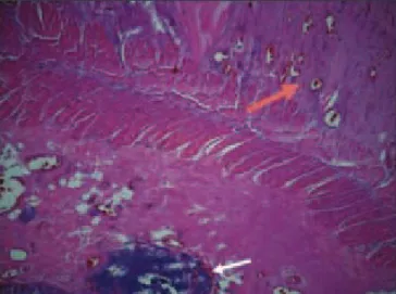
Histology revealed ischemic changes in a para-sitic (serosal) leiomyoma with areas of localized serositis (Figure 1). Adjacent ileum and colon showed no inlam-matory bowel disease, dysplasia, or neoplasia.
Case report 2
A 30-year old female had a laparoscopic myomec-tomy for multiple large ibroids. The procedure was quite eventful. There was prolonged operating time (eight hours), heavy blood loss, and a signiicant persistent post-operative ileus (eight days). The patient presented to our hospital 11 days after the procedure and had complaints of generalized abdominal pain and high-grade fever, with chills and rigors.
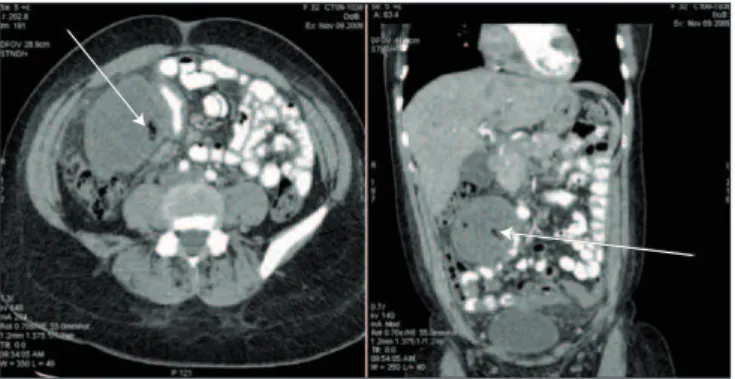
On examination, the patient was tachycardiac (110/minutes) and had an elevated temperature (38.6ºC). Her abdominal examination revealed generalized tender-ness with peritonism and a tender right upper quadrant mass. The patient also had a leukocytosis (WBC count of 16,000) along with a left axis shift. A CT scan revealed free luid in the pelvis and an enhancing mass in the right upper quadrant, suggestive of intra-abdominal abscess as there was air within it (Figure 2).
A diagnostic laparoscopy revealed approximately 100 mL of pus in the pelvis and a mobile mass in the right upper quadrant, which was intimately related to the hepatic lexure of the colon and several loops of small bowel. Separation was not possible due to serosal tears.
A conversion to upper midline laparotomy was done, and a right hemicolectomy was performed with a primary ileocolic anastomosis and ‘en bloc’ resection of the mass with the specimen. Grossly after separa-tion of the mass (now recognized to be a ibroid) from the bowel, there was signiicant friability and bleeding from the serosal surface (Figure 3).
Postoperatively, the patient’s sepsis resolved with intravenous antibiotics and she had an uneventful recovery. Histology confirmed ischemic changes in a leiomyoma. Adjacent ileum and colon showed no inflammatory bowel disease, pathogens, dysplasia, or neoplasia.
Discussion
Uterine leiomyomas are benign tumors of smooth muscle origin with protean symptomatology, and they are the most common gynecological tumors in women of reproductive age. Very rarely, benign uterine leio-myomas display bizarre growth patterns with associated extrauterine benign-appearing smooth muscle tumors, which are similar to those found in a uterine ibroid. Leiomyomas occur infrequently outside the uterus. Although they are histologically benign, extrauterine leiomyomas may mimic malignant tumors at imaging and may present a diagnostic challenge. The clinical symptoms and imaging features depend on the location of the lesion and on its growth pattern. These tumors may have arisen from uterus and underwent separation. The spectrum of smooth-muscle tumors arising from the uterus ranges from benign leiomyoma to malignant leiomyosarcoma, but also includes a variety of lesions
with growth patterns that extend outside the uterus, including parasitic leiomyoma, intravenous leiomyo-matosis, disseminated peritoneal leiomyoleiomyo-matosis, and benign metastasizing leiomyoma. These benign tumors resemble typical uterine leiomyomas at both gross and microscopic levels2.
Parasitic leiomyomas are presented as peritoneal benign smooth-muscle masses separate from the uterus. A parasitic leiomyoma likely originates as a peduncu-lated subserosal leiomyoma that twists and torts from its uterine pedicle. It becomes free in the peritoneal cavity and it survives by recruiting neovascularization from adjacent structures. In the era of laparoscopy, where many procedures are done with morcellation techniques to remove the specimen, it is not dificult to fathom the possible genesis of this rare condition3,4.
In the cases presented, one of the patients had a true parasitic myoma, while the other one may have had a retained myoma, which had already started to recruit a blood supply and may have presented later on as a parasitic myoma.
Parasitic leiomyomas, though infrequent, are most commonly located in the broad ligament, pelvic peritoneum, cul-de-sac, and omentum2. In both cases,
these parasitic tumors seem to have a predilection for the recesses of the right side of the abdomen. In the case of the latter, the ibroids are usually placed in the right side of the abdomen, in order to allow an easier localization when they are ready to be morcel-lated and removed. One of the ibroids was left inside the abdomen inadvertently. Even so, in the early phase of the laparoscopic myomectomy, it was not unusual to leave the excised ibroid in the abdomen allowing for self-digestion.
The literature is limited in its description of parasitic leiomyoma since this is a rarely diagnosed condition. It is likely that this condition may be under-diagnosed since many of these tumors, if present, may remain asymptomatic for a number of years and may only
Figure 2. Computed tomography scan showing mass in the right upper quadrant with intramural air (arrow).
become evident on routine scanning for other causes. The two cases presented may be the irst description to our knowledge of parasitic leiomyomas presenting as generalized abdominal sepsis.
Kho and Nezhat4 have one of the largest series in
the literature of parasitic myomas, they attempted to determine their cause, associations, and risk factors. However, in this retrospective review, they were only able to identify 12 cases of parasitic myomas from 2000 to 2008. Laparoscopy was used as a diagnostic tool, which conirmed the presence of intra/retroperitoneal myomas distinct from the uterus in the 12 cases. Ten of the 12 patients had prior abdominal surgery. Eight had prior morcellation procedures; six performed lapa-roscopically, two performed by laparotomy. This clearly suggests that prior surgery for uterine ibroid was a risk factor for the development of parasitic leiomyoma4.
Within the last 15 years, laparoscopic myomectomy has gained popularity and has shown clear advantages over traditional open techniques. It is usually indicated in patients with uterine ibroids numbering less than three to four with a maximum size of 8 to 9 cm5. However,
these indications have been called into question, and it has been shown that in the hands of experienced laparoscopic surgeon, size, number, and location do not matter6. Despite the controversies regarding the
indica-tion, laparoscopic myomectomy has been shown to be a feasible option that boasts a conversion rate as low as 1 to 3%, fertility rates after laparoscopic myomectomy of more than 50% and successful myomectomy rate of more than 80%5. As surgeons’ dexterity with
the pro-cedure improved and the associated standardization of the technique accomplished, operative intervention time has become acceptable and blood loss became less important. In addition, laparoscopic myomectomy is also associated with all of the advantages of laparoscopy with respect to postoperative adhesion, decreased post-operative pain, short hospital stay, and early return to normal function7.
Despite the optimism regarding laparoscopic myomec-tomy and its superior safety proile, there are a number of case reports in the literature describing parasitic myomas after laparoscopic myomectomy. One of the early cases was described by Hutchins and Reinoehl8, in 1998, in
which a 6 cm ibroid was lost at the time of a laparoscopic assisted supracervical hysterectomy and resulted in severe abdominal pain. The ibroid was eventually removed by laparotomy. In this case, the ibroid was located in the
right upper quadrant of the abdomen at laparotomy similar to Case 28.
Cucinella et al.9 recently reported, on their series of
parasitic myomas, that they identiied four cases over a three-year period. Electric morcellators were used in 423 cases and the overall prevalence of developing parasitic myoma was reported at 0.9 and 1.2% for those who had laparoscopic myomectomies9. There are also cases in the
literature in which parasitic myomas have been found attached to the bowel and anterior abdominal wall after laparoscopic myomectomy10,11. There were no cases in the
literature in which parasitic myomas were the primary cause or associated with generalized abdominal sepsis.
Parasitic myomas are rare occurrences, which can prove to be a diagnostic dilemma and may even be pres-ent in the acute setting or even mimic a malignancy. There are increasing reports of this condition being recognized after laparoscopic myomectomy especially in those cases where tumors are morcellated for removal. It is likely that primary parasitic myomas will continue to be rare occurrences, however, parasitic myomas, which are iatrogenically caused, may be diagnosed more commonly in this the laparoscopic era.
More parasitic myomas may be iatrogenically created after surgery, particularly surgeries using morcellation techniques, and within the recent literature there is the emergence of iatrogenic parasitic leiomyomas as a new class of myomas12.
With increasing rates of laparoscopic procedures, surgeons should become aware of the potential for iatrogenic parasitic myoma formation and take the necessary intraoperative precautions to minimize their occurrences.
Some authors recommend a thorough inspection and washing of the abdominal-pelvic cavity at the end of the surgery, in an attempt to prevent this rare complication and in patients with a history in which morcellation techniques were used in a previous operation and who have now presented with abdominal masses, iatrogenic parasitic myomas should be considered in the differential diagnosis9,13. The literature on parasitic
1. Van Katwijk C, Peeters LLH. Clinical aspects of pregnancy after the age of 35 years: a review of the literature. Hum Reprod Update. 1998;4(2):185-94.
2. Fasih N, Prasad Shanbhogue AK, Macdonald DB, Fraser-Hill MA, Papadatos D, Kielar AZ, et al. Leiomyomas beyond the uterus: unusual locations, rare manifestations. Radiographics. 2008;28(7):1931-48.
3. Hameed N, Ali MA. Recent trends in Laparoscopic myomectomy. J Ayub Med Coll Abbottabad. 2004;16(1):58-63.
4. Kho KA, Nezhat C. Parasitic myomas. Obstet Gynecol. 2009;114(3):611-5.
5. Malartic C, Morel O, Akerman G, Tulpin L, Clément D, Barranger E. Laparoscopic myomectomy in 2007: state of the art. J Gynecol Obstet Biol Reprod (Paris). 2007;36(6):567-76.
6. Sinha R, Hegde A, Mahajan C, Dubey N, Sundaram M. Laparoscopic myomectomy: do size, number, and location of the myomas form limiting factors for laparoscopic myomectomy? J Minim Invasive Gynecol. 2008;15(3):292-300.
7. Sizzi O, Rossetti A, Malzoni M, Minelli L, La Grotta F, Soranna L, et al. Italian multicenter study on complications
of laparoscopic myomectomy. J Minim Invasive Gynecol. 2007;14(4):453-62.
8. Hutchins FL Jr, Reinoehl EM. Retained myoma after laparoscopic supracervical hysterectomy with morcellation. J Am Assoc Gynecol Laparosc. 1998;5(3):293-5.
9. Cucinella G, Granese R, Calagna G, Somigliana E, Perino A. Parasitic myomas after laparoscopic surgery: an emerging complication in the use of morcellator? Description of four cases. Fertil Steril. 2011;96(2):e90-6.
10. Pezzuto A, Serboli G, Ceccaroni M, Ferrari B, Nardelli GB, Minelli LL. Two case reports of bowel leiomyomas and review of literature. Gynecol Endocrinol. 2010;26(12):894-6.
11. Moon HS, Koo JS, Park SH, Park GS, Choi JG, Kim SG. Parasitic leiomyoma in the abdominal wall after laparoscopic myomectomy. Fertil Steril. 2008;90(4):1201.e1-2.
12. Nezhat C, Kho K. Iatrogenic myomas: new class of myomas? J Minim Invasive Gynecol. 2010;17(5):544-50.
13. Larraín D, Rabischong B, Khoo CK, Botchorishvili R, Canis M, Mage G. “Iatrogenic” parasitic myomas: unusual late complication of laparoscopic morcellation procedures. J Minim Invasive Gynecol. 2010;17(6):719-24.