High Risk HPV E6/E7 Oncoprotein Expression in
Women with High Grade Squamous
Intraepithelial Lesion
Expressão de oncoproteinas E6/e7 do HPV de alto risco em
Mulheres com Lesão Intraepitelial Escamosa de alto grau
Jefferson Elias Cordeiro Valença
1Ana Katherine Gonçalves
2Ismael Dale Cotrim Guerreiro da Silva
3José Eleutério Junior
4Terezinha Tenório da Silva
1Danyelly Bruneska
5Ricardo Arraes de Alencar Ximenes
61Gynecology and Obstetric Department, Universidade Federal de Pernambuco - UFPE, Recife, PE, Brazil
2Gynecology and Obstetric Department, Universidade Federal do Rio Grande do Norte, Natal, RN, Brazil
3Gynecology and Obstetric Department, Universidade Federal de São Paulo, São Paulo, SP, Brazil
4Gynecology and Obstetric Department, Universidade Federal do Ceará, Fortaleza, CE, Brazil
5Biochemistry Department, UFPE, Recife, PE, Brazil 6Tropical Medicine Department, UFPE, Recife, PE, Brazil
Rev Bras Ginecol Obstet 2016;38:154–159.
Address for correspondence Jefferson Elias Cordeiro Valença, MD, PhD, Hospital das Clínicas da UFPE, Av. Prof. Moraes Rego, s/n, Bl. A, Cidade Universitária, Recife, PE, Brazil 50670-901
(e-mail: [email protected]).
Keywords
►
colposcopy
►
HPV
►
mRNA
►
Pap smear
►
women
Abstract
Purpose
To correlate the expression of high-risk HPV E6 mRNA with pap smear,
colposcopy, and biopsy results in women with high grade squamous intraepithelial
lesion (HSIL).
Methods
A cross-sectional study was performed on women referred for primary care
services after cytological diagnosis of HSIL. We evaluated the expression of E6/E7
mRNA of HPV types 16,18,31,33, and 45 and correlated the results with those of Pap
smear, colposcopy, and biopsy. For ampli
fi
cation/detection of mRNA E6 / E7 we used
NucliSENSEasyQ kit to detect HPV mRNA by polymerase chain reaction with primers/
probes for HPV types 16, 18, 31, 33, and 45.
Results
Out of 128 valid tests, the results of 30 (23.4%) tests were negative and 98
(70%) tests were positive. Only one type of HPV was detected in 87.7% of the E6/E7
mRNA positive cases. HPV16 was detected in 61.2% of the cases, followed by HPV33
(26.5%), HPV31 (17.3%), HPV18 (10%), and HPV45 (4.08%). Pap smear tests revealed
that the E6/E7 test was positive in 107 (83.8%) women with atypical squamous cells
–
high grade (ASC-H), HSIL, or higher. The E6/E7 test was positive in 69 (57.5%) specimens
presenting negative cytology results. When analyzing the association with colposcopy
results, the frequency of positive E6/E7 results increased with the severity of the injury,
ranging from 57.1% in women without colposcopy-detected injury to 86.5% in those
received
September 21, 2015 accepted
December 17, 2015 published online March 21, 2016
DOI http://dx.doi.org/ 10.1055/s-0036-1580713. ISSN 0100-7203.
Copyright © 2016 by Thieme Publicações Ltda, Rio de Janeiro, Brazil
Introduction
Frequent occurrence of cervical cancer has been reported despite the use of cytological cervical screening (Papani-colau test).1Persistent infection by high-risk human pap-illoma virus (HPV) is an important factor associated with this neoplasia,2 which displays well-defined precursor lesions that may become invasive after variable periods. Thus, if the precursor lesions are detected at the initial or pre-invasive stages, the progression of cervical cancer can be interrupted.3
Oncotic cytology, introduced in 1943, is the most widespread secondary prevention program ever intro-duced for the screening of cervical cancer. However, the sensitivity is limited and varies between 30 and 87% in differentlaboratories.4Recent studies defended its use in conjunction with molecular tests to detect HPV DNA, with a substantial increase in sensitivity and improved reproducibility.5,6
Nevertheless, the detection of DNA only indicates the presence of HPV7 and few infections are associated with lesions that progress toward malignancy. For this to occur there should be an overexpression of the E6 and E7 genes from the genome of an integrated HPV. Thus, the E6 and E7 transcripts could be useful as markers of disease progres-sion.8 The detection of the HPV E6/E7 messenger RNA (mRNA) allows the monitoring of the oncogenic activity of the virus, by detecting the active transcription of viral DNA.7 The levels of E6/E7 mRNA increase according to the severity of the lesion.8,9Thus, E6/E7 mRNA detection can have a high prognostic value and can improve the specificity and positive predictive value when compared with the HPV-DNA test. According to a previous study, E6/E7 HPV-mRNA is detected in more than 90% of the cervical intraepithelial lesion CIN grades 2 and 3, or cervical cancer.7 Thus, the objective of this study was to evaluate the expression of E6/ E7 mRNA of HPV types 16, 18, 31, 33, and 45 among women with atypical cytological diagnosis, and correlate E6/E7
with higher levels of colposcopy
fi
ndings. Of the 111 women who underwent biopsy
and E6/E7 testing, the E6/E7 test was positive in 84.7% of the women who presented
with lesions of cervical intraepithelial neoplasia (CIN) grade 2 or higher. Finally, 41.2% of
women with a negative biopsy presented a positive E6/E7 test.
Conclusions
E6/E7 mRNA expression was higher in women with HSIL and CIN grade 2
or higher.
Resumo
Objetivo
Correlacionar a expressão mRNAE6/E7 do HPV de alto risco com os exames
de Papanicolau, colposcopia e biópsia em mulheres com lesão intraepitelial escamosa
de alto grau (HSIL).
Métodos
Estudo transversal com mulheres encaminhadas aos serviços de atenção
primária com diagnóstico citológico de HSIL. Foi avaliada a expressão do mRNAE6/E7
dos tipos de HPV 16,18,31,33 e 45, correlacionando-se a expressão com os exames de
Papanicolau, colposcopia e biópsia. Para a ampli
fi
cação/detecção de mRNA de E6/E7
foi usado o kit NucliSENS EasyQ® HPV que detecta mRNA do HPV por meio da reação
em cadeia da polimerase com primers/probes HPV dos tipos 16, 18, 31, 33 e 45.
Resultados
Foram obtidos 128 testes válidos. Destes: 30 (23,4%) foram negativos e
98 (70%) dos testes foram positivos. Foi encontrado apenas um tipo de HPV em 87,7%
dos positivos. O HPV16 foi o mais encontrado em 61,2%, seguido pelos HPV33 (26,5%);
HPV31 (17,34%); HPV 18 (10,0%) e HPV (45 4,0%). Quanto ao exame de Papanicolau, o
teste E6/E7 foi positivo em 107 (83,8%) das mulheres com ASC-H, HSIL ou superior,
enquanto em citologia negativa foi encontrado um resultado positivo em 69 (57,5%)
colposcopia. A frequência de teste E6/E7 positivo aumentou com a gravidade da lesão,
detectada na colposcopia variando de 57,1% em mulheres sem lesão identi
fi
cada em
colposcopia até 86,5% naqueles com achado de colposcopia de grau maior. Das 111
mulheres que se submeteram a biópsia e o teste E6/E7, o teste foi positivo em 84,7%
das que apresentaram lesão igual ou superior a NIC 2 (neoplasia intraepitelial cervical) e
41,2% daqueles com biópsia negativa.
Conclusões
A expressão de E6, E7 RNAm ocorreu com maior frequência em lesões de
alto grau citológica e em casos com biópsias de NIC2 ou maior.
Palavras-chave
mRNA expression with the results of colposcopy-guided biopsy.
Methods
A prospective cross-sectional study was conducted be-tween July 2010 and November 2013. Women who at-tended the Department of Pathology of the Lower Genital Tract and Colposcopy of the Gynecology Department in the Hospital das Clínicas of the Federal University of Pernambuco (UFPE) - Brazil were included. All women referred to primary care services were cytologically diag-nosed with HSIL. After being informed about the objec-tives of the study, women who agreed to participate in the study signed an informed consent form and answered a standard questionnaire. The study was approved by the Ethics Committee in Research with Humans (CEPSH) of the CCS (Health Sciences Center), UFPE.
The exclusion criteria were pregnancy, positive serology for human immunodeficiency virus (HIV), and use of immunosuppressants.
After the introduction of the speculum, a new ecto and endocervical harvest was performed for smear cytology (CO) by the conventional method, using an Ayre spatula and
cytobrush. The cytological report was based on the nomen-clature of the Bethesda System.10
The brush used to collect samples for oncotic cytology, was used to harvest new cervical material with the bristle side placed in a 2 mL Eppendorf™tube containing 1.5 mL of buffered methanol, and the cervical samples were subse-quently stored at -20°C until the HPV RNA extraction. The amplification and detection of the E6/E7 mRNA was per-formed with the NucliSENSEasyQ® HPV kit (BIOMÉRIEUX, Marcy l'Etoile, France), using the polymerase chain reaction (PCR) with primers/probes for HPV types 16, 18, 31, 33, and 45. In addition to the negative and positive controls, all reactions were processed with the mRNA of the U1A gene as an internal control, to ensure the integrity of the RNA and the reagents. The preparation of the reagent mix and addi-tion of the samples followed the protocol recommended by the manufacturer. The protocol was concluded by placing the plate in the NucliSENSEasyQ® equipment for the mRNA detection and amplification.
After collecting the materials for oncotic cytology and the E6/E7 test, colposcopy examination was performed and findings were described according to the nomenclature of the International Federation of Colposcopy and Cervical Pathology.11 In cases of suspected lesion, the biopsy was
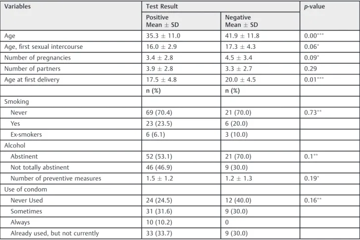
Table 1 E6/E7 test results according to the epidemiological characteristics of women referred to a primary care service with Pap test compatible with high-grade squamous intraepithelial lesion (HSIL)
Variables Test Result p-value
Positive MeanSD
Negative MeanSD
Age 35.311.0 41.911.8 0.00
Age,first sexual intercourse 16.02.9 17.34.3 0.06
Number of pregnancies 3.42.8 4.53.4 0.09
Number of partners 3.92.8 3.32.7 0.29
Age atfirst delivery 17.54.8 20.04.5 0.01
n (%) n (%)
Smoking
Never 69 (70.4) 21 (70.0) 0.73
Yes 23 (23.5) 6 (20.0)
Ex-smokers 6 (6.1) 3 (10.0)
Alcohol
Abstinent 52 (53.1) 21 (70.0) 0.1
Not totally abstinent 46 (46.9) 9 (30.0)
Number of preventive measures 1.51.2 1.21.3 0.19
Use of condom
Never Used 24 (24.5) 12 (40.0) 0.16
Sometimes 31 (31.6) 9 (30.0)
Always 10 (10.2) 0
Already used, but not currently 33 (33.7) 9 (30.0)
Abbreviation: SD, standard deviation.
performed with Gaylor-Medina forceps. The fragments were placed in a vial with 10% buffered formalin and sent for histopathological examination.
In the pathology laboratory, the fragments were processed and embedded in paraffin and subsequently sliced to obtain 5-µm-sections, which were stained with hematoxylin-eosin and analyzed by optical microscopy. The reports were in accordance with the international nomenclature of pathology for cervical intraepithelial neoplasia and cervical cancer.12
The responses to the questionnaire and the results of the examinations and procedures were entered in the EPI-INFO program. The chi-square test and the chi-square test for trend were used to compare categorical variables and the t-test for comparison of means. The Mann-Whitney test was used for the variables that did not present a normal distri-bution. The differences were considered statistically signifi -cant whenp<0.05. All analyzes were performed using the Stata (version 10.1) statistical software.
Results
Samples were obtained from the 140 women presenting with HSIL to detect E6/E7 mRNA of HPV types 16, 18, 31, 33, and 45. Out of all cases, 8.6% (12/140) presented invalid data, 21.4% (30/140) were negative, and 70% (98/140) positive.
Among the 128 valid tests, 23.4% (30/128) were negative for E6/E7 mRNA. Among the 98 positive cases, only one type of HPV was detected in 87.7% of the samples (86/98).
The comparison of socio-demographic characteristics between the patients with negative and positive results showed no significant difference (►Table 1).
HPV16 was the most common type detected, 61.2% (60/ 98), followed by HPV33, 26.5% (26/98), HPV31, 17.3% (17/98), HPV18, 5.1% (5/98), and HPV45, 4.1% (4/98).
Among the 12.2% (12/98) cases presenting multiple in-fections, the most frequently detected type was also HPV16 (83.3%), followed by HPV33 (75%), HPV45 (25%), HPV31 (16.7%), and HPV18 (8.3%).
The HPV18 type was detected in one biopsy displaying adenocarcinoma, in the only cytology that displayed
adeno-carcinomain-situ(AIS), and in one of the two cytology with adenocarcinoma; in the other cytology, HPV16 was identified. When the cytology was repeated, the following results were observed: unsatisfactory, two (1.4%); negative, 23 (16.4%); atypical squamous cells of undetermined signifi -cance (ASC-US), eight (5.7%); low-grade squamous intra-epithelial lesion (LSIL), 23 (16.4%); atypical squamous cells in which HSIL cannot be excluded (ASC-H), 6 (4.3%); HSIL, 70 (50.0%); HSIL without exclusion of microinvasion, one (0.7%); squamous cancer, four (2.9%); atypical squamous cells, one (0.7%); adenocarcinoma, two (1.4%).
The E6/E7 test was positive in 83.8% of women with ASC-H, HSIL, or greater, while, in women presenting a negative cytology, E6/E7 test was positive in 57.9% of the cases (►Table 2).
Of the 140 colposcopy examinations, 11.4% of the cases presented no lesion and 6.3% presented unspecific or mis-cellaneous changes. Abnormal low-grade colposcopy fi nd-ings were identified in 42.9% of the cases and higher gradecolposcopy findings in 39.3%. The squamocolumnar junction was not visualized in 37.9% of the cases.
Among the 128 women who were subjected to colposcopy and for which the E6/E7 test was valid, 82% presented abnormal colposcopy findings and 18% showed no lesion or nonspecific lesion. The frequency of E6/E7 positivity ranged from 57.1% in women without colposcopy-detected lesions and up to 86.5% in those with higher-grade colposco-py-detected lesions.
Histopathological examination of 124 biopsy samples showed the following results: negative, 13.6%; CIN1, 15.7%; CIN2, 19.3%; CIN3, 35.7%; microinvasion, 0.7%; squamous cancer, 0.7%; adenocarcinoma, 0.7%; CIN ungraded, 1.4%; and inadequate sample, 0.7%.
Among the 111 women who underwent a biopsy and presented a valid E6/E7 test, the E6/E7 test was positive in 84.72% of the women presenting with neoplasia equal or more serious than CIN2 and in 41.18% of women present-ing with negative biopsy. The sensitivity of the test for CIN2 or higher was 84.7%, while the specificity was 30.8%. The positive predictive value was 69.3%. The negative
Table 2 Relationship between colposcopy-detectedfindings and positivity of HPV E6/E7 mRNA test in women referred to a primary care service with a Pap test compatible with high-grade squamous intraepithelial lesion (HSIL)
Cytology Test Result Total
Positive Negative
N % N % N %
Negative 11 57.8 8 42.1 19 15.4
ASC-US / LSIL 22 73.3 8 26.6 30 24.3
ASC-H /HSIL 62 83.7 12 16.2 74 60.1
Total 95 77.2 28 22.7 123 100.0
Abbreviation: ASC-H, atypical squamous cells of undetermined significance in which high-grade squamous intraepithelial lesion cannot be excluded; ASC-US, atypical squamous cells of undetermined significance possibly non-neoplastic; HSIL, high-grade squamous intraepithelial lesion; LSIL, low-grade squamous intraepithelial lesion.
Pvalue¼0.02127; x2trend value¼5.30481.
predictive value was 52.2%, and the accuracy was 65.7% (►Table 3).
Discussion
The E6/E7 mRNA test of HPV types 16, 18, 31, 33, and 45 was positive in 76% of women who presented with a cytological diagnosis of HSIL and in 83% of the women whose diagnosis was confirmed by new cytology tests performed for the study. There was an increase in the percentage of positivity proportional to the grading of the cytological and histopath-ological changes. HPV16 was detected with higher frequency, followed by HPV 33, 31, 18, and 45. Suchfindings are similar to those of Dixon et al,13who observed positive results in 80% of HSIL samples. The cytological, colposcopic, and histologi-cal correlations confirmed the observations of other authors.8,9,14
Among the cases with negative cytology, ASC-H cases had 57.9% with an E6/E7 positive test, while the≥HSIL cases had 83.8%. Cases in which the cytology was negative and the E6/ E7 test was positive suggest the possibility of false negative cytology or viral integration without morphological le-sions.15,16According to Li and Kristensen,7when the cytolo-gy examination reveals negative result and the E6/E7 test is positive, one could consider a latent infection. Negative E6/ E7 tests observed in women with high-grade colpocytologic findings could be due to the presence of other types of HPV, not screened in this study.7 However, previous studies suggest caution in these cases due to the higher risk of other importantfindings in the follow-up.17
The HPV E6/E7 mRNA test was positive in 85% of the biopsies with CIN2 or higher, in agreement with the variation described by others, who detected HPV E6/E7 mRNA be-tween 63% and 92% of CIN2, 3, or cervical cancer.7,18,19Such findings demonstrated an interesting sensitivity for the identification of high-grade and invasive lesions and are in agreement with the literature.20
In this study, HPV16 was identified as the most frequent type (61%), in agreement with other studies, taking into account the regional variability of other types.21–26 The HPV18 type, related with glandular lesions,27–29was associ-ated with confirmed adenocarcinoma.
The performance of the test was studied in relation to CIN2 or higher grade, considering the histopathologic diag-nosis as the gold standard.30,31The sensitivity of the E6/E7 test, 84% for CIN2/3, was greater than that observed by
Jeantet et al,32 who determined a 73.6% sensitivity for CIN2 and 82.2% for CIN3. These numbers were lower than that obtained by Pierry et al,33who observed a 89% sensitiv-ity for CIN2 and 100% for CIN3. These numbers led some researchers to suggest the use of the E6/E7 test as a screening test in cases with abnormal cytology or HPV-DNA tests.34
Given the results, one can conclude that the expression of E6/E7 mRNA was observed the most among patients with HSIL and CIN2 or higher grades in the biopsy, which suggests an important correlation that could be inferred as a prog-nostic factor. Nevertheless, despite the tendency to use E6/E7 mRNA screening test due to its prognostic value, new and more well-designed studies are needed to confirm the effectiveness of this test.
Referências
1 Fitch MI, Greenberg M, Cava M, Spaner D, Taylor K. Exploring the barriers to cervical screening in an urban Canadian setting. Cancer Nurs 1998;21(6):441–449
2 zurHausen H. Papillomaviruses in the causation of human cancers -a brief historic-al -account. Virology 2009;384(2):260–265 3 Boicea A, Pătraşcu A, Surlin V, Iliescu D, Schenker M, Chiuţu L.
Correlations between colposcopy and histologic results from colposcopically directed biopsy in cervical precancerous lesions. Rom J MorpholEmbryol 2012;53(3, Suppl)735–741
4 Nanda K, McCrory DC, Myers ER, et al. Accuracy of the Papanico-laou test in screening for and follow-up of cervical cytologic abnormalities: a systematic review. Ann Intern Med 2000;132-(10):810–819
5 International Agency for Research on Cancer Working Group on the Evaluation of Carcinogenic Risks to Humans [Internet]. Hu-man papillomaviruses. Lyon: IARC; 2005. (IARC monographs on evaluation of carcinogenic risks to humans, 90) [cited 2015 Jul 19]. Available from: http://monographs.iarc.fr/ENG/Monographs/ vol90/mono90.pdf
6 Wright TC Jr. HPV DNA testing for cervical cancer screening. FIGO 26th Annual Report on the Results of Treatment in Gynecological Cancer. Int J GynaecolObstet 2006;95(Suppl 1): S239–S246
7 Lie AK, Kristensen G. Human papillomavirus E6/E7 mRNA testing as a predictive marker for cervical carcinoma. Expert Rev Mol-Diagn 2008;8(4):405–415
8 Cattani P, Zannoni GF, Ricci C, et al. Clinical performance of human papillomavirus E6 and E7 mRNA testing for high-grade lesions of the cervix. J ClinMicrobiol 2009;47(12):3895–3901
9 Argyri E, Tsimplaki E, Daskalopoulou D, et al. E6/E7 mRNA expression of high-risk HPV types in 849 Greek women. Antican-cer Res 2013;33(9):4007–4011
Table 3 E6/E7 mRNA test results according to histopathological results in women referred to a primary care service with a Pap test compatible with high-grade squamous intraepithelial lesion (HSIL)
E6/E7 Test CIN2 (%) <CIN2 (%) Total (%)
Positive 61 (69.3) 27 (30.6) 88 (79.2)
Negative 11 (47.8) 12 (52.1) 23 (20.7)
Total 72 (64.8) 39 (35.1) 111 (100.0)
10 Sellors JW, Sankaranarayanan R. Colposcopy and treatment of cervical intraepithelial neoplasia: a beginner’s manual. Lyon: IARC; 2004. Chapter 1: An introduction to the anatomy of the uterine cervix [cited 2015 Jul 19]. Available from:<
http://screen-ing.iarc.fr/doc/colpochapter01.pdf>
11 International Federation for Cervical Pathology and Colposcopy [Internet]. IFCPC colposcopic terminology of the cervix. 2011 [cited 2014 May 06]. Available from: <http://www.ifcpc.org/
images/docs/nomenclature7-11.pdf>
12 Sellors JW, Sankaranarayanan R. Colposcopy and treatment of cervical intraepithelial neoplasia: a beginner’s manual. Lyon: IARC; 2004 [cited 2015 Jul 19]. Chapter 2: Introduction to cervical intraepithelial neoplasia (CIN). Available from:<http://screening.
iarc.fr/doc/colpochapter02.pdf>
13 Dixon EP, King LM, Adams MD, et al. Isolation of RNA from residual BD SurePath liquid-based cytology specimens and detection of HPV E6/E7 mRNA using the PreTectt HPV-Proofer assay. J Virol Methods 2008;154(1–2):220–222
14 Galarowicz B, Jach R, Kidzierska J, et al. The role of mRNA E6/E7 HPV high oncogenic risk expression in colposcopy of cervical intraepithelial neoplasia (CIN). PrzeglLek 2012;69(9):651–657 15 Cernescu EC, Anton G, RuţăS, Cernescu C. The effectiveness of
cytological rescreening in the reduction of false negative/ positive Pap reports. Roum Arch MicrobiolImmunol 2013; 72(2):93–104
16 Poomtavorn Y, Himakhun W, Suwannarurk K, Thaweekul Y, Maireang K. Cytohistologic discrepancy of high-grade squamous intraepithelial lesions in Papanicolaou smears. Asian Pac J Cancer Prev 2013;14(1):599–602
17 Giorgi Rossi P, Benevolo M, Vocaturo A, et al. Prognostic value of HPV E6/E7 mRNA assay in women with negative colposcopy or CIN1 histology result: a follow-up study. PLoS ONE 2013;8(2): e57600
18 Massad LS, Einstein MH, Huh WK, et al; 2012 ASCCP Consensus Guidelines Conference. 2012 updated consensus guidelines for the management of abnormal cervical cancer screening tests and cancer precursors. J Low Genit Tract Dis 2013;17(5, Suppl 1): S1–S27
19 Halfon P, Benmoura D, Agostini A, et al. Relevance of HPV mRNA detection in a population of ASCUS plus women using the NucliSENSEasyQ HPV assay. J ClinVirol 2010;47(2):177–181
20 Reid JL, Wright TC Jr, Stoler MH, et al. Human papillomavirus oncogenic mRNA testing for cervical cancer screening: baseline and longitudinal results from the CLEAR study. Am J ClinPathol 2015;144(3):473–483
21 Kraus I, Molden T, Ernø LE, Skomedal H, Karlsen F, Hagmar B. Human papillomavirus oncogenic expression in the dysplastic portio; an investigation of biopsies from 190 cervical cones. Br J Cancer 2004;90(7):1407–1413
22 Muñoz N, Bosch FX, Castellsagué X, et al. Against which human papillomavirus types shall we vaccinate and screen? The interna-tional perspective. Int J Cancer 2004;111(2):278–285
23 Ali-Risasi C, Verdonck K, Padalko E, Vanden Broeck D, Praet M. Prevalence and risk factors for cancer of the uterine cervix among women living in Kinshasa, the Democratic Republic of the Congo: a cross-sectional study. Infect Agent Cancer 2015;10:20 24 de Mendonça VG, Guimarães MJ, de Lima Filho JL, et al. [Human
papillomavirus cervical infection: viral genotyping and risk factors for high-grade squamous intraepithelial lesion and cervix cancer]. Rev Bras GinecolObstet 2010;32(10):476–485Portuguese 25 Kirschner B, Schledermann D, Holl K, et al. HPV-genotypes in
high-grade intraepithelial cervical lesions in Danish women. ActaObstetGynecolScand 2013;92(9):1032–1040
26 Mu-Mu-Shwe, Harano T, Okada S, et al. Prevalence of high-risk human papillomavirus (HR-HPV) infection among women with normal and abnormal cervical cytology in Myanmar. Acta Med Okayama 2014;68(2):79–87
27 Molden T, Kraus I, Karlsen F, Skomedal H, Nygård JF, Hagmar B. Comparison of human papillomavirus messenger RNA and DNA detection: a cross-sectional study of 4,136 women>30 years of age
with a 2-year follow-up of high-grade squamous intraepithelial lesion. Cancer Epidemiol Biomarkers Prev 2005;14(2):367–372
28 Rabelo-Santos SH, Derchain SF, Villa LL, et al. Human papilloma-virus-specific genotypes in cervical lesions of women referred for smears with atypical glandular cells or adenocarcinoma in situ. Int J GynecolPathol 2009;28(3):272–278
29 Kim JY, Nam BH, Lee JA. Is human papillomavirus genotype an influencing factor on radiotherapy outcome? Ambiguity caused by an association of HPV 18 genotype and adenocarcinoma histology. J GynecolOncol 2011;22(1):32–38
30 Andersson S, Mints M, Wilander E. Results of cytology and high-risk human papillomavirus testing in females with cervical adenocarcinoma in situ. OncolLett 2013;6(1):215–219
31 Aidé S, Almeida G, Val I, Vespa Junior N, Campaner AR. Neoplasia intraepitalial cervical. DST J Bras Doenças Sex Transm 2009;21(4): 166–170
32 Jeantet D, Schwarzmann F, Tromp J, et al. NucliSENSEasyQ HPV v1 test - Testing for oncogenic activity of human papillomaviruses. J ClinVirol 2009;45(Suppl 1):S29–S37
33 Pierry D, Weiss G, Lack B, Chen V, Fusco J. Intracellular human papillomavirus E6, E7 mRNA quantification predicts CIN 2þin cervical biopsies better than Papanicolaou screening for women regardless of age. Arch Pathol Lab Med 2012;136(8):956–960