Langerhans cell histiocytosis: 37 cases in a single Brazilian institution
1
Hematology Service, Hospital das Clínicas, Universidade Federal de Minas Gerais – UFMG,
Belo Horizonte, MG, Brazil 2
Pediatrics Department, Universidade Federal de Minas Gerais – UFMG, Belo Horizonte, MG, Brazil
3
Pathological Anatomy and Legal Medicine Department, Universidade Federal de Minas Gerais – UFMG, Belo Horizonte, MG, Brazil
Luciana Terra Babeto1
Benigna Maria de Oliveira2
Lúcia Porto Fonseca de Castro3
Márcia Kanadani Campos1
Maria Thereza Macedo Valadares2
Marcos Borato Viana2
Objectives: To improve the level of 'definitive' diagnosis of Langerhans cell histiocytosis by immunohistochemical investigation of the CD1a surface antigen and to compare outcomes in respect to age, gender, stage of the disease, treatment response and level of diagnostic accuracy.
Methods: A retrospective study was carried out of 37 children and adolescents with possible Langerhans cell histiocytosis between 1988 and 2008. The diagnoses were revisited using immunohistochemical investigations for CD1a, S-100 and CD68 in an attempt to reach definitive diagnoses for all cases.
Results: Before the study, only 13 of 37 patients (35.1%) had a 'definitive' diagnosis; by the end of the study, this number rose to 25 patients (67.6%). All reviewed cases were positive for the CD1a antigen. Overall survival was 88.5%. Multisystem disease (Stage 2; n=19) and absence of response at the 6th week of therapy (n=5) were associated to significantly lower overall survival (p-value = 0.04 and 0.0001, respectively). All deaths occurred in patients with multisystem disease and organ dysfunction at diagnosis. Other potential prognostic factors were not significant. Reactivation episodes occurred in 75% of the patients with multisystem disease. Diabetes insipidus was the most common sequel (21.6%).
Conclusion: The level of diagnostic accuracy was increased through immunohistochemistry. The overall survival rate was similar to international multicentric studies. Multisystem disease and absence of response at six weeks of treatment were the most important unfavorable prognostic factors. The frequency of reactivation for patients with multisystem disease was higher than described in the literature, probably because maintenance chemotherapy was used only in two cases.
Keywords: Histiocytosis; Langerhans-cell; Pathology; Prognosis; Diabetes insipidus; Otitis
Introduction
Langerhans cell histiocytosis (LCH) is a rare disease that mainly affects children. Of unknown etiology, it is caused by clonal proliferation of Langerhans cells and may involve any organ or tissue in the body. The clinical manifestations are a result of this involvement and can range from isolated lesions that evolve into spontaneous remission to multisystemic and severe forms leading to death. The sites most commonly affected are the bones, skin and lymph nodes. Frequent recurrences can lead to significant morbidity and severe sequelae may therefore result. Depending on the clinical presentation, treatment can range from watchful waiting, surgical excision of lesions, steroid therapy, chemotherapy and
bone marrow transplantation.(1)
'Presumptive' diagnosis is considered when conventional histology of the lesions is characteristic; 'probable', when, in addition, two of the following histochemical reactions are positive: S100 protein, mannosidase, ATPase or peanut lecithin. A 'definitive' diagnosis is reached when, associated with a suggestive clinical picture, immunohistochemical investigation of suspected lesions test positive for the CD1a antigen or when Birbeck
granules are found under electron microscopy.(2) Recently, positive staining with Langerin
(CD207) has also been considered a definitive diagnosis.(1,3) The main risk factors related to
death are multisystem disease, risk-organ involvement and the absence of response by the 6th week of therapy.(4-6)
Only 7 out of 33 patients included in a previous study from the same group of
investigators had definitive diagnoses for the disease(7) when, ideally, all cases should
have one.(4,5,8)
The main aim of the current study was to establish definitive diagnoses for those patients as well as the evaluation of four new cases. Disease outcomes were compared
according to age, gender, disease stage, response at the 6th week of therapy and diagnostic
certainty.
Conflict-of-interest disclosure: The authors declare no competing financial interest
Submitted: 3/10/2011 Accepted: 7/4/2011
Corresponding author:
Benigna Maria de Oliveira
Departamento de Pediatria e Hospital das Clínicas da Universidade Federal de Minas Gerais – UFMG
Avenida Professor Alfredo Balena, 190 sala 267
30130-100 – Belo Horizonte, MG, Brazil Phone: 55 31 3409-9772
www.rbhh.org or www.scielo.br/rbhh
Methods
The study was retrospective (1988-2008) and analyzed all patients with ages at diagnosis of less than 18 years old between June 1988 and December 2008. The patients were diagnosed and treated at a single university hospital in Brazil. Clinical data were obtained through a review of medical records.
Criteria for histological diagnosis were based on the
recommendations by the Histiocyte Society.(2) 'Presumptive'
diagnosis was established on conventional histological
findings; 'probable' diagnosis, when these findings were
associated with positive staining for protein S-100 by
immunohistochemistry. A 'definitive' diagnosis was reached
based on electron microscopy (presence of Birbeck granules) or CD1a positive immune staining. The paraffin blocks and slides from previous biopsies of all cases without definitive diagnoses were retrieved as were three cases for which a definitive diagnosis had previously been reached. Immunohistochemical investigations for CD1a, S-100, and CD68 were carried out for all available blocks. The corresponding histological slides were analyzed by two of the involved investigators.
Patients were retrospectively classified(6) as:
– Stage 1: Only one organ or system involved. The involvement can be unifocal (1a) or multifocal (1b);
– Stage 2: Multisystem disease with more than one organ or system involved, with or without organ dysfunction. Patients were retrospectively divided into 3 categories, according to the response observed at the 6th week of therapy:(6)
– "Better" response: total resolution or continued regression of disease;
– Intermediate response: stable disease or regression in some sites with new lesions in others;
– Lack of response ("worse"): disease progression. Reactivation was defined as the appearance of new lesions or organ dysfunction after remaining stable for three
months.(9) The Kaplan-Meier method was used for survival
analyses. Event free survival (EFS) was estimated considering both death and reactivation as events. Patients without any events on the day of statistical analysis (December 15, 2008) were censored. The Logrank test was
used to compare survival curves. A p-value ≤ 0.05 was
considered statistically significant.
The study was approved by the Universidade Federal de Minas Gerais Research Ethics Committee (Protocol ETIC 271/07).
Results
For the period of 1988-2008, 37 patients were followed up, 20 of which were female (54%). Age at diagnosis varied from 1 month to 16.9 years old (median 2.4 y). Time of follow up varied from 1 month to 20.9 years (median 6.2 y).
The general epidemiological data of patients are summarized in Table 1.
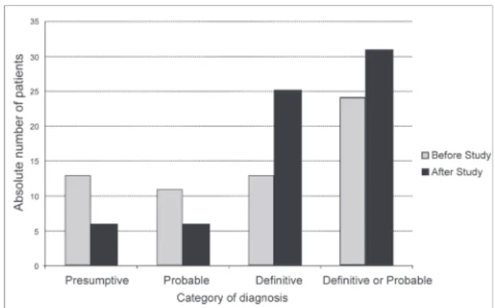
Thirteen (35.1%) of the 37 patients had had 'definitive'
diagnoses before the study. Of the 24 patients without a 'definitive' diagnosis, 11 (29.7%) had previous 'probable'
diagnoses and 13 (35.1%) had 'presumptive' diagnoses. Of
the 11 patients with 'probable' diagnoses, 5 samples were
found for review. Of the 13 patients with 'presumptive'
diagnoses, 7 samples were found. We also reviewed 3
randomly selected cases that previously had 'definitive'
diagnoses, (2 with previous CD1a positivity and 1 with electron microscopy evidence). All 15 reviewed samples tested positive for CD1a.
At the end of the study, 25 patients (67.6%) had 'definitive' diagnoses and 31 (83.8%) had 'probable' or 'definitive' diagnosis (Figure 1).
classification of the Histiocyte Society. Eleven patients (29.7%) had unifocal (Stage 1a) and seven (18.9%) multifocal disease (Stage 1b).
The most commonly used treatment initially was prednisone with vinblastine (17 out of 37 patients; 45.9%). Three patients were submitted to the treatment protocol proposed by the Histiocyte Society International Study Protocol (LCH III - Arm A). Of the 17 remaining patients, six received etoposide, three received prednisone alone, three underwent surgical resection, two received no treatment (watchful waiting) and three received other types of treatment. Only two patients received maintenance chemotherapy as part of the Histiocyte Society LCH-III protocol. A third patient, also treated with this protocol, had not reached the maintenance phase at the time of censoring for this report.
In relation to response by the 6th week of therapy, 27
(73%) had a "better" response, 4 (10.8%) an intermediate response and 5 (13.5%) a lack of response. It was impossible to evaluate the response in one case for lack of data. This patient was excluded from the survival analysis for this variable.
At the end of the study, 29 patients were in complete remission: eight had been discharged and 21 were still in follow-up. Four children had died. Clinical follow-up was lost for four patients: one was in remission at the time of his last visit (5 years from diagnosis), but had presented an episode of reactivation in the seventh month of observation. This event was considered in the analysis of EFS; for overall survival (OS), the patient was censored on that date. Two patients who dropped out were in continuous clinical remission and were censored at the date of their last visit with 4 and 5 years of follow-up. The fourth patient had not achieved clinical remission and abandoned her follow-up after one and a half years (Table 1).
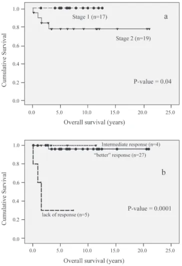
OS for the whole group was 88.5% (95% confidence interval - 95% CI: 72.1-95.5%). As previously mentioned, four patients died. All deaths occurred among patients with multisystem disease and organ dysfunction at diagnosis. OS was significantly higher for patients with single-system (100%) when compared to those with multisystem disease (77.2%; 95% CI: 49.7-90.8%; p-value = 0.04; Figure 2a).
OS probability was significantly lower for patients with no response at the 6th week of treatment (30%; 95% CI: 1.2-71.9%) when compared to patients with 'better' (95.8%; 95% CI: 73.9-99.4%) or 'intermediate' response (100%; p-value = 0.0001; Figure 2b). There was no statistically significant difference in OS in terms of age younger or older than 2 years old (p-value = 0.14), age younger or older than 3 years old (p-value = 0.47), gender (p-value = 0.96) or degree of diagnostic certainty (p-value = 0.71).
The probability of EFS at 10 years for the whole group was 32.5% (95% CI: 17.9-48.1%). Of the 33 who entered into remission, 20 children (60.6%) had at least one reactivation episode. When only patients with multisystem disease are considered, a higher rate (75%) of reactivation was recorded. The majority of first reactivation episodes (60%) happened within the first two years of follow-up.
The probability of EFS was 47.1% (95% CI: 23-68%) for patients with single-system disease and 14.1% (95% CI: 26-35.1%) for patients with multisystem disease, a difference that was not statistically significant (p-value = 0.12). When patients with complete response were added to those with intermediate response, the probability of EFS was 38.9%. Patients unresponsive at the sixth week of treatment had 0% probability of EFS, a difference that was statistically significant (p-value = 0.0001). There was no statistically significant difference regarding gender (p-value = 0.8) or age younger or older than two years (p-value = 0.81).
There was no statistically significant difference in the probability of OS and EFS with respect to the degree
of diagnostic certainty (p-value = 0.71 and 0.39, respectively). The presence of one or more permanent sequelae occurred in 15 of the 37 patients (40.5%) with diabetes insipidus, detected in eight cases (21.6%), being the most common. Of the 15 patients with sequelae, 9 (60%) had multisystem disease and of the total of patients with multisystem disease, 47.4% presented with at least one permanent sequel.
One patient developed acute promyelocytic leukemia two years and nine months after etoposide had been given as treatment for disease relapse. Prednisone with vinblastine without radiation was the initial treatment for this patient.
Discussion
Diagnostic confirmation of LCH is very important in particular in dubious cases – such as in cases in which the
disease is confined to lymph nodes(10) – and also to compare
results with those reported in international studies that only
include patients with definitive diagnoses.(4,5,8) Despite the
limitations of a retrospective study, the main aim of the present study was attained with an increase in the proportion of patients with definitive diagnoses (from 35% to 67.5%). All the reviewed tests were positive for CD1a. This shows the importance of carrying out conventional histological examinations routinely by professionals with experience at diagnosing LCH. Moreover, this demonstrates how effective and practical the use of CD1a detection can be in confirming the disease: paraffin blocks obtained as early as 1988 stained positively, for instance. Langerin was not used because it was unavailable in Brazil when this study was carried out.
Some studies also include patients with 'probable'
diagnoses, in addition to those with 'definitive'
diagnoses.(11,12) At the end of this study, 31 of the 37 patients
(83.8%) had 'probable' or 'definitive' diagnoses. Of the 6
patients who remained with 'presumptive' diagnoses, 4 presented clinical manifestations highly suggestive of the disease, such as diabetes insipidus and multisystem disease. Lymphadenomegaly was the single clinical manifestation in two patients and only in these, a diagnosis of LCH might be considered dubious. For this reason, every case was included in the statistical analyses.
Clinical manifestations at diagnosis were very similar
to international reports.(13,14) Isolated or combined osteolytic
lesions are by far the commonest reported manifestation and were recorded in two thirds of our patients. Lymphadenomegaly and cutaneous or mucocutaneous involvement are also very common and were present in almost half of our patients. A quarter of patients had involvement of the external auditory canal at diagnosis. This finding is relevant in the general pediatric practice as there is a hypothesis that LCH should be considered in children with persistent otitis refractory to usual treatments, associated with dermatitis of the outer ear.
The estimated OS was similar to that found in
international multicenter studies.(4-6) Likewise, bad prognostic
factors in the present study were multisystem disease with risk-organ dysfunction and the absence of response by the
6th week of therapy.(4-6) Results of the LCH II study showed
that therapy intensification is of crucial importance for this type of patient.(6)
The majority of patients who had remission at some point presented at least one episode of disease reactivation. Most reactivation episodes occurred within the first two years
of follow-up which is in agreement with previous reports.(9,15)
Also similar to reports in the literature, the majority of patients with permanent sequelae presented multisystem
disease.(9,16,17) The frequency of reactivations in the group
with multisystem disease was considerably higher than that
verified in international studies.(4-6) A number of treatment
protocols were used during the studied period, which makes it impossible to compare treatment of this study with others. Remarkably, however, only two patients received maintenance
Figure 2 – Overall survival by groups (Kaplan-Meier's method); small marks over the curves represent censored patients;
a: Curves according to disease stage at diagnosis; Stage 1 (n = 17): involvement of one organ or system with unifocal or multifocal lesions; Stage 2 (n = 19): multisystem disease (p-value = 0.04);
therapy. With the added knowledge of more recent
publications,(1) we can state that many patients who had LCH
reactivation could probably have benefited from an increase in therapy intensity and/or duration to reduce reactivations, and possibly, sequelae.
References
1. Histiocyte Society. Langerhans cell histiocytosis. Evaluation and treatment guidelines. Histiocyte Society; April 2009.
2. The Clinical Writing Group of the Histiocyte Society. Histiocytosis syndromes in children. Lancet. 1987;1(8526): 208-9.
3. Valladeau J, Ravel O, Dezutter-Dambuyant C, Moore K, Kleijmeer M, Liu Y, et al. Langerin, a novel C-type lectin specific to Langerhans cells, is an endocytic receptor that induces the formation of Birbeck Granules. Immunity. 2000;12(1):71-81. 4. Minkov M, Grois N, Heitger A, Pötschger U, Westermeier T, Gadner
H. Treatment of multisystem Langerhans cell histiocytosis. Results of the DAL-HX 83 e DAL-HX 90 studies. Klin Padiatr. 2000;212 (4):139-44.
5. Gadner H, Grois N, Arico M, Broadbent V, Ceci A, Jakobson A, et al. A randomized trial of treatment for multisystem Langerhans cell histiocytosis. J Pediatr. 2001;138(5):728-34. Comment in: J Pediatr. 2002;140(2):280. Erratum in: J Pediatr. 2001;139 (1):170.
6. Gadner H, Grois N, Pötschger U, Minkov M, Aricò M, Braier J, et al. Improved outcome in multisystem Langerhans cell histiocytosis is associated with therapy intensification. Blood. 2008; 111 (5):2556-62. Comment in: Blood. 2008;112(8):3527; author reply 3528.
7. Campos MK, Viana MB, Oliveira BM, Ribeiro DD, Silva CM. Langerhans cell histiocytosis: a 16-year experience. J Pediatr (Rio J). 2007;83(1):79-86.
8. Narula G, Bhagwat R, Arora B, Banavali S, Pai S, Nair C, et al. Clinico-Biologic profile of Langerhans cell histiocytosis: a single institutional study. Indian J Cancer. 2007;44(3):93-8.
9. Pollono D, Rey G, Latella A, Rosso D, Chantada G, Braier J. Reactivation and risk of sequelae in Langerhans cell histiocytosis. Pediatr Blood Cancer. 2007;48(7):696-9.
10. Khadilkar UN, Rao AT, Sahoo KK, Pai MR. Langerhans cell histiocytosis of mediastinal node. Indian J Pediatr. 2008;75 (3):294-6.
11. Morimoto A, Ikushima S, Kinugawa N, Ishii E, Kohdera U, Sako M, Fujimoto J, Bessho F, Horibe K, Tsunematsu Y, Imashuku S; Japan Langerhans Cell Histiocytosis Study Group. Improved outcome in the treatment of pediatric multifocal Langerhans cell histiocytosis: Results from the Japan Langerhans Cell Histiocytosis Study Group-96 Protocol Study. Cancer. 2006;107(3):613-9.
12. Bernstrand C, Sandstedt B, Ahström L, Henter JI. Long-term follow-up of Langerhans cell histiocytosis: 39 years' experience at a single centre. Acta Paediatr. 2005;94(8):1073-84.
13.Aricò M, Egeler R. Clinical aspects of Langerhans cell histiocytosis. Hematol Oncol Clin North Am. 1998;12(2):247-58.
14.Salotti JA, Nanduri V, Pearce MS, Parker L, Lynn R, Windebank KP. Incidence and clinical features of Langerhans cell histiocytosis in the UK and Ireland. Arch Dis Child. 2009;94(5):376-80.
15. Minkov M, Steiner M, Pötschger U, Aricò M, Braier J, Donadieu J, Grois N, Henter JI, Janka G, McClain K, Weitzman S, Windebank K, Ladisch S, Gadner H; International LCH Study Group. Reactivations in multisystem Langerhans Cell Histiocytosis: data of the international LCH registry. J Pediatr. 2008;153(5):700-5.
16.Arceci RJ. The Histiocytosis: the fall of the Tower of Babel. Eur J Cancer. 1999;35(5):747-67; discussion 767-9
17.Lau LM, Stuurman K, Weitzman S. Skeletal Langerhans cell histiocytosis in children: permanent consequences and health-related quality of life in long-term survivors. Pediatr Blood Cancer. 2008;50(3):607-12.