ABSTRACT
BACKGROUND AND OBJECTIVES:Temporomandibular
disorders are diseases causing pain and dysfunction in joints and muscles controlling mandibular movements. heir etiology is multifactorial and multidisciplinary approaches are needed to reach a diferential diagnosis and an adequate management plan. his case report proposes a management protocol, with monthly sodium hyaluronate iniltrations, with diferent mo-lecular weights, to control such changes and promote improve-ment of temporomandibular joint biomechanics and pain. CASE REPORT:his study describes a case of a 48-year old patient with a 10-year history of temporomandibular pain with function loss since 2001. Patient has classiied her pain as 9 according to analog visual scale. In addition, history and detailed physical evaluation have shown diferent signs and symptoms, such as localized pain (right side), and right temporomandibular joint arthralgia with noise. Diagnosis was disc displacement with reduction and possible synovitis/ capsulitis to the right. Right temporomandibular joint osteo-arthritis was also diagnosed by cone beam CT-scan. Initially, a lat upper splint with total coverage and contact with all an-tagonist teeth was used. In the attempt to decrease temporo-mandibular arthralgia, non-steroid anti-inlammatory drugs and muscle relaxants were used. Since right temporomandib-ular joint pain was not efectively managed, we decided to use intra-joint sodium hyaluronate injections with diferent mo-lecular weights, per month, in a total of four applications. At treatment completion, clinical evaluation has shown normal
Sequential infiltration of sodium hyaluronate in the temporomandibular
joint with different molecular weights. Case report
Infiltração sequencial de hialuronato de sódio com diferentes pesos moleculares na articulação
temporomandibular. Relato de caso
Eduardo Grossmann1, Roberta Fonseca2, Camila Almeida-Leite2, Rafael Tardin Gonçalves3, Pedro Gonçalves de Oliveira4,
Eduardo Januzzi3
1. Universidade Federal do Rio Grande do Sul, Porto Alegre, RS, Brasil. 2. Universidade Federal de Minas Gerais, MG, Brasil.
3. Ciodonto, Belo Horizonte, MG, Brasil. 4. Universidade Anhembi, São Paulo, SP. Brasil.
Submitted in July 27, 2015.
Accepted for publication in October 13, 2015. Conlict of interests: none – Sponsoring sources: none.
Correspondence to: Eduardo Grossmann
Rua Coronel Corte Real, 513, Bairro Petrópolis. 90630-080 Porto Alegre, RS,Brasil.
E-mail: [email protected]
© Sociedade Brasileira para o Estudo da Dor
function, no pain with visual analog scale = zero, in addition to adequate interincisal distance.
CONCLUSION:his report has suggested that viscosupple-mentation cycles with sodium hyaluronate of diferent molecu-lar weights may provide excellent results in the long run, to control joint temporomandibular disorder symptoms. hera-peutic beneits were maintained for four years with no need for annual maintenance cycles.
Keywords: Disk displacement with reduction, Sodium hyal-uronate, Temporomandibular joint, Viscosupplementation.
RESUMO
JUSTIFICATIVA E OBJETIVOS: As disfunções temporo-mandibulares são um grupo de doenças que causam dor e dis-função na articulação e nos músculos que controlam os movi-mentos da mandíbula. Sua etiologia é multifatorial e abordagens multidisciplinares são necessárias para chegar a um diagnóstico diferencial e plano de tratamento adequado. Propõe-se um pro-tocolo de tratamento, com iniltrações mensais, empregando hialuronato de sódio, com diferentes pesos moleculares, para controlar essas alterações e promover uma melhoria da função biomecânica da articulação temporomandibular, bem como da sua dor.
RELATO DO CASO: Este estudo descreve um caso de uma paciente de 48 anos que apresentava uma história de 10 anos de dor temporomandibular com perda de função que apare-ceu em 2001. Ela classiicou a intensidade da sua dor como grau 9 de acordo com a escala visual analógica. Além disso, anamnese e exame clínico detalhado mostraram diversos sinais e sintomas, como dor localizada miofascial (lado direito), ar-tralgia da articulação temporomandibular direita com ruído. O diagnóstico foi de deslocamento de disco com redução e possível sinovite/capsulite à direita. Osteoartrose da articulação temporomandibular direita também foi conirmada por tomo-graia computadorizada de feixe cônico. Inicialmente, foi uti-lizada uma placa superior plana de cobertura total com contato com todos os dentes antagonistas. Numa tentativa de diminuir a artralgia temporomandibular, foram empregados anti-inla-matórios não esteroides e um relaxante muscular. Como a dor da articulação temporomandibular direita não foi efetivamente eliminada,optou-se pela utilização de uma injeção intra-articu-lar de hialuronato de sódio, com diferentes pesos molecuintra-articu-lares,
por mês, totalizando quatro aplicações. No inal do tratamento, o exame clínico mostrou normalização da função, a dor havia desaparecido, escala analógica visual=zero, além de uma distân-cia interincisal adequada.
CONCLUSÃO: O presente trabalho sugeriu que ciclos de vis-cossuplementação com hialuronato de sódio, de diferentes pe-sos moleculares, podem proporcionar excelentes resultados, em longo prazo, no controle de sinais e sintomas da disfunção tem-poromandibular de origem articular. Os benefícios terapêuticos foram mantidos por um período de quatro anos, sem a neces-sidade de ciclos anuais de manutenção.
Descritores: Articulação temporomandibular, Deslocamento do disco com redução, Hialuronato de sódio,Viscossuplementação.
INTRODUCTION
Among temporomandibular disorders (TMD), disc displace-ment with or without reduction, osteoarthrosis and osteoar-thritis are the most prevalent diseases in patients that seek for treatment1,2.
Considering therapies which are minimally invasive, some studies have shown that the use of intra-articular iniltration of sodium hyaluronate (SH) in the upper articular cavity and, sometimes, in both cavities, is efective for treating intra-artic-ular abnormalities in patients with TMJ disorders3-5.
Hyaluronic acid (HA), which is usually present in the organism as SH, is a polyionic hydrophilic linear chain glycosaminogly-can of high molecular weight. It is usually found in the extra-cellular matrix of the connective tissue, including the articular cartilage and synovial luid6-8, where SH molecules are mainly
synthesized by sinovial B cells9,10. he metabolic activity of SH
in cell renewal and its combination with glycosaminoglycan, originated from proteoglycans produced by chondrocytes, fa-cilitate the nourishment of avascular zones of the disc and the articular cartilage5-11.
Increases in proteoglycans synthesis, as well as in the produc-tion of metalloproteinase (MP), are observed in pathological conditions. MP acts over collagen and proteoglycans weaken-ing the articular cartilage matrix, generatweaken-ing fragments of col-lagen and proteoglycans, as well as leukotrienes and cytokines, which become disperse in the articular luid. Such process gen-erates an inlammatory response in the synovial membrane and capsular ligament, which may limit the articular movement and cause pain11,12.
herefore, the intraarticular use of SH, according to its mo-lecular weight, may increase its endogenous production by sy-novial cells and improve or normalize mandibular functions, releasing early stages adherence or adhesiveness between the fossa and the articular disc5,13.
Despite the use of SH in viscosupplementation therapy, it is necessary to highlight that very high molecular weight
mol-ecules (between 1 and 6x10⁶Da) are prevented from passing
from the intraarticular environment to the intercellular envi-ronment, being then incapable of acting on synoviocytes and chondrocytes, which would be necessary to reduce synovial in-lammation and restore the natural properties of the synovial
luid, being it recently called “visco-induction”8,10,14.
According to this theory, products with molecular weight be-tween 0.5 and 1x10⁶Da present the best in vivo efects,
be-ing capable of producbe-ing the synthesis of endogenous HA by synoviocytes. Likewise, other researchers established an even narrower molecular weight range (500-730 kDa) as the one capable of acting on the synovial ibroblasts and restoring their capacity of synthesizing hyaluronic acid5,8,12.
TMJ viscosupplementation is a minimally invasive technique that consists in intra-articular injection of HA aiming at elimi-nating or diminishing the pain and improving articulation functional activity, which would improve the qualitative and quantitative quality of the synovial luid5.
Due to HA mechanical and metabolic characteristics, visco-supplementation, isolated or combined with another surgical modalities, such as arthrocentesis and arthroscopy15-20, may be
a therapeutic option to inlammatory conditions and biome-chanical alterations of the TMJ, being it an ideal conservative treatment, as it has been considered minimally invasive and has presented no deleterious efects so far5,6,13.
his paper describes a protocol of treatment that uses sequential iniltration of HA, of diferent molecular weights, for treating intra-articular alterations of TMJ accompanied by functional limitation and pain.
CASE REPORT
his study describes the case of a 48 year-old female patient who presents a 10-year history of temporomandibular pain and impaired function.
Patient described that her irst symptoms appeared in 2001, when she began working, and that they worsened for the next ten years. During that time she felt facial distress, pain, discom-fort and the right TMJ made noises during mastication. She classiied the intensity of her pain as degree 9 according to the visual analog scale (VAS), and reported that nervousness and anxiety made the pain worse. She tried to live with the symp-toms for a number of years, but then they became worse, pain increased and she inally sought for medical care.
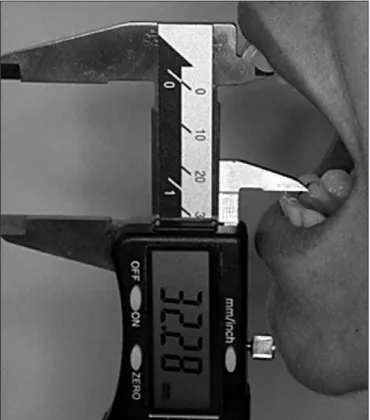
he dentist diagnosed her with temporomandibular disorder. She experienced pain when performing mandibular function-al activities and had her sleep disturbed at night. Further-more, she experienced extreme jaw stifness in the morning with episodes of locking joint and interincisal distance of
32.28mm (measured with digital caliper, Vonder® - 150mm)
(Figure 1).
Further careful anamnesis and detailed clinical examination showed several signs and symptoms, such as localized myofas-cial pain (right side), arthralgia of the right temporomandibu-lar joint (TMJ) with noise. Patient also experienced pain under electric shock, pressure of short duration over the joint and deviation of the mandibular trajectory to the right.
Initially, the treatment aimed at minimizing the overload on the TMJ caused by sleep bruxism. herefore, an upper lat oc-clusal splint with full coverage, thermo-polymerized and bilat-eral contact with antagonist teeth was used. In an attempt of diminishing the temporomandibular arthralgia, a non-steroidal anti-inlammatory was also used for the irst 12 days (Tenoxi-cam, 20mg), at every 12 hours, along with a muscle relaxant (cyclobenzaprine, 5mg) two hours before sleeping. he dose of muscle relaxant was increased to 10mg in an attempt to control myalgia that accompanied arthralgia. After six months using 10mg cyclobenzaprine the dose was reduced to 5mg again, as the pain was reduced to degree 3 in the VAS, and the medicine was gradually withdrawn in alternate days until the end of the seventh month.
A nuclear magnetic resonance was requested before the begin-ning of the treatment to measure the position and morphology of the articular disc and to verify the type and possible location of the efusion. However, for economical reasons, patient could not perform the test.
As pain in right the TMJ was not efectively eliminated, the solution found was to use a minimally invasive therapy with sodium hyaluronate, and the patient received four intra-ar-ticular injections of SH with diferent molecular weights per month. he solution with the lowest molecular weight
(500-730 kDa - Polireumin®) SH was used in the irst and third
months and the SH solution with higher molecular weight (1,000 – 2,000 kDa – Osteonil Mini®) in the second and forth
months.
he procedure was conducted as follows. Patient sat comfort-ably in a chair with an inclination of 45°. She was instructed
Figure 2. The needle was inserted 10mm anterior to the tragus and 2mm below the tragus line – external corner of the orbital cavity. It was directed anteriorly, superiorly, and medially until the tip of it reached the glenoid fossa close to the upper joint space of the right TMJ
Figure 1. Limitation of the interincisal distance
to rotate her head towards the asymptomatic side to facilitate the approach to the symptomatic TMJ. he pre-auricular area
was then disinfected with 70% alcohol and povidone®.
Lido-caine (1.8mL), without vasoconstrictor, was used to block the auricular nerve17. After 3 minutes the area under anesthesia was
tested with a probe n° 5. Afterwards, 1mL/cc syringe BD and gauge needle 0,80x25 mm (21G) were used for the intra-artic-ular joint injection. he needle was inserted 10mm anterior to the tragus and 2mm below the tragus line – external corner of the orbital cavity (Homlund line). he needle was directed an-teriorly, superiorly, and medially until the tip of it reached the glenoid fossa close to the upper joint space (Figure 2).
One milliliter of SHwas injected in the upper joint space and, as procedure went on, the substance was aspirated to verify if it was not being injected into a blood vessel (supericial temporal vein or artery), or in the vascular part located along the retro-discal area. Patient was then instructed to move the lower jaw actively, but without the operator manipulation.
here was a protocol (1st, 2nd, 3rd, 4th month) to be followed the
day following SH application to verify the mandibular ampli-tude and the intensity of the articular pain.
At the end of the treatment, her clinical examination showed the full restoration of the function. Besides, pain had disap-peared, VAS=zero and she presented an adequate interincisal distance (Figure 3).
DISCUSSION
TMJ is a diarthrodial synovial joint with peculiar characteris-tics, as it has three bone components: the condyle, the articular tubercle and the mandibular fossa of the temporal bone. It has also a ibrocartilaginous joint disc located between the condyle and the articular fossa, which divides the articular cavity in up-per and lower cavities. he articular disc in adults is avascular, presents a biconcave shape and is composed by three segments: anterior, intermediate and posterior bands, being the latest the tickest one21,22. Each TMJ is considered a ginglymus joint, as
it allows hinge movements in one axis and sliding movements in another23. he synovial luid that ills the upper and lower
articular cavities is responsible by the nourishment and lubrica-tion of the articular tissues, being its quantity and quality di-rectly related to the articular health and function22. Many
stud-ies have demonstrated changes in the synovial luids of TMJ with disorders, which cause temporomandibular pain23-29. Our
study presented a case where the patient underwent disc dis-placement with reduction associated to efusion. Whenever the patient performed the maneuver of mandibular translation,the anterior area of the condyle pressed the retrodiscal area, dislo-cated from its original innervated and vascularized site, pain was produced, as an electric shock. he second pain was prob-ably caused by the algogenic substances present in the right upper cavity, which generated localized extra pressure when the mandible moved and, consequently, pain. Both algias were not eliminated by the use of the occlusal splint associated to a pain-killer and a muscle relaxant of central action.
he hyaluronic acid is present in the synovial luid and in the
cellular matrix of various connective tissues30. It acts in the
articular lubrication, reducing the friction in the intra-articular spaces and contributing to the diminishment of the adhesive-ness and of inlammatory mediator levels, which would be re-lated to the relief of pain. Its metabolic activity contributes to cell renewal and facilitates the nourishment of avascular areas of the disc and articular cartilage due to its combination to the glycosaminoglycans originated from proteoglycans31,32. HA is
usually found in a concentration of 3 mg/mL in a healthy and normal joint30. However, in pathological conditions, a chain of
events at molecular level takes place and the glycosaminogly-cans are found disintegrated and disperse in the synovial luid, being contained within the synovial cavity33. As a consequence,
the concentration and volume of SH inside the damaged joint may be reduced up to 50%. he synovial luid becomes less viscous and, as a result shocks are not properly absorbed, which
may impair the articular protection and lubrication34. he
presence of inlammatory mediators in the synovial luid has a relevant role in the pathophysiology of articular diseases. Pros-taglandin E2 (PGE2), leukotriene B4 (LTB4), interleukin (IL) 1β, IL-2, IL-8, interferon (IFN) Υ and tumor necrosis factor
TNF-α were identiied in the synovial luid of patients with
pain, which suggests that these mediators are related to TMJ osteoarthritis24. We believe this may have happened in this case,
however we cannot precise which mediators were present nor where, once no laboratorial analysis was performed. he inan-cial problems faced by the patient did not allow the examina-tion of TMJ by nuclear magnetic resonance. If such imaging test could have been performed in T1 and mostly T2 weighted imaging, we might have visualized the presence of these inlam-matory changes in one or both cavities, with a mild, moder-ate or severe hyper signal. Such information would give us an idea of the articular efusion level and its location regarding the TMJ. If a new magnetic resonance of the TMJ could have been obtained after the sequential iniltration with sodium hyaluro-nate, we would have better understood what had happened with the disc and if there had been any morphological or po-sitioning changes and if the efusion remained, diminished or disappeared, once the intra-articular pain, the reciprocal noise and jaw deviation were eliminated.
Studies analyzing knee joint diseases observed an association between increase of the TNF-α and IL-6 serum values and lev-els of pain and function limitation35.herefore it is evident the
role of inlammation and its mediators in the pathophysiology, signals and symptoms and evolution of articular diseases, being TMD included.
he viscosupplementation may improve lubrication (qualita-tively and quantita(qualita-tively), biomechanics and the elimination or reduction of pain, since it aims at restoring the rheological properties of the synovial luid, having it a mechanical, analge-sic, anti-inlammatory and chrondroprotective objective32. he
articular iniltration with sodium hyaluronate, as performed in this case, increased the concentration of hyaluronic acid in the synovial luid, facilitating the releasing of adherence areas. It probably promoted articular mobility and diminished the secondary joint wear and it also allowed the synovial luid to
better circulate, which helped nutrients and metabolites dif-fusion into a vascular tissues. HA may also reduce inlamma-tory mediators levels, contributing to the relief of pain in the articulations3,33. Good results have been obtained with this
in-tervention in various TMD due to mechanical and metabolic characteristics of HA7,36-38.
he present paper is a case report, and regardless of its low evi-dence, it generates a hypothesis.
Results found in a meta-analysis study38 suggested that HA
ap-plication can improve TMD clinical signals in comparison with placebo. However, results are inconclusive due to the absence of well designed and controlled randomized clinical trials. Se-lected studies indicated some positive evidences, however some methodological problems and incomplete reports inluenced their validity and reproducibility. Furthermore, diferent doses of SH with distinct molecular weights and diferent cycles of treatment were used, compromising the selection of trials to the systematic review.
he patient of the present study received a sequence of alter-nate iniltrations of SH that aimed at improving viscosupple-mentation and posterior visco-induction, generating a possible synergism between the diferent substances applied into the articulation. Another relevant aspect of this strategy of sequen-tial iniltrations is that the inisequen-tial application of the lower mo-lecular weight SH possibly contributed with its interaction at a molecular/cellular level and has led to the decrease of pain. When the higher molecular weight SH was applied our aim was to diminish the action of phospholipase A2 (PLA2), which is secreted by synoviocytes, chondrocytes and osteoblasts into the synovial luid, and may lead to a degenerative process. As the inhibition of (PLA2) is dose dependent, the higher the mo-lecular weight and HA concentration used the higher will be (PLA2) inhibition40.herefore, the exogenous use of such
vis-coelastic substance, with such molecular weight, probably con-tributed to the relief of pain and reestablished the homeostasis of the synovial luid and the biomechanics of the TMJ. he last dose, with a higher molecular weight, was used as viscoelastic reinforcement. he most common side efect associated with viscosupplementation is the occurrence of local reaction at the injection site, such as transient pain and swelling41-44. he
pa-tient described in this paper did not present any side efects, neither local nor systemic, during the monthly applications. he monthly frequency of our applications seemed to work in an efective and safe way in our research. However, the lit-erature suggests weekly and fortnightly cycles7,18,36,45. he
pres-ent study performed four cycles with an application a month, which generates a higher cost in the treatment. Such aspects should be taken into consideration and discussed with patients when the treatment is agreed with them, taking into consider-ation the costs/beneits aspects of it at the moment.
Randomized clinical trials should be conducted to verify what is the best cycle and what would be the impact of using irst the higher molecular SH followed by the lower molecular weight one, or even comparing this modality of treatment with iso-lated arthrocentesis, arthrocentesis and viscosupplementation regarding the beneits of the diferent treatments over time.
CONCLUSION
he present study suggests that cycles of viscosupplementation with SH of diferent molecular weights may present excellent long term results in the control of signals and symptoms of TMD of articular origin, as the therapeutic beneits were kept for a period of four years, without the need for annual main-tenance cycles.
REFERENCES
1. Locker D, Grushka M. Prevalence of oral and facial pain and discomfort: preliminary results of a mail survey. Community Dent Oral Epidemiol. 1987;15(3):169-72. 2. Wilkes CH. Structural and functional alterations of the temporomandibular joint.
Northwest Dent. 1978;57(5):287-94.
3. Escoda-Francolí J, Vázquez-Delgado E, Gay-Escoda C. Scientiic evidence on the use-fulness of intraarticular hyaluronic acid injection in the management of temporoman-dibular dysfunction. Med Oral Patol Oral Cir Bucal. 2010;15(4):e644-8.
4. Li C, Zhang Y, Lv J, Shi Z. Inferior or double joint spaces injection versus superior joint space injection for temporomandibular disorders: a systematic review and meta--analysis. J Oral MaxillofacSurg. 2012;70(1):37-44.
5. Grossmann E, Januzzi E, Iwaki Filho L. O uso do hialuronato de sódio no tratamento das disfunções temporomandibulares articulares. Rev Dor. 2013;14(4):301-6. 6. Yeung RW, Chow RL, Samman N, Chiu K. Short-term therapeutic outcome of
intra--articular high molecular weight hyaluronic acid injection for nonreducing disc displa-cement of the temporomandibular joint. Oral Surg Oral Med Oral Pathol Oral Radiol Endod. 2006;102(4):453-61.
7. Guarda-Nardini L, Rossi A, Ramonda R, Punzi L, Ferronato G, Manfredini D. Efec-tiveness of treatment with viscosupplementation in temporomandibular joints with or without efusion. Int J Oral Maxillofac Surg. 2014;43(10):1218-23.
8. Ghosh P, Guidolin D. Potential mechanism of action of intra-articular hyaluronan therapy in osteoarthritis: are the efects molecular weight dependent? Semin Arthritis Rheum. 2002;32(1):10-37.
9. Fraser JR, Laurent TC, Laurent UB. Hyaluronan: its nature, distribution, functions and turnover. J Intern Med. 1997;242(1):27-33.
10. Asari A, Miyauchi S, Matsuzaka S, Ito T, Kominami E, Uchiyama Y. Molecular wei-ght-dependent efects of hyaluronate on the arthritic synovium. Arch Histol Cytol. 1998;61(2):125-35.
11. Bertolami CN, Gay T, Clark GT, Rendell J, Shetty V, Liu C, et al. Use of sodium hya-luronate in treating temporomandibular joint disorders: a randomized, double-blind, placebo-controlled clinical trial. J Oral Maxillofac Surg. 1993;51(3):232-42. 12. Migliore A, Procopio S. Efectiveness and utility of hyaluronic acid in osteoarthritis.
Clin Cases Miner Bone Metab. 2015;12(1):31-3.
13. Kwiecinski JJ, Dorosz SG, Ludwig TE, Abubacker S, Cowman MK, Schmidt TA. he efect of molecular weight on hyaluronan’s cartilage boundary lubricating ability alone and in combination with proteoglycan 4.Osteoarthritis Cartilage. 2011;19(11):1356-62. 14. Smith MM, Ghosh P. he synthesis of hyaluronic acid by human synovial ibroblasts
is inluenced by the nature of the hyaluronate in the extracellular environment. Rheu-matol Int. 1987;7(3):113-22.
15. Kopp S, Akerman S, Nilner M. Short-term efects of intra-articular sodium hyalu-ronate, glucocorticoid, and saline injections on rheumatoid arthritis of the temporo-mandibular joint. J Craniomandib Disord. 1991;5(4):231-8.
16. Xinmin Y, Jian H. Treatment of temporomandibular joint osteoarthritis with viscosu-pplementation and arthrocentesis on rabbit model. Oral Surg Oral Med Oral Pathol Oral Radiol Endod. 2005;100(3):e35-8.
17. Grossmann E. O uso de artrocentese e da lavagem articulação temporomandibular em pacientes com deslocamento anterior do disco sem redução. Rev Dor. 2001;3(3):97-102. 18. Aktas I, Yalcin S, Sencer S. Prognostic indicators of the outcome of arthrocentesis with and
without sodium hyaluronate injection for the treatment of disc displacement without reduc-tion: a magnetic resonance imaging study. Int J Oral Maxillofac Surg. 2010;39(11):1080-5. 19. Zhang ZK, Ma XC, Gao S, Gu ZY, Fu KY. Studies on contributing factors in
tempo-romandibular disorders. Chin J Dent Res. 1999;2(3-4):7-20.
20. Millon-Cruz A, Martín-Granizo R, Encinas A, Berguer A. Relationship between intra-articular adhesions and disc position in temporomandibular joints: magnetic resonance and arthroscopic indings and clinical results. J Craniomaxillofac Surg. 2015;43(4):497-502.
21. Grossmann E, Munerato MC. Aspectos anátomo-isiológicos da articulação temporo-mandibular. Rev Fac Odonto Univers Passo Fundo. 1996;1(2):11-20.
22. Aiken A, Bouloux G, Hudgins P. MR imaging of the temporomandibular joint. Magn Reson Imaging Clin N Am. 2012;20(3):397-412.
23. de Leeuw R, Klassser GD. Orofacial pain: guidelines for classiication, assessment, diagnosis, and management. 5thed. Chicago: Quintessence publ. Co. 2013. 301p.
25. Kubota E, Imamura H, Kubota T, Shibata T, Murakami K. Interleukin 1 beta and stromelysin (MMP3) activity of synovial luid as possible markers of osteoarthritis in the temporomandibular joint. J Oral Maxillofac Surg. 1997;55(1):20-7.
26. Takahashi T, Tominaga K, Takano H, Aryioshi W, Habu M, Fukuda J, Maeda H. A decrease in the molecular weight of hyaluronic acid in synovial luid from patients with temporomandibular disorders. J Oral Pathol Med. 2004;33(4):224-9. 27. Fujimura K, Kobayashi S, Yoshitake Y, Tsuruoka N, Kaneyama K, Segami N.
Elec-trophoretically separation of the synovial luid proteins in rabbit temporomandibular arthritis induced by mechanical loading. J Oral Pathol Med. 2005;34(9):546-51. 28. Matsumoto K, Honda K, Ohshima M, Yamaguchi M, Nakajima I, Micke P, et al.
Cytokine proile in synovial luid from patients with internal derangement of the tempo-romandibular joint: a preliminary study. Dentomaxillofac Radiol. 2006;35(6):432-41. 29. Suzuki T, Segami N, Nishimura M, Nojima T. Coexpression of interleukin-1β and
tumor necrosis factor α in synovial tissues and synovial luids of temporomandibular joint with internal derangement: comparison with histological grading of synovial inlammation. J Oral Pathol Med. 2002;31(9):549-57.
30. Clegg TE, Caborn D, Maufrey C. Viscosupplementation with hyaluronic acid in the treatment for cartilage lesions: a review of current evidence and future directions. Eur J Orthop Surg Traumatol. 2013;23(2):119-24.
31. Swann DA, Radin EL, Nazimiec M, Weisser PA, Curran N, Lewinnek G. Role of hyaluronic acid in joint lubrication. Ann Rheum Dis. 1974 ;33(4):318-26.
32. Rezende MU, de Campos GC. Viscosuplementation. Rev Bras Ortop. 2012;47(2):160-4.
33. Hepguler S, Akkoc YS, Pehlivan M, Ozturk C, Celebi G, Saracoglu A, et al. he eicacy of intra-articular sodium hyaluronate in patients with reducing displaced disc of the temporomandibular joint. J Oral Rehabil. 2002;29(1):80-6.
34. Watterson JR, Esdaile JM. Viscosupplementation: therapeutic mechanisms and clini-cal potential in osteoarthritis of the knee. J Am Acad Orthop Surg. 2000;8(5):277-84. 35. Stannus OP, Jones G, Blizzard L, Cicuttini FM, Ding C. Associations between serum
levels of inlammatory markers and change in knee pain over 5 years in older adults: a
prospective cohort study. Ann Rheum Dis. 2013;72(4):535-40.
36. Guarda-Nardini L, Rossi A, Arboretti R, Bonnini S, Stellini E, Manfredini D. Sin-gle- or multiple-session viscosupplementation protocols for temporomandibular joint degenerative disorders: a randomized clinical trial. J Oral Rehabil. 2015; 42(7):521-8. 37. Li C, Long X, Deng M, Li J, Cai H, Meng Q. Osteoarthritic changes after superior and inferior joint space injection of hyaluronic acid for the treatment of temporomandibu-lar joint osteoarthritis with anterior disc displacement withoutreduction: a cone-beam computed tomographic evaluation. J Oral Maxillofac Surg. 2015;73(2):232-44. 38. Lu J, Long X, Deng M, Cheng Y, Li B. [Treatment of temporomandibular jointdisc
perforation with injection of hyaluronic acid or disc repair]. Zhonghua Kou Qiang Yi Xue Za Zhi. 2014;49(8):476-80. Chinese.
39. Shi Z, Guo C, Awad M. WITHDRAWN: Hyaluronate for temporomandibular joint disorders. Cochrane Database Syst Rev. 2013;8;10:CD002970.
40. Tanaka E, Detamore MS, Tanimoto K, Kawai N. Lubrication of the temporomandi-bular joint. Ann Biomed Eng. 2008;36(1):14-29.
41. Altman RD, Moskowitz R. Intraarticular sodium hyaluronate (Hyalgan) in the treat-ment of patients with osteoarthritis of the knee: a randomized clinical trial. Hyalgan Study Group. J Rheumatol. 1998;25(11):2203-12.
42. Kolarz G, Kotz R, Hochmayer, I. Long-term beneits and repeated treatment cycles of intra-articular sodium hyaluronate (Hyalgan) in patients with osteoarthritis of the knee. Semin Arthritis Rheum. 2003;32(5):310-9.
43. Neustadt DH. Long-term eicacy and safety of intra-articular sodium hyaluro-nate (Hyalgan) in patients with osteoarthritis of the knee. Clin Exp Rheumatol. 2003;21(3):307-11.
44. Phiphobmongkol V, Sudhasaneya V. he efectiveness and safety of intra-articular injection of sodium hyaluronate (500-730 kDa) in the treatment of patients with painful knee osteoarthritis. J Med Assoc hai. 2009;92(10):1287-94.