Arq. NeuroPsiquiatr. vol.73 número3
Texto
Imagem
Documentos relacionados
Substantia Nigra (SN) echogenic area of patients with Parkinson’s disease and healthy controls examined by Transcranial Sonography... Arq
The spectrum of neurological abnormalities reported in children born after TTTS pregnancies is similar to that observed in low birthweight (LBW) babies follow-up 12.. With this in
Measures of physical performance assessments: Self-Paced Walk Test (SPWT), Stair Climb Test (SCT), Six-Minute Walk Test (6MWT), Chair Stand Test (CST), Timed Up and Go (TUG), Sock
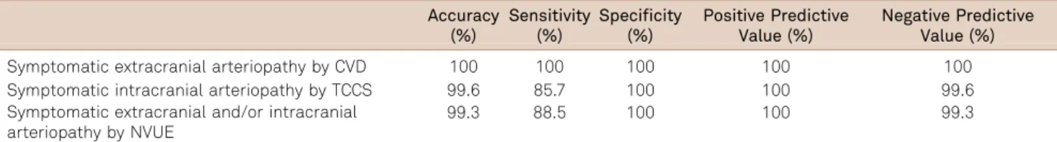
In order to examine preoperative stenosis, different sonographic and angiographic methods were used (e.g. color-coded duplex sonography, intra-arterial subtraction
We present a case of suture granuloma that developed at the inguinal region after orchiec- tomy, and define the sonography, color Doppler sonography and real-time
The purpose of this study is to assess the renal arterial hemo- dynamic changes in obstructive uropathy using duplex sonography, and to evaluate the ability of this modality
METHOD : Between February and September 2005, 206 patients underwent duplex scan examination of carotid vessels, and the intima-media thickness of 407 common carotids were measured
OBJECTIVES: A duplex ultrasound study was performed to investigate morphological and hemodynamic patterns of carotid stenoses treated by endarterectomy with patch closure
