Pandemic influenza A/H1N1: comparative analysis
of microscopic lung histopathological findings
Influenza pandêmica A/H1N1: análise comparativa
de alterações histopatológicas pulmonares
Roberta Marchiori1, Carla Sakuma de Oliveira Bredt2, Marcos Menezes Freitas de Campos1,
Fábio Negretti1, Péricles Almeida Delfino Duarte1
Study carried out at Hospital Universitário do Oeste do Paraná – HUOP, Cascavel (PR), Brazil.
1 Hospital Universitário do Oeste do Paraná – HUOP, Cascavel (PR), Brazil. 2 Universidade Estadual do Oeste do Paraná – UNIOESTE, Cascavel (PR), Brazil.
Corresponding author: Roberta Marchiori – Avenida Tancredo Neves, 3.224 – Santo Onofre – Zip code: 85806-470 – Cascavel (PR), Brazil – Phone: (55 45) 3321-5151 – E-mail: [email protected] Received on: Jan 11, 2012 – Accepted on: May 2, 2012
Conflict of interest: none.
ABSTRACT
Objective: To analyze the histopathological lung findings of four fatal cases of the 2009 H1N1 influenza pandemic and their correlation with clinical and epidemiological characteristics. Methods: Descriptive data from medical records of four patients who died in the Intensive Care Unit of a university hospital in 2009. Nasopharyngeal aspirate specimens were collected from the patients and were analyzed by real-time polymerase chain reaction. Lung biopsy was performed
post mortem; a score of intensity for pathological changes was
applied. Results: Three patients had positive real-time polymerase chain reaction (although all of them had a clinical diagnose of influenza H1N1). The main histopathological changes were: exudative diffuse alveolar damage with atelectasis; varying degrees of alveolar hemorrhage and edema, necrosis and sloughing of the respiratory epithelium in several bronchioli; and thrombus formation. One of the patients (the pregnant one) presented histopathological findings of cytomegalic inclusion. Conclusion: The pulmonary histopathological findings in patients with fatal 2009 H1N1 influenza pandemic disclosed intense alveolar damage and hemorrhage and severe bronchiolitis. A co-infection with cytomegalovirus was described in the pregnant patient.
Keywords: Influenza A virus, H1N1 subtype; Pandemics; Intensive care units
RESUMO
Objetivo: Analisar as alterações histológicas pulmonares de quatro casos fatais de influenza pandêmica H1N1, correlacionando-os a características clínico-epidemiológicas. Métodos: Estudo retrospectivo e descritivo de dados de prontuários de quatro pacientes que faleceram por influenza H1N1 na Unidade de Terapia Intensiva de um hospital universitário, em 2009. Os pacientes haviam sido submetidos
a aspirado de nasofaringe e as amostras foram analisadas pelo método de reação em cadeia da polimerase em tempo real. Biópsia pulmonar foi realizada no dia do óbito; um escore de intensidade das alterações histopatológica foi aplicado. Resultados: Três pacientes apresentaram reação em cadeia da polimerase em tempo real com resultado positivo (embora todos tivessem diagnóstico de influenza H1N1). As principais alterações histológicas identificadas foram: dano alveolar difuso exsudativo, com atelectasia de alvéolos; graus variáveis de hemorragia e edema alveolar; necrose e descamação do epitélio respiratório de vários bronquíolos; e formação de trombos. Uma das pacientes (gestante) apresentou, à histopatologia, achado de inclusão citomegálica. Conclusão: Os achados histopatológicos pulmonares em pacientes com influenza H1N1 fatal revelaram dano alveolar grave, com hemorragia alveolar e bronquiolite. Foi descrita uma coinfecção com citomegalovírus em paciente gestante.
Descritores: Vírus da influenza A subtipo H1N1; Pandemias; Unidades de terapia intensiva
INTRODUCTION
Influenza pandemics are unforeseeable and associated to high morbidity and mortality, as well as to economic and
social turmoil(1). Influenza viruses have a strong capacity
for mutation; their antigenic variation, frequently make the virus unrecognizable to the immunological system and every new subtype becomes a threat, being able to cause a
serious epidemic or even a new pandemic(2).
pandemic(3). Firstly identified in Mexico and in the United States, it spread globally in few weeks via international flights(4,5).
Brazil was particularly hit: more than 45 thousand people were infected in the epidemiological year of 2009, when 2052 deaths occurred. The country south
and southwestern regions were the most affected(6,7).
In Paraná state, in 2009, 63 patients with the diagnose of H1N1 influenza were admitted in 11 Intensive Care Units (ICU) in a period of 45 days presenting a
high mortality rate(8). In 2010, another 1623 cases of
influenza A/H1N1 still occurred, according to the data of the Secretaria da Saúde do Paraná(9).
OBJECTIVE
To analyze the histopathological lung findings of four fatal cases of the 2009 H1N1 influenza pandemic and their correlation with clinical and epidemiological characteristics.
METHODS
This is a retrospective study reviewing the medical records of four patients clinically diagnosed with H1N1 virus infection that were admitted to the ICU of
Hospital Universitário do Oeste do Paraná (HUOP) of Cascavel city, in 2009, had death as outcome and were submitted to lung biopsy.
The following data were collected: gender, age, origin of the patients, the presence of co-morbidities, clinical follow-up, laboratory tests and ventilator parameters.
The four patients belonged to the female gender. One of them (patient A) was an AIDS patient; one other case (patient B) presented a story of neurodevelopmental retardation; a third patient (patient C) was pregnant with a gestational age of 34 weeks; and the last patient (patient D) had heart insufficiency, liver dysfunction and chronic kidney disease. All patients came from cities of the western region of Paraná state, near the Paraguayan and Argentinian frontiers.
Samples of nasopharyngeal aspirate were analyzed at the Central State Laboratory in Curitiba (Paraná) by the real time polymerase chain reaction (RT-PCR)
method. The SuperScriptTM III Platinum® One-Step
Quantitative RT-PCR System (Invitrogen, Carlsbad, EUA) kit was used to process the samples.
The patients were submitted to post-mortem lung
biopsy. The samples were fixed in 10% buffered formalin, embedded in paraffin blocks, and then stained by hematoxylin-eosin, Grocott
methenamine-silver, and Ziehl-Neelsen techniques for fungal and acid- fast bacilli identification. Sample culture was not performed.
A histological semi-quantitative evaluation was made by attributing a score from 0 to 3 (0=absent; 1=light; 2=moderate; 3=severe) for the following histological parameters: granulation tissue, hyaline membrane, alveolar hemorrhage, intra alveolar edema, interstitial inflammation, cytopathic effect, perivasculitis, microthrombus, thromboembolism, besides highlighting the presence of bacteria, acid-fast bacilli or fungus (criteria adapted from Mauad et al.(10)). After the score was attributed by the pathologist in a blind way, the total sum was registered showing a maximum score for each sample.
The present study was approved by the Research
Ethics Committee of the Universidade Estadual do
Oeste do Paraná (UNIOESTE).
RESULTS
The patient’s mean age was 35.5 years (21 to 65 years). A post mortem surgical removal of lung specimens was performed in all patients. The microscopic findings are shown in table 1.
The main clinical findings and their frequency were the following: fever and dyspnea (present in 100%); myalgia (in 50%); cough (in 75%); vomits (in 25%). None of the cases presented diarrhoea. All cases needed mechanical ventilation. Acute kidney insufficiency developed in two cases but dialysis was not necessary.
All patients that were RT-PCR positive presented the acute respiratory distress syndrome (ARDS). This diagnose was established by the presence of a
Table 1. Lung histopathological findings. Each item is graded from 0 (absent) to 3 (severe)
Histopathological findings
Patients
A B C D
Granulation tissue 1 2 3 2
Hyaline membrane 3 3 1 2
Alveolar hemorrhage 2 3 1 0
Intra alveolar edema 1 3 1 2
Interstitial inflammation 3 2 3 3
Cytopathic effect 0 0 0 0
Perivasculitis 3 3 1 1
Microthrombus 3 3 2 0
Thromboembolism 3 3 2 0
Total 19 23 14 10
Acid-fast bacilli or fungus Negative Negative Negative Negative
-radiological bilateral lung infiltrate and the relation between arterial oxygen partial pressure/fraction of
inspired oxygen in inspired air (PaO2/FiO2) ≤200.
Patient A presented hemoptysis and had low platelet count since her hospital admission. Patient C, the pregnant one, presented the same complication but had a normal platelet count. All patients were treated with oseltamivir for at least five days with the recommended dosage according to the Ministry of Health protocol. Corticotherapy was also used in all patients as well as vasoactive drugs. Table 2 shows the clinicopathological data of the four patients.
All patients presented various degrees of alveolar damage. Severe alveolar hemorrhage occurred in one patient, moderate in another, and light in a third. Pneumonitis was found in various degrees, moderate interstitial inflammation in one patient and severe in three of them. Two patients presented low grade
perivasculitis. The presence of microthrombus and thromboembolism was severe in two cases and moderate in one. No cytopathic effect was found nor infection either by acid-fast bacilli or fungus (Table 1).
Patient B presented a severe degree of alveolar hemorrhage and edema receiving the highest score, and clinically had the worst respiratory clinical course.
Figure 1 shows the histopathological findings of one of the patients (patient C) who was RT-PCR positive. Her bronchoalveolar epithelium showed a cytomegalic inclusion which was compatible with a concurrent cytomegalovirus infection.
Patients A and B showed severe perivasculitis and thromboembolism. All cases presented severe necrosis and scaling of the respiratory epithelium in various bronchioli. A few alveoli were expanded and the pulmonary tissue among these alveoli showed atelectatic alveoli.
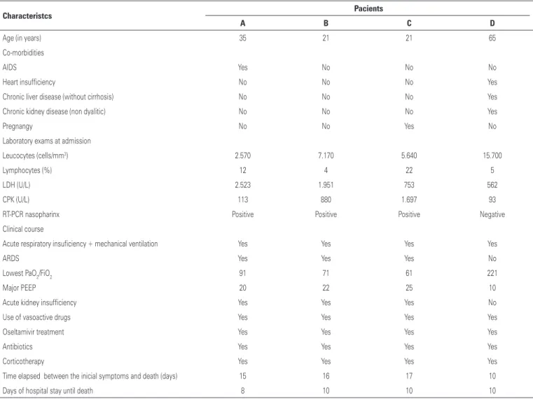
Table 2. Clinical, epidemiological and laboratorial aspects
Characteristcs Pacients
A B C D
Age (in years) 35 21 21 65
Co-morbidities
AIDS Yes No No No
Heart insufficiency No No No Yes
Chronic liver disease (without cirrhosis) No No No Yes
Chronic kidney disease (non dyalitic) No No No Yes
Pregnangy No No Yes No
Laboratory exams at admission
Leucocytes (cells/mm3) 2.570 7.170 5.640 15.700
Lymphocytes (%) 12 4 22 5
LDH (U/L) 2.523 1.951 753 562
CPK (U/L) 113 880 1.697 93
RT-PCR nasopharinx Positive Positive Positive Negative
Clinical course
Acute respiratory insuficiency + mechanical ventilation Yes Yes Yes Yes
ARDS Yes Yes Yes No
Lowest PaO2/FiO2 91 71 61 221
Major PEEP 20 22 25 10
Acute kidney insufficiency Yes Yes Yes No
Use of vasoactive drugs Yes Yes Yes Yes
Oseltamivir treatment Yes Yes Yes Yes
Antibiotics Yes Yes Yes Yes
Corticotherapy Yes Yes Yes Yes
Time elapsed between the inicial symptoms and death (days) 15 16 17 10
Days of hospital stay until death 8 10 10 10
DISCUSSION
All cases presented severe acute respiratory insufficiency related to the 2009 influenza pandemic virus H1N1, were admitted to the ICU and needed orotracheal intubation.
The four patients belonged to the female gender and the three confirmed cases occurred in young adults. In a Brazilian study involving 21 patients with confirmed
2009 influenza pandemic the mean age was 34 years(10).
Co-morbidities were possibly related to the disease severity and the patients’ worst course. In a USA study in 2009, involving 272 patients, presenting a confirmed
H1N1 diagnose, 73% had at least one co-morbidity(3,11).
Among the patients of the present study, there were no obese ones, different from what was found in patients of a ICU cohort from Australia and New Zealand, in 2009. From the 722 patients studied, 601 had available data for weight and 28.5% had body mass índex >35; while in California, among 268 hospitalized
patients, 58% were obese(12).
All patients in this study had fever, dyspnea and severe respiratory insufficiency that required mechanical ventilation. Myalgia occurred in two patients and vomiting in one. None had diarrhoea. In the Mexican epidemics cough, fever and dyspnea were the prevailing
clinical signs(13). In an American study involving 642
patients, dyspnea was not reported, 94% cases presented fever and 92% had cough. However, in this American study not all patients were hospitalized(14).
The patients’ fatal course in the present study may be explained by the risk factors that were present favoring the worst prognosis, such as pregnancy and immunossupression (the AIDS patient).
As related to the laboratory tests (Table 2), lymphopenia was present in all 2009 H1N1 influenza pandemic patients and a low platelet count was found in patient A, probably related to AIDS. There was an
important high level of lactate dehydrogenase (LDH) in the three confirmed cases and a high level of creatine phosphokinase (CPK) in two cases. Patient B presented levels over 1.000U/l LDH, with the worst histological picture and respiratory course as compared to the others implying in a severe degree of pulmonary injury.
Patient A was an AIDS case presenting severe respiratory insufficiency and an associated clinical hypothesis of a pneumocystic infection which could explain the considerable increase in the LDH level.
Increased CPK level over 1.000U/l was found in the pregnant patient. High CPK and LDH levels as well as lymphopenia were described in Mexican patients with respiratory insufficiency due to the 2009 influenza
pandemic H1N1(13).
The confirmed cases presented various degrees of alveolar hemorrhage, edema and diffuse alveolar damage on histopathology, similarly to previous
influenza pandemics(15). Patient B presented the worst
respiratory course obtaining also the worst pathological score according to the histological classification, besides presenting ARDS (the same fact that occurred with the other two confirmed cases). She also required prone positioning on two occasions, probably associated to the severe alveolar edema and hemorrhage as shown by the biopsy. In order to improve ARDS, a few mechanisms must remain active such as the reabsorption of pulmonary edema from the alveolar spaces. A decrease in the capacity of the alveolar epithelial barrier to remove liquids was demonstrated in patients with
acute lung injury(16). Some studies have demonstrated
that the alveolar deficiency in transporting liquids is directly related to the severity of the clinical picture in
patients with ARDS(17,18). It was also observed in mice
infected by influenza A virus that this virus promotes a clearance blockage of alveolar fluid directly
compromising gaseous exchanges(18) .
The prevalence of infection by the pandemic H1N1 virus in pregnancy has been described in the literature
as being between 7% and 9%(3,11). The pregnant
patient (patient C) presented histological findings compatible with the presence of cytomegalovirus in the bronchoalveolar epithelium, a concurrent infection with the H1N1 disease, which may have contributed to the severe interstitial inflammation found in this case. Cytomegalic pneumonitis is an uncommon complication and its diagnose was based on the direct visualization of a cytomegalic inclusion and its cytopathic effect on the
pulmonary tissue(19). The description of this co-infection
was not found in the literature. It is reported that the majority of pregnant women have been previously infected by cytomegalovirus; however, approximately
Figure 1. Histopathological findings. (A) Pneumonitis. (B) Hyaline membrane/ septal thickening. (C) Vascular thrombus. (D) Cytomegalic inclusion
A
B
15% of them are susceptible to primary infection
during pregnancy(20,21). In this patient, microthrombus
and moderate thromboembolism (according to the previously referred score) were found in the histological preparation. This finding could be related to a gestational hypercoagulable state but also to 2009 H1N1 virus itself, as reported by the Pathology Department of São Paulo University (USP)(10,22) .
Patient A, an AIDS carrier, presented severe perivasculitis and thromboembolism that may be related to the extensive pleural effusion (caused by hydrostatic pressure changes) that needed thoracic closed drainage. Besides, a severe interstitial inflammation was present, as in patient C the one suspected to have a concurrent cytomegalovirus infection. However, in patient A, it might have been the result of a clinically suspected infection by
Pneumocystis jirovecii despite the negative search in the
tracheal secretion and the fact that P. jirovecii was not
visualized on the histological preparations that were performed.
Patient D was PCR negative for H1N1 in nasopharynx and oropharynx aspirates. However, a specimen from the inferior respiratory tract was not collected, which could bring a weighty evidence for the diagnose as it
is described in the literature(23). Up to 10% of severe
H1N1 influenza cases (admitted to ICUs) showing a RT-PCR positive test in the tracheal secretion have negative exams in the nasopharynx and oropharynx
secretions(24). Nevertheless, the patient’s clinical course
differed from the other ones. She presented leukocytosis during her entire hospital stay; she also presented acute respiratory insufficiency but it was only on the third admission day that she required orotracheal intubation and did not course with ARDS (the other patients needed mechanical ventilation immediately before ICU admission).
ICUs patients when RT-PCR positive present a worse clinical outcome when compared to those with
a negative result(8). So it is possible to speculate that
the H1N1 infection in this patient (older and with co-morbidities) could not have been so severe but the outcome was inauspicious because of her age and the co-morbidities. The main histological lung change in this patient was interstitial inflammation which is not specific. Alveolar hemorrhage and the presence of thrombus that were found in the other patients with positive tests were absent in this case.
Patient B had the worst histological score, but she did not have any co-morbidities that could justify such an ominous course; so maybe a more virulent viral mutation could have infected this patient being capable of promoting the histological changes that were found with greater intensity.
This article has limitations, including the fact that it is a retrospective one. Cultures to identify bacteria were not performed in the patients’ respiratory specimens because of technical problems in the laboratory as well as cultures were not performed in the lung biopsies because the specimens were processed in formaldeyde. The case that had a probable concurrent infection with cytomegalovirus did not have all laboratory tests for its confirmation, because such an hypothesis was not raised by the time the patient was being cared for.
CONCLUSION
These four fatal cases of pandemic influenza H1N1 with lung histopathology showed as the main findings exudative alveolar damage with alveolar atelectasis; various degrees of alveolar hemorrhage and edema; necrosis and scaling of the bronchioli epithelium. A rare concurrent infection with cytomegalovirus in one patient was also described.
REFERENCES
1. World Health Organization. Avian influenza: assessing the pandemic threat [Internet]. 2005 [cited 2011 Jul 26]. Available from: http://whqlibdoc.who. int/hq/2005/WHO_CDS_2005.29.pdf
2. Salomon R, Webster RG. The influenza virus enigma. Cell. 2009;136(3):402-10. 3. Jain S, Kamimoto L, Bramley AM, Schmitz AM, Benoit SR, Louie J, Sugerman DE, Druckenmiller JK, Ritger KA, Chugh R, Jasuja S, Deutscher M, Chen S, Walker JD, Duchin JS, Lett S, Soliva S, Wells EV, Swerdlow D, Uyeki TM, Fiore AE, Olsen SJ, Fry AM, Bridges CB, Finelli L; 2009 Pandemic Influenza A (H1N1) Virus Hospitalizations Investigation Team. Hospitalized patients with 2009 H1N1 influenza in the United States, April-June 2009. N Engl J Med. 2009;361(20):1935-44.
4. Khan K, Arino J, Hu W, Raposo P, Sears J, Calderon F, et al. Spread of a novel influenza (H1N1) virus via global airline transportation. N Engl J Med. 2009;361(2):212-4.
5. Trifonov V, Khiabanian H, Rabadan R. Geographic dependence, surveillance, and origins of the 2009 influenza A (H1N1) virus. N Engl J Med. 2009;361(2): 115-9.
6. Brasil. Ministério da Saúde. Secretaria de Vigilância em Saúde. Nota técnica nº 15/2010. Decreta o início da fase pós-pandêmica do vírus influenza pandêmica (H1N1) 2009 [Internet]. 2010 [citado 2012 Set 12]. Disponível em: http://portal.saude.gov.br/portal/arquivos/pdf/nt_15_fase_pos_pand_ virus_influ_a_10082010.pdf
7. Brasil. Ministério da Saúde. Secretaria de Vigilância em Saúde. Situação epidemiológica da Influenza Pandêmica (H1N1) 2009 no mundo e no Brasil, até a semana epidemiológica 47 de 2009. Informe Epidemiol. 2009 [citado 2012 Set 12];(11): [cerca de 11p]. Disponível em: http://portal.saude.gov.br/ portal/arquivos/pdf/boletim_influenza_se_47.pdf
8. Duarte PA, Venazzi A, Youssef NC, Oliveira MC, Tannous LA, Duarte CB, et al. Pacientes com infecção por vírus A (H1N1) admitidos em unidades de terapia intensiva do Estado do Paraná, Brasil. Rev Bras Ter Intensiva. 2009;21(3):231-6. 9. Paraná (Estado). Secretaria do Estado de Saúde. Situação epidemiológica
10. Mauad T, Hajjar LA, Callegari GD, Silva LF, Schout D, Galas FR, et al. Lung pathology in fatal novel human influenza A (H1N1) infection. Am J Respir Crit Care Med. 2010;181(1):72-9.
11. ANZIC Influenza Investigators, Webb SA, Pettilä V, Seppelt I, Bellomo R, Bailey M, et al. Critical care services and 2009 H1N1 influenza in Australia and New Zealand. N Engl J Med. 2009;361(20):1925-34.
12. Louie JK, Acosta M, Winter K, Jean C, Gavali S, Schechter R, et al. Factors associated with death or hospitalization due to pandemic 2009 influenza A(H1N1) infection in California. JAMA. 2009; 302(17):1896-902.
13. Perez-Padilla R, de la Rosa-Zamboni D, Ponce de Leon S, Hernandez M, Quiñones-Falconi F, Bautista E, Ramirez-Venegas A, Rojas-Serrano J, Ormsby CE, Corrales A, Higuera A, Mondragon E, Cordova-Villalobos JA; INER Working Group on Influenza. Pneumonia and respiratory failure from swine-origin influenza A (H1N1) in Mexico. N Engl J Med. 2009;361(7):680-9. 14. Novel Swine-Origin Influenza A (H1N1) Virus Investigation Team, Dawood
FS, Jain S, Finelli L, Shaw WM, Lindstrom S, et al. Emergence of a novel swine-origin influenza A (H1N1) virus in human. N Engl J Med. 2009;360(25): 2605-15.
15. Taubenberger JK, Morens DM. The pathology of influenza virus infections. Annu Rev Pathol. 2008;3:499-522.
16. Matthay MA. Alveolar fluid clearance in patients with ARDS: does it make a difference? Chest. 2002;122(6 Suppl):340S-343S.
17. Sartori C, Matthay MA. Alveolar epithelial fluid transport in acute lung injury: new insights. Eur Respir J. 2002;20(5):1299-313.
18. Wolk KE, Lazarowski ER, Traylor ZP, Yu EN, Jewell NA, Durbin RK, et al.
Influenza A virus inhibits alveolar fluid clearance in BALB/c mice. Am J Respir Crit Care Med. 2008;178(9):969-76.
19. Huang L, Crothers K. HIV-associated opportunistic pneumonias. Respirology. 2009;14(4):474–85.
20. Figueiró-Filho EA, Senefonte FR, Lopes AH, Morais OO, Souza Junior VG, Maia TL, et al. Freqüência das infecções pelo HIV-1, rubéola, sífilis, toxoplasmose, citomegalovírus, herpes simples, hepatite B, hepatite C, doença de Chagas e HTLV I/II em gestantes, do Estado de Mato Grosso do Sul. Rev SocBras Med Trop. 2007;40(2):181-7.
21. Serra FC, Machado J, Nicola MH, Jorge MC, Cruz LE, Giordano MV, et al. Soroprevalência de citomegalvírus em gestantes brasileiras de classe socioeconômica favorecida. DST J Bras Doenças Sex Transm. 2009;21(1): 12-5.
22. Shieh WJ, Blau DM, Denison AM, Deleon-Carnes M, Adem P, Bhatnagar J, et al. 2009 pandemic influenza A (H1N1): pathology and pathogenesis of 100 fatal cases in the United States. Am J Pathol. 2010;177(1):166-75. 23. World Health Organization. Clinical management of human infection with
pandemic (H1N1) 2009: revised guidance [Internet] [cited 2012 Set 12]. Available from: http://www.who.int/csr/resources/publications/swineflu/ clinical_management_h1n1.pdf