DOI: 10.5935/2359-4802.20170014
Abstract
The objective of this study is to make a review of the narrative of coronary artery tortuosity (CAT) approaching several situations in clinical practice where tortuosity can have a relevant role, and also evaluate if tortuosity can be related to the presence of myocardial ischemia in patients without coronary obstruction using scientific evidences in medical literature. Textbook of applied Physiology in Cardiology with study of coronary circulation, theoretical articles with studies of Hemodynamics, Fluid and Mechanical Dynamic, and experimental articles with simulation in computers were used as support to answer this last question.
Introduction
Coronary circulation
There are two basic types of coronary vessels: conductance and resistance vessels. The epicardial arteries, right and left, and its main and major branches that emerge in acute angle of relative large caliber work as vessels of conductance offering in the diastole minimum resistance to the blood flow. The deep perforators that originate in a right angle of the epicardial arteries penetrate deeply in the myocardial walls and nurture sub endocardial layers offering great resistance to the flow mainly in the ventricular systole. They are responsible for the coronary flow autoregulation maintaining it adequately in broad spectrum of the pressure variation and increasing the flow in exercising situations mainly through the
local metabolic regulation. Microcirculation is part of the coronary circulation constituted by arterioles and capillaries responsible for regulating the oxygen supply to the myocardium.
The heart is a highly aerobic organ, but it depends almost exclusively on the oxidation of the substrates to generate energy which will move it and has almost no oxygen reserve. It receives about 5% of cardiac output and it is a little perfused organ, but it is the organ that has the highest oxygen extraction of the organism.
Taking into consideration Fick’s equation (oxygen consumption = coronary flow X arteriovenous difference of oxygen), we verified that the physiological determinants of the coronary flow are the same that command the demand and consumption of oxygen: blood pressure, heart rate, ventricular wall tension, dP/dt maximum.
According to Poiseuille’s law, the flow in any vessel system is directly proportional to the difference of pressure in its extremities, and inversely proportional to the resistance of the system, which is in turn proportional to the length of the tube, viscosity of the fluid and inversely proportional to the fourth power of the radius (most important factor). Influences of vasomotility of the autonomous nervous system, of drugs, mainly of local autoregulation, determine variations in the flow. Due to cyclical pressure and variations and myocardium tension, the coronary flow, in the systole represents from 25% to 30% of the total, and from 70% to 75% in the diastole.
Factors that can affect myocardial consumption of oxygen and consequently the coronary flow can be divided into 3 groups:
a) Factors that affect consumption (demand): intraventricular tension, heart rate, myocardial contractile state and electrical activation, and cardiac metabolism.
REVIEW ARTICLE
Mailing Address: André Pereira Duque Estrada
Rua Coronel Moreira César, 376/602. Postal Code: 24230064. Niterói, Rio de Janeiro, RJ – Brazil E-mail: [email protected]
Coronary tortuosity and its role in myocardial ischemia in patients with no
coronary obstructions
André Pereira Duque Estrada,1 Rosane de Oliveira Lopes2 Humberto Villacorta Junior2
Hospital Federal dos Servidores do Estado1; Universidade Federal Fluminense2, Niterói, RJ – Brazil
Manuscript received August26, 2016; revised manuscript September 30, 2016; accepted November 21, 2016.
Coronary Vessels; Coronary Circulation; Myocardial Ischemia; Hypertension; Aging.
b) Factors that affect oxygen supply (circulation): perfusion pressure (aortic pressure minus left atrial pressure), anatomical condition of arterial bed, arterial
PaO2 and %HbO2, dissociation of O2 from hemoglobin,
transmyocardial distribution of flow with capillary opening and microcirculation alterations.
c) Factors that compromise oxygen supply to the myocardium (arterial caliber): neurovegetative and humoral action, local regulation (nitric oxide,
adenosine, pH and PCO2), coronary flow reserve
and drugs.1
Coronary flow regulation, coronary flow reserve, and myocardial ischemia
Regardless of extra-coronary factors, the contraction or relaxation of the arteries and arterioles are influenced by muscular, neurovegetative, and humoral factors acting on vessel walls. There are, at least, four main systems: the myogenic control reflects intrinsic property of the vascular muscle to react to pressure distension on the wall vessel (possibly by channels activated by distension), autonomous control with catecholamine, adrenaline, and noradrenaline, endothelial control with major importance of nitric oxide- a potent vasodilator released by mechanical forces of flow friction on the endothelium – and the main – the metabolic control by the partial drop of the pressure of the local oxygen.
Small partial pressure variation of oxygen (decrease) can be sufficient to cause vasodilatation and flow increase, balancing the demand and supply until a new imbalance occurs.
The mechanism of this active dilatation results from a direct effect of hypoxia on coronary artery smooth muscle, and/or the increase of metabolic vasodilators by the effect of hypoxia in the cells, mainly the adenosine.
The concept of the coronary flow reserve is related to the maximum ability of coronary vessels to increase flow in response to myocardial demand and this capacity is 5 times greater in relation to the flow at rest.
When compensation mechanisms are exhausted the process of myocardial ischemia occurs, with metabolic contractility and electrocardiographic changes in the
moment that the coronary flow decreases from 40 ml/min.1
Development
Coronary arteries tend to be more tortuous than other arteries and accompany repetitive movements
of flexion and relaxation that occur during the cardiac cycle. Intra luminal traction and pressure are two forces that try to stretch the vessel and opposing to this there is the force of retraction that depends basically of elastin. The degeneration of elastin in the coronary wall can occur due to pathological processes or related to old age and
lead to aneurysmal dilatations and to tortuosity.2
Postmortem angiographies of 145 patients without coronary obstruction showed that coronary tortuosity is positively related to age and negatively related to the heart weight in equal importance, and, in a smaller degree, positively to the caliber of the vessel.
Moreover coronary tortuosity is positively related to arterial hypertension and to the female gender, and
negatively related to the process of atherosclerosis.3
In Turkey, in 2013, 148 male patients that underwent coronariography in a period of three months were selected. Exclusion criteria included patients with a history of revascularization surgery or coronary angioplasty. These patients were divided into two groups: with and without coronary tortuosity, and the relation between of tortuosity with coronary obstruction was studied. A negative correlation between the employed tortuosity score and severe obstructive coronary disease was found. It was questioned if coronary tortuosity could represent a genetic geometric factor of protection against obstructive coronary disease
or a mechanism of coronary remodeling.4
Patients with stable angina and no coronary obstruction, but with coronary tortuosity, present an increased calcium score in relation to those without tortuosity, suggesting an association with subclinical atherosclerosis. A similar finding was found in another study that evaluated patients with retinal arteries tortuosity, and with coronary tortuosity that presented an increase of the mid-intimal thickness in the carotids. In this last study, it was verified an association with females and individuals of short stature. They are more commonly found in the circumflex followed by the anterior descending artery and lastly
right coronary.5 The latter can be described as S or C
shaped and the S shape presents less frequent obstructive
atherosclerotic disease.6,7
A statistical analysis of an angiographic study with 52 patients in which were selected 32 left coronary arteriography with no obstruction, and 35 right coronary arteriography with no obstruction showed that, in the left coronary, tortuosity was larger in its distal portion and, in
pci D
C separation
B
C v1
1
v2 3 v3
pco
B
A
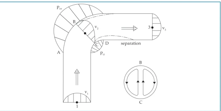
Figure 1 – Separation of blood flow from the coronary wall in the location of the curve
Pathophysiology
Coronary tortuosity leads to coronary flow alterations with reduction of the distal perfusion pressure and, lastly, the appearance of myocardial ischemia. Decomposition of force vectors with great loss of kinetic energy and the presence of curves extend the blood path to the myocardium.
There are two causes for this pressure reduction: friction, due to shear stress that can be calculated by
Poiseuille’s law (Efr = 32 nlv/d2, in which n = absolute
blood viscosity; l = artery length; v = velocity; and d = diameter); and the other is the centrifugal effect. The curves lead to extra energy loss largely caused by blood swirling, which occurs due to the change in flow direction in the curve, with separation of the blood flow from the coronary wall in the curve location.
Figure 1 shows that in the AB section (part outside the curve) there is an increase in pressure, and, in the CD section (part inside the curve) there is pressure reduction,
which creates a swirling area and loss of kinetic energy.2
Studies with computer simulations
Chinese researchers have been delving into this theme. A Chinese study by Yang Li et al. with numeric simulation evaluated the impact of coronary tortuosity on pressure distribution inside coronary circulation.
They idealized 21 models varying in tortuosity angle and quantity, and verified that these two factors influenced pressure loss in coronary circulation, and the greater the severity of the tortuosity (measured by these two factors), the greater the pressure drop, which, in more severe
cases, may lead to myocardial ischemia.9
The impact of coronary tortuosity on coronary
circulation was assessed by Xie X et al.10 through the
computational fluid dynamic technique. They selected two models of tortuous anterior descending arteries of different patients, and reconstructed the arteries, in a three-dimensional model, without the presence of tortuosity. After that, simulations of rest and exercise of the models were carried out, in appropriate conditions, and it was verified that tortuosity has a smaller influence on coronary circulation at rest; however, during exercise, tortuosity may represent greater resistance to blood flow, in such a way that compensatory mechanisms of flow adjustment may not be enough to keep an adequate
flow and lead to myocardial ischemia.10
Xie et al.11,12 conducted a study with three-dimensional
causing coronary autoregulation to fail. This study also suggests that tortuosity may constitute a risk factor for atherosclerosis, since it can lead to the appearance of a region with low, wavering shear stress in the internal wall of the curve’s descent, when the angle of the curve
is greater than 120 degrees.11,12 Outro estudo recente
confirma que a tortuosidade leva a queda da pressão e
pode levar a déficit de perfusão.13
Prevalence
A retrospective study from 2009 carried out in West Virginia with 1221 patients, who had undergone catheterization in the previous 8 months, identified 12.45% of patients with coronary tortuosity and showed a higher occurrence in women and lower incidence of obstructive coronary disease, but it did not find predictors of coronary tortuosity among the following conditions: systemic arterial hypertension, diabetes mellitus, advanced age, dyslipidemia, smoking, and family history of obstructive
coronary disease.14
Another retrospective study carried out at the Zhongda Hospital of Southeast University in Nanjing, China, with 1010 patients who had undergone coronarography due to anginal complaints, separated these patients into four groups according to the presence or absence of coronary tortuosity and presence or absence of coronary obstructions, and did a 2 to 4-year follow-up with these patients. The prevalence of tortuosity was 39.1% and was significantly higher in women and in patients with systemic arterial hypertension. It was negatively related
to the presence of coronary obstructions.5
Age-related alterations
Among the modifications in coronary circulation that occur with the aging process, are the increase of coronary tortuosity with minimal atherosclerotic lesions,
calcification and,3,15 at least in animals,16 an imbalance
between the extension of the capillary network and myocyte hypertrophy. Maximum oxygen consumption is progressively reduced. In advanced age rats, it has also been demonstrated a higher deviation angle of secondary branches in relation to the main branch.
Studies with hypertensive individuals
There is a strong correlation between the presence
of hypertension and coronary tortuosity.17 In 1981,
Sanchez Torres G. et al.17 studied, in Mexico, a group
of 46 hypertensive patients, who were divided into three subgroups: Group 1 – angina pectoris; Group 2 – left ventricular hypertrophy in the electrocardiogram; and Group 3 – asymptomatic and without left ventricular hypertrophy in the electrocardiogram. In the coronarography, Group 1 presented coronary obstructions in 28% and tortuosity in 94.8%; Group 2 presented tortuosity in 74.9%; and Group 3 in 69.1%. Groups 2 and 3 did not present cases of
coronary obstruction.18
In 1982, Sanchez Torres G. et al.18 continued to study
the presence of myocardial ischemia in hypertensive patients, this time with patients who met the criteria of left ventricular hypertrophy in the electrocardiogram. In a group of 70 patients who all underwent ergometric test, coronarography, and left ventriculography, and 10 patients who also underwent a study with myocardial scintigraphy, describe "corkscrew tortuosity" in coronary angiography (without associated obstructions) in 83.7% of patients and suggest that subendocardial ischemia may be related to the increase of ventricular mass with
a lower coronary reserve.19
Relation to ventricular relaxation
An echocardiographic study with 104 patients (50 with coronary tortuosity and 54 without) has shown that coronary tortuosity is related to the
worsening of ventricular relaxation20 evaluated by the
decrease of the E/A relation of the transmitral flow, increase of the E-wave deceleration time, increase of isovolumetric relaxation time, and greater thickness of the interventricular septum and of the left ventricular
posterior wall.21
Relation to coronary dissection
to a higher risk of spontaneous dissection (RR = 3.29, confidence interval of 0.99 – 8.29; p = 0.05). The presence of tortuosity markers, such as multi-arterial symmetrical tortuosity and corkscrew aspect were positively related to the presence of extra-coronary vasculopathy such as
fibromuscular dysplasia (p < 0.05).22
Relation to collateral vessels
Coronary tortuosity and vessel diameter can be considered indicative of a coronary artery’s occlusion time after acute myocardial infarction, if this vessel originated collaterals to the occluded vessel. Thus, identification of a tortuous artery may suggest that it functions as a collateral channel, and the enlarged vessel caliber suggests how long this channel has been used. This is due to the fact that the increase in length of the vessel that produces the tortuosity is originated from the same mean relaxation stress that
induces dilatation as a result of blood flow increase.23
Arterial tortuosity syndrome
Cases of coronary tortuosity during childhood, including cases of early death, have been described in literature and are related to the malformation of the artery wall. They compose a poorly defined systemic syndrome with prolongation of the arteries, tortuosity
and thinning of the arterial wall.24-26
A case described in 1969 shows a patient who died at 17 months of age due to coronary insufficiency and multiple peripheral pulmonary stenosis. Postmortem exams showed that pathological alterations were restricted to elastic arteries and to the first part of muscular arteries. Aortic and pulmonary artery walls were thinned and with an increase of elastic fibers. Coronary walls were thinned and with a reduction of arterial light. Major muscular artery walls presented thinning of the intima with elastic fibers hyperplasia
and degeneration of the internal elastic membrane.27
Hyperextensibility of the skin, hypermobility of the joints, and elongated facies have been described in some patients, suggesting an alteration in collagen or elastin synthesis. Connective tissue diseases such as Ehlers-Danlos syndrome, Marfan syndrome, cutis laxa, and Menkes disease make a differential diagnsosis with
the syndrome.28
A case of quadruplets, whose parents were blood relatives, with the syndrome, suggests that it is an
autosomal recessive disease.29
Coronary tortuosity and coronary angioplasty
Coronary tortuosity is a predictor of inaccuracy in the evaluation of a lesion’s obstruction degree through
angiotomography.30-32 Coronary tortuosity is a challenging
problem during coronary angioplasty, and it is related to several complications, such as vessel dissection, stent
loss, and even acute arterial occlusion.33 Its presence is
also related to a larger quantity of radiation during the
procedure,34 and a difficulty to use adjunct methods in
coronary angioplasty, such as intracoronary ultrasound, optical coherence tomography, and fractional flow reserve measurement. It is a predictor of failure in thrombus aspiration during primary angioplasty and recanalization of chronic occlusions.
Adequate preparation of the segment to be treated is also a problem due to the difficulty to advance cutting
balloons and rotablator.35
Several techniques are used to solve this problem, such as the use of more delicate catheters (soft delivery catheters), quick-cross support catheters (e.g. Guideliner), deep guide intubation, and “mother and child” catheter. Specific studies to evaluate catheter performance are carried out by evaluating tortuosity parameters in specific coronary platforms.
Meticulous vessel preparation with pre-dilatation, use of short stents with thinner structures, guidewires with magnetic navigation systems, use of a second guidewire for material progression (“buddy wire technique and “crooked buddy technique”), and use of the “over
the wire”36-41 system are employed. Once the lesion is
overpassed with the guidewire, it may be difficult to recognize the location to implant the stent due to the rectification of the tortuous segment and appearance of
phantom lesions – Concertina effect.42
Relation to myocardial ischemia
Some articles suggest that there is a correlation between coronary tortuosity and the presence of myocardial ischemia in patients without coronary obstructions, although the definition of coronary tortuosity is not the same in every article. Thus, Sova SH
and Lebedieva43 EO demonstrated that 89% of a group
Figure 2 – Angle of the coronary tortuosity
Chart 1 – Main studies correlating coronary tortuosity to myocardial ischemia
Country Year Study Complementary method
Number of patients
Inclusion of myocardial bridge
Relation to myocardial ischemia
Gaibazzi
N et al.44 Italy 2011 Retrospective Echocardiogram 96 Yes Yes
LI Y et al.45 China 2012 Prospective Myocardial
scintigraphy 48 No Yes
Table 1 shows us two of the most important articles on the theme, which used the same definition of coronary tortuosity: one or more coronary with, at least, three consecutive curvatures with an angle < 90° (Figure 2).
An Italian team retrospectively reviewed a subgroup of patients from the study SPAM (stress-echo Parma Mestre). In two centers, 400 patients presenting chest pains of probable coronary origin were selected. These patients underwent stress echocardiogram with dipyridamole, and researchers evaluated the degree of contractility of left ventricular walls and myocardial perfusion, and performed a Doppler study of the left coronary flow reserve before patients underwent coronarography.
They then selected 96 patients without coronary obstructions and searched for two findings: myocardial bridge and coronary tortuosity. The patients were divided into two groups: those with perfusion defects in the echocardiogram, called false positive (37 patients),
and those without, called true negatives (59 patients). These two groups were compared in relation to clinical and demographic variables and angiographic and echocardiographic characteristics.
A total of 16 patients with coronary tortuosity and six with myocardial bridge were identified. There was no statistically significant difference in clinical and demographic variables between the two groups. The prevalence of myocardial bridge (p < 0.05) and coronary tortuosity (p < 0.001) was seven times higher in the false positive group. These patients also had more angina crises (p < 0.05).44
1. Beuren AJ, Hort W, Kalbfleisch H, Müller H, Stoermer J. Dysplasia of the systemic and pulmonary arterial system with tortuosity and lengthening of the arteries. A new entity, diagnosed during life, and leading to coronary death in early childhood. Circulation. 1969;39(1):109-15.
2. Hutchins GM, Bulkley BH, Miner MM, Boitnott JK. Correlation of age and heart weight with tortuosity and caliber of normal human coronary arteries. Am Heart J. 1977;94(2):196-202.
3. Hutchins GM, Miner MM, Bulkley BH. Tortuosity as an index of the age and diameter increase of coronary collateral vessels in patients after acute myocardial infarction. Am J Cardiol. 1978;41(2):210-5.
4. Sánchez Torres G, Trevethan Craviotto S, Bialostozky D, Gutiérrez Fuster E, Olvera Cruz S. [Clinical and coronary angiographic characteristics of hypertensive angina]. Arch Inst Cardiol Mex. 1981;51(6):541-7.
5. Trevethan S, Sánchez Torres G, Martínez Ríos MA, Medrano GA, Cuarón A, Cruz Ayala G. [Myocardial ischemia in hypertension heart disease (author’s transl)]. Arch Inst Cardiol Mex. 1982; 52(3):229-35.
6. LaVeau PJ, Remetz MS, Cabin HS, Hennecken JF, McConnell SH, Rosen RE, et al. Predictors of success in percutaneous transluminal coronary angioplasty of chronic total occlusions. Am J Cardiol. 1989;64(19):1264-9.
7. Carbonin P, Cocchi A, Zuccalà G, Menichelli P. [Heart aging and its clinical implications]. Recenti Prog Med. 1990;81(4):215-20.
8. Kimball BP, Bui S, Dafopoulos N. Angiographic features associated with acute coronary artery occlusion during elective angioplasty. Can J Cardiol. 1990;6(8):327-32.
9. Ellis SG, De Cesare NB, Pinkerton CA, Whitlow P, King SB 3rd, Ghazzel ZM, et al. Relation of stenosis morphology and clinical presentation to the procedural results of directional coronary atherectomy. Circulation. 1991;84(2):644-53.
10. Dagianti A, Rosanio S, Luongo R, Dagianti A, Fedele F. [Coronary morphometry in essential arterial hypertension]. Cardiologia. 1993;38(8):497-502.
11. Brinkman AM, Baker PB, Newman WP, Vigorito R, Friedman MH. Variability of human coronary artery geometry: an angiographic study of the left anterior descending arteries of 30 autopsy hearts. Ann Biomed Eng. 1994;22(1):34-44.
12. Pimentel CX, Schreiter SW, Gurbel PA. The use of the Tracker catheter as a guidewire support device in angioplasty of angulated and tortuous circumflex coronary arteries. J Invasive Cardiol. 1995;7(3):66-71.
13. Topol EJ, Nissen SE. Our preoccupation with coronary luminology. The dissociation between clinical and angiographic findings in ischemic heart disease. Circulation. 1995;92(8):2333-42.
14. Jakob M, Spasojevic D, Krogmann ON, Wiher H, Hug R, Hess OM. Tortuosity of coronary arteries in chronic pressure and volume overload. Cathet Cardiovasc Diagn. 1996; 38(1):25-31.
15. Pletcher BA, Fox JE, Boxer RA, Singh S, Blumenthal D, Cohen T, et al. Four sibs with arterial tortuosity: description and review of the literature. Am J Med Genet. 1996;66(2):121-8.
16. Al Fadley F, Al Manea W, Nykanen DG, Al Fadley A, Bulbul Z, Al Halees Z. Severe tortuosity and stenosis of the systemic, pulmonary and coronary vessels in 12 patients with similar phenotypic features: a new syndrome? Cardiol Young. 2000;10(6):582-9.
17. Goel PK, Agarwal A, Kapoor A. "Concertina" effect during angioplasty of tortuous right and left coronary arteries and importance of using over-the-wire system: a case report. Indian Heart J. 2001;53(1):87-90.
18. Ward MR, Smits P, Herity NA, Jeremias A, Fitzgerald PJ, Yeung AC, et al. No relationship between compensatory arterial remodeling of focal stenotic atherosclerotic lesions and tortuosity of the arterial segment involved. Arterioscler Thromb Vasc Biol. 2001;21(8):1383.
19. Ambrozic J, Zorman D, Noc M. Severe coronary ectasia and tortuosity— an unpleasant finding during percutaneous revascularization in acute coronary syndrome. Wien Klin Wochenschr. 2003;115(3-4):132-4.
20. Shamoon FE, Younan SK, Chakhtoura EY. "Buddy wire" technique to overcome proximal coronary tortuosity during rotational atherectomy. J Invasive Cardiol. 2005;17(11):E30-2.
References
The prevalence of coronary tortuosity was 37.5%, and 8.3% of the total were multi-arterial. It was more frequent in women than in men (66.7% vs 35.7%) and, after multiple
regression analysis (p = 0.011 and OR = 5.732),45 it was
related to defects visible in myocardial perfusion.
Conclusion
The few existing studies correlated to the presence of coronary tortuosity with myocardial ischemia suggest that more attention should be given to the angiographic aspect of coronary circulation and not only to the degree of obstruction of epicardial coronary arteries.
Proving that coronary tortuosity, by itself, is a cause of myocardial ischemia is of great practical importance, but coronary tortuosity has not been considered a cause of myocardial ischemia. Great value has been given to the degree of coronary obstruction, but other factors, in addition to obstruction, may hinder oxygen supply to the myocardium.
Author contributions
Conception and design of the research: Estrada APD. Writing of the manuscript: Estrada APD. Critical revision of the manuscript for intellectual content: Villacorta H, Lopes RO.
Potential Conflict of Interest
No potential conflict of interest relevant to this article was reported.
Sources of Funding
There were no external funding sources for this study.
Study Association
21. Tsapaki V, Magginas A, Vano E, Kottou S, Papadakis E, Dafnomili P, et al. Factors that influence radiation dose in percutaneous coronary intervention. J Interv Cardiol. 2006;19(3):237-44.
22. Hoop R, Steinmann B, Valsangiacomo Buechel ER. Cardiovascular findings in arterial tortuosity syndrome. Eur Heart J. 2006;27(17):2045.
23. Subramanyan R, Narayan R, Maskeri SA. Familial arterial tortuosity syndrome. Indian Heart J. 2007;59(2):178-80.
24. Zegers ES, Meursing BT, Zegers EB, Oude Ophuis AJ. Coronary tortuosity: a long and winding road. Neth Heart J. 2007;15(5):191-5.
25. Ramcharitar S, Patterson MS, van Geuns RJ, van der Ent M, Sianos G, Welten GM, et al. A randomised controlled study comparing conventional and magnetic guidewires in a two-dimensional branching tortuous phantom simulating angulated coronary vessels. Cathet Cardiovasc Interv. 2007;70(5):662-8.
26. Turgut O, Yilmaz A, Yalta K, Yilmaz BM, Ozyol A, Kendirlioglu O, et al. Tortuosity of coronary arteries: an indicator for impaired left ventricular relaxation? Int J Cardiovasc Imaging. 2007;23(6):671-7.
27. Saeed B, Banerjee S, Brilakis ES. Percutaneous coronary intervention in tortuous coronary arteries: associated complications and strategies to improve success. J Intervent Cardiol. 2008; 21(6):504-11.
28. Subramanyan R, Sridhar A, Cherian K. Arterial tortuosity syndrome. Pediatr Cardiol. 2009;30(4):555-6.
29. Zhu H, Ding Z, Piana RN, Gehrig TR, Friedman MH. Cataloguing the geometry of the human coronary arteries: a potential tool for predicting risk of coronary artery disease. Int J Cardiol. 2009;135(1):43-52.
30. Groves SS, Jain AC, Warden BE, Gharib W, Beto RJ 2nd. Severe coronary tortuosity and the relationship to significant coronary artery disease. W V Med J. 2009; 105(4):14-7.
31. Turgut O, Tandogan I, Yalta K, Yilmaz MB, Dizman R. Geodesic pattern of coronary arteries as a predictor for cardiovascular risk: clinical perspectives. Int J Cardiol. 2010;142(3):e38-9.
32. Chism BS, Lee RW, Sweeney JP, Fortuin FD. The crooked buddy technique: use of a Wiggle wire alongside an extra support wire to improve device deliverability. J Invasive Cardiol. 2010;22(8):377-81.
33. Li Y, Shen C, Ji Y, Feng Y, Ma G, Liu N. Clinical implication of coronary tortuosity in patients with coronary artery disease. PloS One. 2011;6(8):e24232.
34. Gaibazzi N, Rigo F, Reverberi C. Severe coronary tortuosity or myocardial bridging in patients with chest pain, normal coronary arteries, and reversible myocardial perfusion defects. Am J Cardiol. 2011;108(7):973-8.
35. Li Y, Shi Z, Cai Y, Feng Y, Ma G, Shen C, et al. Impact of coronary tortuosity on coronary pressure: numerical simulation study. PloS One. 2012;7(8):e42558.
36. Dahdouh ZS, Roule V, Sabatier R, Grollier G. An alternative approach in tortuous coronary artery and distal stenosis during transradial percutaneous coronary intervention: deep engagement by a 5-Fr guiding catheter. Turk Kardiyol Dern Ars. 2012;40(2):159-61.
37. Li Y, Liu NF, Gu ZZ, Chen Y, Lu J, Feng Y, et al. Coronary tortuosity is associated with reversible myocardial perfusion defects in patients without coronary artery disease. Chin Med J (Engl). 2012;125(19):3581-3.
38. Xie X, Wang Y, Zhu H, Zhou H, Zhou J. Impact of coronary tortuosity on coronary blood supply: a patient-specific study. PloS One. 2013;8(5):e64564.
39. Sova SH, Lebedieva EO. [The coronary arterial tortuosity in workers of vibro-noisy professions and its role in the ischemic damage of the myocardium]. Lik Sprava. 2013;(5):121-7.
40. Xie X, Wang Y, Zhou H. Impact of coronary tortuosity on the coronary blood flow: a 3D computational study. J Biomech. 2013;46(11):1833-41.
41. Helmberger M, Pienn M, Urschler M, Kullnig P, Stollberger R, Kovacs G, et al. Quantification of tortuosity and fractal dimension of the lung vessels in pulmonary hypertension patients. PloS One. 2014;9(1):e87515.
42. Parekh P, Agrawal N, Vasavada A, Vinchurkar M. Extreme coronary artery tortuosity in association with tortuosity of the systemic arteries: a rare and challenging situation for the interventionist. BMJ Case Rep. 2014;2014.
43. Eleid MF, Guddeti RR, Tweet MS, Lerman A, Singh M, Best PJ, et al. Coronary artery tortuosity in spontaneous coronary artery dissection: angiographic characteristics and clinical implications. Circ Cardiovasc Interv. 2014;7(5):656-62.
44. Xie X, Wang Y, Zhu H, Zhou J. Computation of hemodynamics in tortuous left coronary artery: a morphological parametric study. J Biomech Eng. 2014;136(10):101006.
45. Lebedeva EO, Lazoryshynets VV, Beshliaga VM, Grusha MM. [Diagnosis of ischemic heart disease caused by tortuosity of coronary arteries]. Lik Sprava. 2015;(1-2):38-43.
46. Christopoulos G, Kandzari DE, Yeh RW, Jaffer FA, Karmpaliotis D, Wyman MR, et al. Development and Validation of a Novel Scoring System for Predicting Technical Success of Chronic Total Occlusion Percutaneous Coronary Interventions: The PROGRESS CTO (Prospective Global Registry for the Study of Chronic Total Occlusion Intervention) Score. JACC Cardiovasc Interv. 2016;9(1):1-9.
47. Finn R, Morris L. An experimental assessment of catheter trackability forces with tortuosity parameters along patient-specific coronary phantoms. Proc Inst Mech Eng H. 2016;230(2):153-65.
48. El Tahlawi M, Sakrana A, Elmurr A, Gouda M, Tharwat M. The relation between coronary tortuosity and calcium score in patients with chronic stable angina and normal coronaries by CT angiography. Atherosclerosis. 2016;246:334-7.