Evaluation of sublingual microcirculation in children
with dengue shock
Daniella Mancino da Luz Caixeta,I,IVFernanda Moraes Daniel Fialho,IZina Maria Almeida Azevedo,IPaulo Ferrez Collett-Solberg,IVNivaldo Ribeiro Villela,II,IIIEliete BouskelaIV
IFundac¸a˜o Oswaldo Cruz (FIOCRUZ), Instituto Fernandes Figueira, Rio de Janeiro/RJ, Brazil.IIUniversidade Federal do Rio de Janeiro, Hospital Universita´rio Clementino Fraga Filho, Rio de Janeiro/RJ, Brazil.IIIUniversidade do Estado do Rio de Janeiro, Servic¸o de Anestesiologia, Rio de Janeiro/RJ, Brazil. IVUniversidade do Estado do Rio de Janeiro, Laborato´rio de Pesquisas Clı´nicas e Experimentais em Biologia Vascular (BioVasc), Centro Biome´dico, Rio de Janeiro/RJ, Brazil.
OBJECTIVE: To report the sublingual microcirculation observed using Sidestream Dark Field imaging in two children with dengue shock.
METHOD:Two children, aged 9 and 10 years, were admitted to the pediatric intensive care unit with dengue shock and multiple organ dysfunction. Sublingual microcirculation was assessed in each patient on the first and second days of shock and was assessed a final time when the patients were no longer in shock (on the day prior to extubation) using Sidestream Dark Field technology. The De Backer score and microvascular flow index were used for the analyses.
RESULTS: Both patients had reduced perfused small vessel density in the first two days and showed predominantly intermittent or no microcirculation flow, as demonstrated by a low microvascular flow index. The blood flow in the large vessels was not affected. Prior to the extubation, the microvascular flow index had increased, although the perfused small vessel density remained diminished, suggesting persistent endothelial dysfunction.
CONCLUSIONS:Severe microcirculation changes may be involved in the pathophysiological mechanisms that lead to the final stages of dengue shock, which is frequently irreversible and associated with high mortality rates. Microcirculatory monitoring may help elucidate the physiopathology of dengue shock and prove useful as a prognostic tool or therapeutic target.
KEYWORDS: Dengue Shock; Microcirculation; Children; Sidestream Dark Field Imaging.
Caixeta DM, Fialho FM, Azevedo ZM, Collett-Solberg PF, Villela NR, Bouskela E. Evaluation of sublingual microcirculation in children with dengue shock. Clinics. 2013;68(7):1061-1064.
Received for publication onFebruary 6, 2013;First review completed onFebruary 14, 2013;Accepted for publication onFebruary 14, 2013 E-mail: [email protected]
Tel.: 55 21 2334-0703
& INTRODUCTION
Dengue infection is considered a worldwide health problem and is endemic in areas of Asia, Africa, South America, and Central America. Its incidence rates have increased 30-fold, and there is a possibility of a pandemic crisis in the future (1).
Dengue fever has a large spectrum of clinical manifesta-tions, varying from asymptomatic infection to severe cases. Dengue shock is the most severe presentation and has a mortality rate above 20% in pediatric populations (2).
Some characteristics of Dengue shock differentiate it from septic shock. Dengue shock is characterized by hypovolemic shock, increased vascular permeability during its critical phase, and intermittent plasma leakage. Although the mean arterial blood pressure is normal or elevated in the beginning, hypotension occurs later because of the increased concentra-tions of inflammatory cytokines. Cardiac dysfunction may occur in more severe cases because of the direct effects of the virus and/or inflammatory mediators on the cardiac tissue (3). Sidestream Dark Field technology uses pulsatile light with a wavelength of 530 nm, which is absorbed by the hemoglobin and allows the indirect observation of blood vessels (capillaries and venules) through erythrocyte visua-lization inside superficial vessels. A movie is recorded, and the images are analyzed to determine the number of perfused capillaries, proportion of perfused vessels, and amount of flow (4).
Recent studies reporting on the microcirculatory changes in children with septic shock (5) and meningococcal disease (6) have shown decreased functional capillary density and Copyrightß2013CLINICS– This is an Open Access article distributed under
the terms of the Creative Commons Attribution Non-Commercial License (http:// creativecommons.org/licenses/by-nc/3.0/) which permits unrestricted non-commercial use, distribution, and reproduction in any medium, provided the original work is properly cited.
No potential conflict of interest was reported.
DOI:10.6061/clinics/2013(07)26
RAPID COMMUNICATION
microvascular flow index. However, there have been no reports concerning the microcirculatory changes in patients with dengue shock.
& CASE PRESENTATIONS
The parents of the two children provided signed informed consent prior to the microcirculation evaluations.
Patient 1
A 10-year-old male who presented with a 5-day history of fever, abdominal pain, diarrhea, and vomiting. The diagnosis of dengue was made (positive immunoglobulin M serology), and the patient was transferred to the pediatric intensive care unit because of shock during the critical phase (according to the World Health Organization [WHO] classifications) with respiratory distress, altered levels of consciousness, cold and poorly perfused extremities, and anuria.
The patient was intubated, mechanically ventilated, resuscitated with 6% hydroxyethyl starch (20 ml/kg) and placed on a dobutamine regimen. Sodium nitroprusside and milrinone were also prescribed because of arterial hyperten-sion and a low cardiac index (,3 L/min/m2), as determined by transesophageal Doppler. Sublingual microcirculation was evaluated after the initial hemodynamic stabilization. Table 1 shows the clinical parameters and medications at the time of analysis. The child was considered without shock on the 6thday of admission.
Patient 2
A 9-year-old female presented with a history of fever, odynophagia and abdominal pain for 10 days. Dengue infection was diagnosed based on positive immunoglobulin M serology, and she was transferred to the pediatric intensive care unit with tachydyspnea, coma, and signs of shock during the critical phase (according to the WHO classification). The patient was intubated, mechanically ventilated, and delivered 35 ml/kg of Ringer’s lactate and dobutamine because of a low cardiac index, as determined by transesophageal Doppler. Sublingual microcirculation was evaluated after an initial hemodynamic stabilization. The patient parameters and medications at the time of the analysis can be seen in Table 1. The patient was considered without shock on the 4thday of admission.
Microcirculatory analysis
Sublingual microcirculation was visualized in these children on the first two days of shock and again on the
day before extubation, when they were considered without shock. Microcirculation was evaluated using a Microscan (Microvision Medical, Amsterdam, The Netherlands) with Sidestream Dark Field technology. De Backer scores and microvascular flow indices were calculated for analysis, as recommended by the 2007 consensus (7).
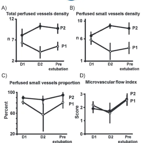
Both patients showed reduced perfused small vessel (,20mm) density, a lower proportion of perfused small
vessels (Figures 1 and 2), and changes in predominant flow (assessed by microvascular flow index) during the first day of shock. On the second day, patient 1 had a decreased vessel density and proportion of perfused small vessels despite the infusion of vasodilators. Patient 2 had an increased number of perfused vessels; however, this increase did not improve the tissue perfusion because the blood flow was arrested or intermittent. The blood flow in the large blood vessels was normal throughout the period of shock. Both patients showed improvements in their mea-sured parameters after the shock subsided.
The microvascular flow indices were low in both patients on the first day, likely because of a predominance of vessels with intermittent or no flow. The indices decreased on the second day but improved on the day prior to extubation, although neither patient achieved normalization of micro-circulatory flow.
& DISCUSSION
Even though the majority of dengue cases present as an acute febrile illness, some patients develop a more severe form. Little is known about the pathophysiological mechan-isms involved in developing severe forms, although the main characteristic is an increase in endothelial permeability with leakage into the interstitium.
Effects of the dengue virus on the endothelium have been documented.In vitrostudies using infected endothelial cells
have shown increased permeability after 48 hours and cytological effects after 72 hours (8). Studies with serum from patients with dengue demonstrated increased levels of intercellular adhesion molecule-1 (ICAM-1) and vascular cell adhesion molecule-1 (VCAM-1) (9), both of which are endothelial activation markers.
Severe forms of dengue infection affect the microcircula-tion, as demonstrated by the dengue virus directly infecting endothelial cells or activating inflammatory markers, such as interleukins 6 and 8 and tumor necrosis factor alpha (10). Microcirculatory visualization in patients with severe dengue could help to elucidate the disease mechanisms, stratify patients, and monitor the disease evolution.
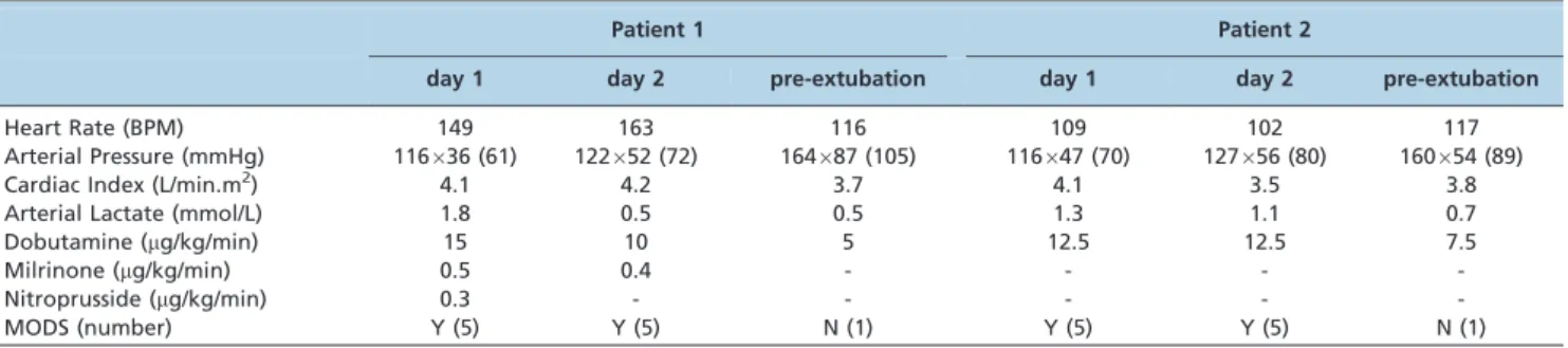
Table 1 -The clinical and laboratory parameters and the drugs used at the moment when the microcirculatory analysis was performed using Sidestream Dark Field technology.
Patient 1 Patient 2
day 1 day 2 pre-extubation day 1 day 2 pre-extubation
Heart Rate (BPM) 149 163 116 109 102 117
Arterial Pressure (mmHg) 116636 (61) 122652 (72) 164687 (105) 116647 (70) 127656 (80) 160654 (89)
Cardiac Index (L/min.m2) 4.1 4.2 3.7 4.1 3.5 3.8
Arterial Lactate (mmol/L) 1.8 0.5 0.5 1.3 1.1 0.7
Dobutamine (mg/kg/min) 15 10 5 12.5 12.5 7.5
Milrinone (mg/kg/min) 0.5 0.4 - - -
-Nitroprusside (mg/kg/min) 0.3 - - - -
-MODS (number) Y (5) Y (5) N (1) Y (5) Y (5) N (1)
MODS, multiple organ dysfunction syndrome, yes/no (the number of dysfunctional organs). Microcirculatory dysfunction in dengue shock
Caixeta DM et al. CLINICS 2013;68(7):1061-1064
Here, we used a Microscan, a portable device that allows direct microcirculatory visualization at a patient’s bedside. The Microscan is limited by the presence of severe bleeding of the oral mucosa, which prevents visualizing the blood
vessels, and by the need for operator training to avoid artifacts, such as pressure and movements.
Using the Microscan, we detected severe microcirculatory changes in two children with dengue shock and multiple organ dysfunction in the first two days of shock; these changes were reflected in a decreased number and propor-tion of perfused small vessels, despite the administrapropor-tion of vasodilators. The predominant feature of microvascular flow was intermittent or no flow. Both patients showed improvement after the resolution of shock, but microcircu-lation was still not completely normalized.
When compared with the published data from healthy children (6), these two patients had severe microcirculatory dysfunction. Furthermore, when compared with children with septic shock (5) and meningococcal disease (6), two conditions known to be accompanied by microcirculation changes, these patients showed more severe microcircula-tory alterations, as demonstrated by a low number of perfused vessels and proportion of perfused small vessels. Additionally, the predominant features of microvascular flow were intermittent or no flow, as established by the low microvascular flow index.
Although biochemical analyses of the markers of endothelial dysfunction were not performed, the micro-circulatory changes visualized with Sidestream Dark Field technology in these patients supported the published data on Figure 1 -The evolution of microcirculatory analysis. The measurements were performed on the first and second days and on the day prior to extubation. A) Number of perfused vessels. The number of perfused vessels that cross the horizontal and vertical equidistant lines drawn on the screen divided by the total length of the lines, according to the De Backer score. B) Number of perfused small vessels. The number of perfused small vessels (,20mm) that cross the horizontal and vertical equidistant lines drawn on the screen
divided by the total length of the lines, according to the De Backer score. C) The proportion of perfused small vessels can be calculated as follows: 100 x (total number of vessels - [no flow vessels+intermittent flow vessels])/total number of vessels. D) Microvascular flow index based on the predominant flow in the four screen quadrants. The flow is characterized as absent (0), intermittent (1), sluggish (2), or normal (3). The final result is the average of the four quadrants.
Figure 2 - Images of the sublingual microcirculation in both patients on the first day and the day prior to extubation.
CLINICS 2013;68(7):1061-1064 Microcirculatory dysfunction in dengue shock
Caixeta DM et al.
dengue virus infections (8,9). To the best of our knowledge, these microcirculatory alterations have never been described in association with dengue.
Microcirculatory monitoring of patients with severe forms of dengue infection could help clarify the pathophysiology of this disease and serve as a prognostic marker or therapeutic target to optimize tissue perfusion and avoid the progression to multiple organ dysfunction.
Severe microcirculation changes may be involved in the pathophysiological mechanisms that lead to the final stages of dengue shock, which is frequently irreversible and has high mortality rates. Microcirculatory monitoring could be an important tool to evaluate the evolution of this type of shock, but more studies are required to answer these questions.
& ACKNOWLEDGMENTS
This study was supported by grants from the following governmental granting agencies: the National Research Council of Brazil (CNPq) and the Rio de Janeiro State Foundation for Research Support (FAPERJ).
& AUTHOR CONTRIBUTIONS
Caixeta DM, Fialho FM and Azevedo ZM collected and analyzed the data. Villela NR designed the study and wrote the final version of the manuscript. Collett-Solberg PF and Bouskela E wrote the final version of the manuscript.
& REFERENCES
1. WHO. Guidelines for diagnosis, treatment, prevention and control. Geneva. World Health Organization, 2009.
2. Ranjit S, Kissoon N. Dengue hemorrhagic fever and shock syndromes. Pediatr Crit Care Med. 2011;12(1):90-100, http://dx.doi.org/10.1097/ PCC.0b013e3181e911a7.
3. Khongphatthanayothin A, Lertsapcharoen P, Supachokchaiwattana P, La-orkhun V, Khumtonvong A, Boonlarptaveechoke C, et al. Myocardial depression in dengue hemorrhagic fever: Prevalence and clinical description. Pediatr Crit Care Med. 2007;8(6):524-9, http://dx.doi.org/ 10.1097/01.PCC.0000288672.77782.D4.
4. De Backer D, Ospina-Tascon G, Salgado D, Favory R, Creteur J, Vincent JL. Monitoring the microcirculation in the critically ill patient: current methods and future approaches. Intensive Care Med. 2010;36:1813-25, http://dx.doi.org/10.1007/s00134-010-2005-3.
5. Top APC, Ince C, Meij N, Dijk M, Tibboel D. Persistent low microcirculatory vessel density in nonsurvivors of sepsis in pediatric intensive care. Crit Care Med. 2011;39(1):8-13, http://dx.doi.org/10. 1097/CCM.0b013e3181fb7994.
6. Paize F, Sarginson R, Makwana N, Baines PB, Thomson APJ, Sinha I, et al. Changes in the sublingual microcirculation and endothelial adhesion molecules during the course of severe meningococcal disease treated in the paediatric intensive care unit. Intensive Care Med. 2012;38(5):863-71, http://dx.doi.org/10.1007/s00134-012-2476-5.
7. De Backer D, Hollenberg S, Boerma C, Goedhart P, Bu¨chele G, Ospina-Tascon G, et al. How to evaluate the microcirculation: report of a round table conference. Crit Care. 2007;11(5):R101, http://dx.doi.org/10.1186/ cc6118.
8. Talavera D, Castillo AM, Dominguez MC, Gutierrez AE, Meza I. IL8 release, tight junction and cytoskeleton dynamics reorganization conducive to permeability increase are induced by dengue vı´rus infection of microvascular endotelial monolayers. J Gen Virol. 2004;85:1801-13, http://dx.doi.org/10.1099/vir.0.19652-0.
9. Koraka P, Murgue B, Deparis X, Van Gorp EC, Setiati TE, Osterhaus AD, et al. Elevation of soluble VCAM-1 plasma levels in children with acute dengue virus infection of varying severity. J Med Virol. 2004;72(3):445-50, http://dx.doi.org/10.1002/jmv.20007.
10. Cardier JE, Marin˜o E, Romano E, Taylor P, Liprandi F, Bosch N, et al. Proinflammatory factors present in sera from patients with acute dengue infection induce activation and apoptosis of human microvascular endotelial cells: Possible role of TNF-ain endothelial cell damage in dengue. Cytokine. 2005;30(6):359-65, http://dx.doi.org/10.1016/j.cyto. 2005.01.021.
Microcirculatory dysfunction in dengue shock
Caixeta DM et al. CLINICS 2013;68(7):1061-1064