Universidade de Trás-os-Montes e Alto Douro
Correlation of Vaginal Cytology and Reproductive
Tract Ultrasonography in Wild Felids for Oestrus Cycle
Stage Determination
Dissertação de Mestrado Integrado em Medicina Veterinária
Sara Martins Dantas Gonçalves
Orientadores:
Professora Doutora Rita Maria Payan Martins Pinto Carreira
Dr. Imke Lueders
DECLARAÇÃO
NOME: Sara Martins Dantas Gonçalves C.C.: 14292859
CORREIO ELETRÓNICO: smdgon@gmail.com
DESIGNAÇÃO DO MESTRADO: Mestrado Integrado em Medicina Veterinária
TÍTULO DA DISSERTAÇÃO DE MESTRADO EM MEDICINA VETERINÁRIA:
Correlation of Vaginal Cytology and Reproductive Tract Ultrasonography in Wild Felids for Oestrus Cycle Stage Determination
ORIENTADOR: Professora Doutora Rita Maria Payan Martins Pinto Carreira CO-ORIENTADOR: Dr. Imke Lueders
ANO DE CONCLUSÃO: 2019
Declaro que esta dissertação de mestrado é resultado da minha pesquisa e trabalho pessoal e das orientações dos meus supervisores. O conteúdo é original e as fontes consultadas estão devidamente mencionadas no texto e na bibliografia final. Declaro ainda que este trabalho não foi apresentado em nenhuma outra instituição parra obtenção de qualquer grau académico.
Vila Real, Outubro de 2019 Sara Martins Dantas Gonçalves
Acknowledgements
To my supervisor, Professor Rita Payan Carreira, for accepting to supervise this dissertation and for all the great advice given both before and during my internship.
To my co-supervisor, Dr. Imke Lueders, who kindly allowed me to access so much valuable information that was of uttermost importance for the elaboration of this dissertation, for giving me the chance to make my dreams a reality, for guiding me through my internships and during the writing of this dissertation and for teaching me so much about wildlife and reproduction physiology and biotechnology. I have found my passion thanks to you.
To Isabel Callealta, for kindly allowing me to use data collected during procedures for her PhD project, for allowing me to assist in her project, for the advices and for all the laughs and good times.
To Dr. Gerhard van der Horst from the Western Cape University, for allowing the access to important data that was key to the completion of this dissertation.
To Ukutula Conservation Centre, the owners Willi and Gill Jacobs and all the staff and volunteers that I met during my time there; for welcoming me into your home and making me part of your “Ukutula Family” and for giving me the best time of my life! A special thanks to my dear friend Pierre for putting up with my early morning grumpiness and for getting me and Isabel coffees and chocolates.
To AllwetterZoo Muenster and all the staff, especially Dr. Carsten Ludwig, for receiving me and teaching me so much about zoo medicine.
To the family I adopted in Vila Real, for all the unforgettable moments that are too many to describe, for all the laughs and despair that this degree brought us and for being one of the best things I take for life.
To my beloved fiancé, Ben, for the unconditional support and love, for helping me with this dissertation when the tasks seemed impossible, for revising it over and over, for not allowing me to give up on my dreams and for showing me that it is possible to follow a different path. And lastly but not least, to my parents, whose sacrifices allowed me to follow my dreams, for always supporting me in their own way. Thank you so much!
Abstract
Felids are top-down predators and keystone species to heathy ecosystems. The combination of human-animal conflicts, territory reduction and fragmentation with low population densities and loss of genetic diversity makes many wild cat species threatened or endangered. In the knowledge that many wild felids reproduce poorly and that reproduction is key to species survival, implementing artificial reproduction into conservation of these species is extremely important. For this to work, a deeper understanding of their reproductive physiology is needed. Assessment of the reproductive cycle in captive animals allows to collect data both for the practical implementation of assisted reproduction protocols and for research purposes.
There are different techniques to assess the reproductive cycle of mammals. This study aimed to describe and correlate two techniques for collection of data for the reproductive cycle assessment in six different species of wild felids for which data was available (Panthera leo,
Panthera pardus kotiya and P. pardus tulliana. Panthera uncia, Catopuma temminckii, Neofelis nebulosa and Acinonyx jubatus) namely using reproductive tract ultrasonography and vaginal
cytology.
The literature review shows that the domestic cat has been used as a model for the wild species of the same Family. It also reveals differences in their reproductive physiology that may be a cause for low success rates in implementing artificial reproduction techniques.
During anaesthesia procedures, vaginal smears and transrectal ultrasound examinations were performed in 29 animals housed in zoos in Europe, Asia and a breeding facility in South Africa. The data was assessed separately and results revealed many commonalities between species and when compared to the literature. A method for identifying the phases of the reproductive cycle was created both for vaginal cytology, using the percentage of cornification of epithelium, and for ultrasonography, assessing the presence and size of follicles and corpora lutea in the ovaries. Then, where sufficient data existed, samples collected from the same animal were compared individually and the phase of the cycle was corrected.
This study showed that a single vaginal cytology allows the differentiation between oestrus and non-oestrus phases in wild felids, but further differentiation is difficult. Transrectal ultrasonography proved to be a finer tool for predicting the cycle stage, however, transitioning phases were still ambiguous. Assessing together the vaginal cytology and ultrasonography allowed a more accurate cycle stage prediction. This was supported by the positive correlation
VI
found between the size of the biggest follicle and percentage of epithelial cornification in African lions (Panthera leo), r = .76, p = .030 (n = 8), and Asiatic golden cats (Catopuma
temminckii), r = .89, p = .042 (n = 5).
Key-words: wild felids, reproduction, vaginal cytology, ultrasonography, reproductive cycle
Resumo
Felinos são predadores de topo de cadeia alimentar e espécies fundamentais para ecossistemas saudáveis. A combinação de conflitos entre humanos e animais, redução e fragmentação de território com densidades populacionais baixas e consequente perda de diversidade genética fazem de muitas espécies de felinos selvagens animais ameaçados de extinção. Sabendo que muitos felinos selvagens têm baixos níveis de reprodução e que a mesma é fundamental para a sobrevivência de espécies, a implementação de técnicas de reprodução assistida na conservação destas espécies é extremamente importante. Para isso, um conhecimento mais profundo da sua fisiologia reprodutiva é necessário. A avaliação do ciclo reprodutivo de animais em cativeiro permite recolher dados tanto para a implementação prática de protocolos de reprodução assistida como para futura investigação científica.
Existem diferentes técnicas para a avaliação do ciclo reprodutivo em mamíferos. Este estudo visou descrever e correlacionar duas técnicas para a recolha de dados para avaliar o ciclo reprodutivo em seis espécies de felinos selvagens para as quais foi possível recolher dados
(Panthera leo, Panthera pardus kotiya e P. pardus tulliana. Panthera uncia, Catopuma temminckii, Neofelis nebulosa e Acinonyx jubatus), nomeadamente o uso de ecografia transretal
do trato reprodutivo e citologia vaginal.
A revisão de literatura mostra que o gato doméstico tem sido usado como modelo para espécies selvagens da mesma Família. Contudo, diferenças na fisiologia reprodutiva entre espécies foram reveladas, que podem ser a causa do baixo sucesso na implementação de técnicas de reprodução assistida.
Durante procedimentos anestésicos, amostras de citologias vaginais e imagens ecográficas por via transretal foram recolhidas em 29 animais alojados em zoos na Europa, Ásia e África do Sul. As amostras foram avaliadas separadamente e resultados mostraram semelhanças entre espécies e quando comparadas com a literatura. Um método para identificar as fases do ciclo reprodutivo foi criado tanto para citologia vaginal, usando a percentagem de queratinização do epitélio, como para ecografia, tendo em conta a presença e tamanho de folículos e corpo lúteo nos ovários. Posteriormente, citologias e imagens ecográficas recolhidas do mesmo animal foram comparadas individualmente e a fase do ciclo foi corrigido.
Este estudo mostrou que uma única amostra de citologia vaginal permite diferenciar estro de não-estro em felinos selvagens, embora uma diferenciação mais precisa seja difícil. Ecografia
VIII
transretal revelou-se uma técnica mais precisa para prever a fase do ciclo reprodutivo, embora fases de transição continuam a ser ambíguas. A avaliação em conjunto da citologia vaginal e ecografia transretal permite uma diferenciação e previsão das fases do ciclo mais precisa. Isto foi suportado pela correlação positiva encontrada entre o tamanho do maior folículo e percentagem de queratinização do epitélio vaginal em Leão africano (Panthera leo), r = .76, p = .030 (n = 8), e Gato-bravo-dourado-da-ásia (Catopuma temminckii), r = .89, p = .042 (n = 5).
Palavras chave: felinos selvagens, reprodução, citologia vaginal, ecografia, avaliação do ciclo
Table of contents
Acknowledgements ... III Abstract ... V Resumo ... VII Table of contents ... IX Figure Index ... XITable Index ... XIII
Abbreviations list ... XV
Part I – Literature Review ... 1
1. INTRODUCTION ... 1
2. FELINE REPRODUCTIVE CYCLE ... 3
2.1. Anatomy of female reproductive system ... 4
2.2. The oestrous cycle ... 5
2.2.1. Prooestrus ... 6 2.2.2. Oestrus ... 7 2.2.3. Post-oestrus ... 8 2.2.4. Dioestrus... 8 2.2.5. Anoestrus ... 9 3. ULTRASONOGRAPHY ... 12 4. VAGINAL CYTOLOGY ... 14
5. IDENTIFICATION OF THE FERTILE PERIOD ... 18
Part II – Retrospective study of the association of ultrasonography and vaginal cytology for oestrous cycle assessment... 21
1. MATERIAL AND METHODS ... 21
1.1. Animals... 21
1.2. Data collection... 22
1.2.1. Anaesthesia ...22
1.2.2. Ultrasonography ...22
1.2.3. Vaginal swab collection and smear preparation ...23
X
1.3. Classification of the oestrous cycle phases through ultrasonography and vaginal
cytology ... 24
1.3.1. Ultrasonography ...24
1.3.2. Vaginal cytology ...25
1.2.2.1. Establishing a classification system ...25
1.3. Statistical analysis ... 26
2. RESULTS ... 27
2.1. Ultrasonography ... 27
2.2. Vaginal Cytology ... 33
2.2.1. Cell appearance: ...33
2.2.2. Results as per classification system for vaginal cytologies: ...33
i. heat (oestrus) ...33
ii. transition (prooestrus and immediately after oestrus) ...34
iii. luteal activity (dioestrus or non-pregnant luteal phase) ...34
iv. quiescence (anoestrus) ...35
2.3. Matching Vaginal Cytology and Ultrasound observations ... 36
2.3.1. Establishing a classification system ...36
2.3.2. Determining the correct phase of the oestrous cycle ...37
2.3.3. Correlations ...42
3. DISCUSSION... 45
4. CONCLUSION AND FINAL CONSIDERATIONS ... 51
Figure Index
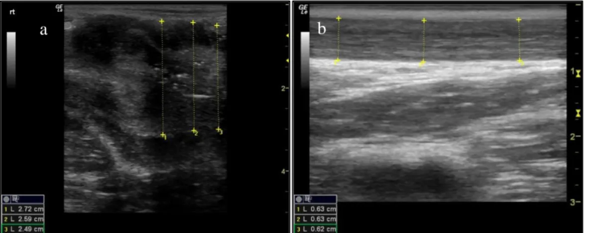
Figure 1. Longitudinal section of the low reproductive tract in the female domestic cat (Felis catus) .. 4 Figure 2. Schematic of the felids oestrous cycle. ... 6 Figure 3. Ultrasound image (longitudinal section) of the (a) Vaginal vestibulum and (b) vagina ...27 Figure 4. Ultrasound image (longitudinal section) of the (a) cervix with measurement and (b) cervix
showing folds/lumen ...28
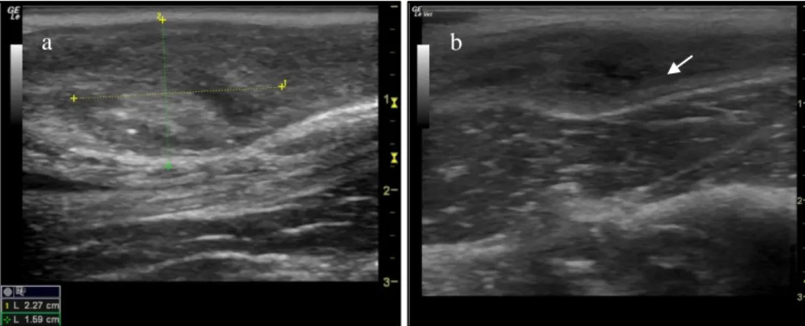
Figure 5. Ultrasound image (longitudinal section) of the (a) uterine body with distinct endometrium and
(b) uterine body and endometrium with urinary bladder appearing ventrally ...29
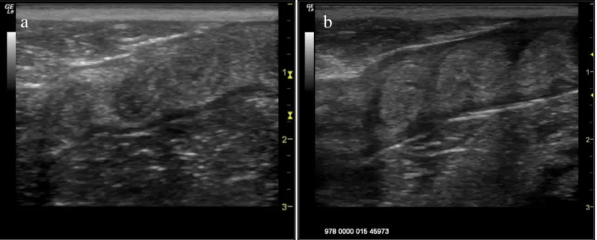
Figure 6. Ultrasound image (longitudinal section) of the uterine horn in a cheetah ...29 Figure 7. Ultrasound image (longitudinal section) of the uterine horns during dioestrus ...30 Figure 8. Ultrasound images of ovaries in different reproductive stages: (a) ovary in prooestrus showing
multiple follicles (b) ovary during oestrus showing big follicles (c) ovary during dioestrus showing a Corpus Luteum (d) ovary during post-oestrus with a luteinised follicle and (e) ovary during anoestrus ...31
Figure 9. Ultrasound image of (a) uterus of a Persian leopard with pyometra; anechoic intrauterine
content is seen and (b) uterine body (with measurements) and cervix of a cheetah with endometritis; thickened hyperechoic mucus membranes are seen ...32
Figure 10. Microscopic image: Overview of an oestrus vaginal cytology, showing superficial nucleated
and superficial enucleated epithelial cells ...33
Figure 11. Microscopic image: Overview of prooestrus/post-oestrus vaginal cytology, showing
parabasal, intermediate and superficial cells and neutrophils...34
Figure 12. Microscopic image: Overview of the vaginal cytologies in early dioestrus, showing parabasal
and superficial epithelial cells and high amount of debris. ...35
Figure 13. Microscopic image: Overview of an anoestrus vaginal cytology, showing parabasal cells and
high amount of debris and mucus. ...35
Figure 14. Graph showing correlation between the size of the biggest follicle measured by
ultrasonography of both ovaries and percentage of cornification of the vaginal epithelium in African lions ...42
Figure 15. Graph showing correlation between the size of the biggest follicle measured by
ultrasonography of both ovaries and percentage of cornification of the vaginal epithelium in Asiatic golden cats ...43
Table Index
Table 1. Summary of reproductive cycles of studied female wild felid species (part 1): conservation
status, geographic range, seasonality, type of ovulation and reproductive behaviour. ...10
Table 2. Summary of reproductive cycles of studied female wild cat species (part 2): characterization
of some reproductive traits. ...11
Table 3. Classification and description of vaginal epithelial cells ...16 Table 4. Summary of examined animals, institutions where the examinations took place and age of
animal at examination ...22
Table 5. Vaginal cytology results as per established classification system. ...36 Table 6. Classification system to match vaginal cytology and ultrasonography results for cycle stage prediction ...37
Table 7. Results as per established classification system using ultrasonography and vaginal cytology in
combination. ...38
Table 8. Visual comparison of ultrasonography and vaginal cytology during the determined stages in
Abbreviations list
AI – Artificial InseminationART – Assisted Reproductive Technology BC – Basal epithelial cells
CH – Corpus haemorrhagicum CL – Corpora Lutea
EEP - European Endangered Species Programmes EIA – Enzyme immunoassay
ESB - European Studbooks ET – Embryo Transfer
GnRH – Gonadotropin-Releasing Hormone IC – Intermediate epithelial cells
IUCN – International Union for the Conservation of Nature LH – Luteinizing Hormone
n.a. – not applicable
PBC – Parabasal epithelial cells SEC – Superficial enucleated cells SNC – Superficial nucleated cells
Part I – Literature Review
1. INTRODUCTION
The Felidae family is composed of 38 species placed into small, medium and large size categories, based on body weight (Brown, 2011). Non-domestic felids are considered top-down predators, which means they control populations of organisms placed lower on the food chain. They are often considered keystone species in their native habitats, influencing the nature and strength of ecosystem functioning. Besides preventing overgrazing and controlling pest populations, felines often pray upon the most vulnerable individuals, promoting robust pray population with decreased vulnerability to disease. The combination of decreased wild felid population densities and reproductive rates with human-animal conflict (felids have relatively high needs for prey animals and roaming territories) is what makes them vulnerable and poorly able to respond to persecution. Because of their ability to structure ecosystems and their vulnerability to extinction, immediate conservation action for wild cat species is exceptionally important (Ripple, et al., 2014).
At present, 28 out of the 38 known wild cat species are threatened or endangered in the red list of the International Union for the Conservation of Nature (IUCN) in at least some part of their habitat. The primary causes for the decline of feline populations in the wild are constant habitat loss and environment fragmentation, human-animal conflict and, for some species, poaching for pelts and traditional medicine (Brown, 2011). Despite the efforts from conservation specialists and organisations, many felid populations are still declining. Fortunately, small populations are maintained under protection ex situ in zoological institutions, sanctuaries and natural reserves all over the world. These reservoirs help not only to protect the living specimens, but also preserve genetic diversity. This allows for selected breeding programs and if correctly managed, these may help repopulating habitats in the future.
The thrive of populations depends not only on their ability to survive but also to reproduce. Despite information derived from a long history in captivity, little is known about their basic reproductive physiology and behaviour, because in the wild, most felids live generally secretively and are difficult to observe (Brown, 2011). Inadequate husbandry, stress, incompatible breeding partners, infectious and/or parasitic diseases, nutritional and behavioural deficiencies are suggested to be the main causes of reproductive failure for wild animals in captivity (Mellen, 1991). Another factor may be the loss of genetic diversity, applying for both
2
in- and ex situ populations, due to habitat fragmentation or low numbers of individuals,
respectively. In the wild, the isolation and fragmentation of habitats results in a decrease on genetic variability for the respective populations, because the number of breeders in each habitat pocket is low and exchange impossible (Paz et al., 2010). This issue could impair natural selection and therefore strong phenotypic characteristics to thrive, which in turn may lead to decreased fertility, higher susceptibility to disease and weaker populations in general.
In captivity, reproduction is highly dependent on husbandry and management. Because most cat species are solitary animals, only meeting occasionally during mating periods, the proposed breeding pair may need to be kept separated for the majority of time. Usually, only during the receptive oestrus period animals are mixed within their enclosures. Selecting a proper breeding partner is however difficult. In addition, identifying oestrus (heat) poses some difficulties in some species such as the cheetah (Wielebnowski and Brown, 1998) and the clouded leopard (Brown, 2011), resulting in animals being put together at non-receptive periods. In result, fatal injuries may derive from individuals fighting (Paz et al., 2010). Therefore, in addition to adequate husbandry for optimized health, behaviour and wellbeing of the animals, close monitoring as well as a proper knowledge and correct assessment of the reproductive cycle becomes extremely important.
Even though fostering genetic diversity is important in any breeding population, at the same time the number of individuals in captivity is limited. Many facilities do not have the capacity to house more than a few individuals. Studbooks have been established to allow exchange animals between facilities. In Europe for example, felid species and subspecies are organised in endangered species breeding programmes such as the European Endangered Species Program (EEP) and the European Studbooks (ESB) (EAZA Population Management Manual, 2012). Still, the problem remains that certain individuals are not represented in the population since they do not reproduce naturally. Because genetic diversity depends on reproduction, the development of alternative strategies for breeding is of utmost importance for the survival of feline species. These strategies may include the development of alternative methods to increase fertility as well as to improve artificial breeding outcomes, namely from assisted reproductive technology (ART), including artificial insemination (AI), in vitro fertilisation (IVF), embryo transfer (ET), and sperm, egg and embryo cryopreservation (Swanson, 2006). These techniques allow the exchange of genetic material between distant populations, as well as its preservation through the creation of biobanks, without the need to relocate animals (Silva et al., 2017). Since Moore et al. (1981) reported a successful AI in a captive puma through a surgical approach,
several advances and successes in ART have been reported but non-surgical successes have been limited to just a few species, such as the tiger, leopard and Asiatic golden cats (Lueders et
al., 2014).
Despite its enormous potential for conservation and the increasing number of papers published so far on this context, still too little knowledge is available, which reflects in the low success rates for artificial insemination (Pelican et al., 2006; Swanson, 2006). Despite many reports on ART attempts in a wide range of feline species exist (Micheletti et al., 2011), the success rate of AI in wild felids remains generally below 20% (Pelican et al., 2006). For one successful birth, several attempts are needed that cannot justify the associated expense, labour and stress caused to the animal by repeated manipulations (Swanson, 2006). For the advanced reproduction biotechnologies developed for humans and domestic animal species to meet their fullest potential in wildlife conservation, it is essential to study the endocrinology and reproductive physiology of the species (including the characteristics of the reproductive cycle, namely seasonality), as well as to describe the behaviour and general regulative mechanisms of reproduction. This information will also be an essential asset in understanding the causes of reproductive failure (Swanson, 2006). To implement assisted breeding, it is fundamental to determine the optimal time for mating and the ideal time to apply ARTs (Silva et al., 2017).
2. FELINE REPRODUCTIVE CYCLE
Felidae have a relatively conserved reproduction biology with basic similarities in the reproduction anatomy and physiology. However, marked inter and intra-species variations have also been documented (Brown, 2011; Andrews et al., 2018), making conservation breeding of wild endangered felines challenging. The ovulation pattern and the effect of seasonality on reproduction are two characteristics that impact both natural and assisted breeding management.
The domestic cat, being easily manipulated, is widely used in reproductive physiology research as a model for wild animal similar species (Kanca et al., 2014). However, the differences between species and the intractability of wild animals makes the application of assisted reproduction protocols difficult (Thongphakdee et al., 2018). Reports on various aspects of the reproduction physiology in a wide array of felid species, ranging from endocrine studies to limited ultrasound or endoscopic observations, exist. Currently, the lion (Panthera leo) is also
4
being studied as a large felid model (Callealta et al., 2018; Callealta 2018, personal communication). Since this particular species lives in groups, contrasting with many solitary roaming felids, it breeds without complication in captivity and is therefore easier to access for reproductive physiology research.
3. Anatomy of female reproductive system
In common with most mammal species, the female feline reproductive system consists of (from posterior to anterior) vulva, vestibulum, vagina, cervix, a bicornuate uterus, two oviducts and two ovaries.
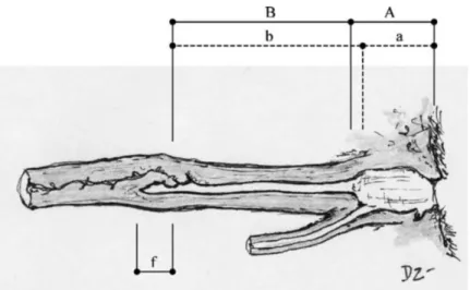
The vulva is the exterior portion of the reproductive tract, located below the anus. It is connected to the vestibulum (or urogenital sinus), the first and widest interior portion, believed to be where the sperm is deposited during mating (Zambelli and Cunto, 2005). The narrow and non-distensible vagina connects the vestibulum to the cervix, which terminates in an extension after the cervical opening, the fornix. Dorsally to the vagina and posterior to the cervix, a prominence called dorsal medial fold had been described (Zambelli and Cunto, 2005). The cervix forms an obtuse angle with the vagina, and separates the uterus from the vagina, working as a physical barrier during the luteal phase and anoestrus (Figure 1).
Figure 1. From Zambelli and Cunto (2005). Longitudinal section of the low reproductive tract in the female domestic cat (Felis catus). Horizontal dashed line: measures taken by Watson and Glover (1993): urogenital sinus (A), vagina (B). Horizontal continuous line: measures taken by Swanson and Godke (1994): vestibule (a), vagina (b). fornix (f).
The uterus, which is suspended dorsally by the mesometrium, starts with a short uterine body that then divides into two uterine horns, each one terminating in an oviduct that is connected to one ovary (Brown, 2011). The ovaries are two small oval structures located in the dorsal
abdomen caudally to the kidneys (Johnston, Kustritz and Olson, 2001b). They are attached proximally by the suspensory ligament and dorsally by the ipsilateral mesovarium. The oviducts and the lateral aspect of the ovaries are covered by the mesosalpinx, forming an open ovarian bursa (Brown, 2011). The blood supply to the ovary and cranial portion of the uterine body is made by the ovarian artery (Johnston, Kustritz and Olson, 2001b). The long uterine horns permit blastocysts to spread evenly after fertilisation, allowing felids to bear litters (Brown, 2011).
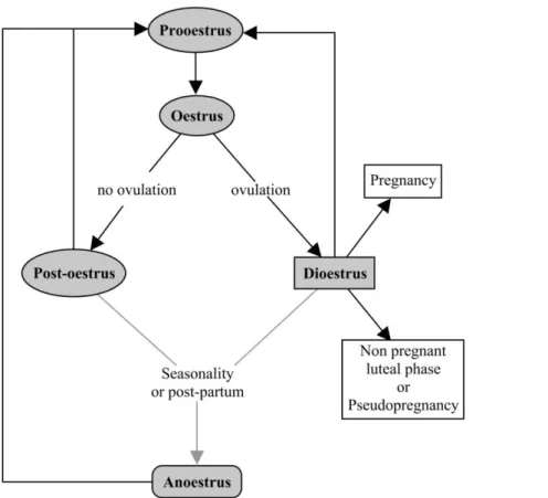
4. The oestrous cycle
The oestrous cycle is defined as the recurring physiological variations induced by hormonal mechanisms controlling the reproductive system of female mammals. To better understand these mechanisms, the cycle is described in different phases, with the prooestrus and oestrus phases being associated with heat (Johnston, Kustritz and Olson, 2001a).
In the Felidae family, the oestrous cycle lengths and some features vary greatly among species, but also within animals of the same species. Most felids show both, ovulatory and anovulatory cycles, which is why they may present different phases after an oestrus, compared to other animal species. An ovulatory cycle leads to a luteal phase – or dioestrus – whereas an anovulatory cycle leads to a new follicular wave starting with a new prooestrus after a short post-oestrous stage (Feldman and Nelson, 2004; Brown, 2011). This mainly is related to cats being generally induced ovulators, only showing ovulation (rupture of dominant follicles) after copulation. Additionally, many cats present seasonal and non-seasonal phases throughout the year, depending on the photoperiod (Shille et at., 1979). The seasonal phase occurs when the cat undergoes consecutively prooestrus, oestrus and post-oestrus or dioestrus. The non-seasonal phase is also called anoestrus and represents a period of acyclicity. The different oestrous cycle phases are represented in Figure 2.
6
Figure 2. Schematic of the felids oestrous cycle (adapted from Johnston, Kustritz and Olson, 2001a).
The domestic cat and most wild species are polyoestrous, meaning they have several oestrous cycles during the breeding season. The only exception known to date are of the animals from Lynx genus (Lynx lynx, Lynx canadensis, Lynx rufus and Lynx pardinus), which have a unique monoestrous cycle (Göritz et al., 2009). The age of sexual maturity varies depending on the species, but individual differences also depend on the current photoperiod, species, breed and weight of the cat (Feldman and Nelson, 2004).
4.1.1. Prooestrus
Usually lasting less than a day, the prooestrus is the phase preceding the oestrus. During this phase, the cat presents behavioural signs of heat - generally increased frequency of vocalisation, territorial marking, rubbing and rolling; occasionally the male shows interest with no copulation (Johnston, Kustritz and Olson, 2001a). In association with the presence of small follicles, containing a developing oocyte, the circulating oestrogens increase rapidly, and the cornification of the vaginal epithelium begins (Brown, 2011). Often, this period is not observed or is difficult to identify, due to species particularity, the little or no external genitalia signs and its rather short time (Shille, Lundström and Stabenfeldt, 1979).
4.1.2. Oestrus
Oestrus is characterised as behavioural receptivity to mating, and it is more commonly designated as heat. In most species, an increased frequency of vocalisation, urination, rubbing, rolling, foot treading and elevation of the perineum with ventral thorax and abdomen touching the floor - a posture named lordosis - may be observed. This phase is associated with advanced ovarian follicular development and peak serum concentrations of oestrogens that induce changes in the vaginal epithelium and cause the labia of the vulva to become slightly oedematous (Johnston, Kustritz and Olson, 2001a; Brown 2011).
During oestrus, ovulation may or may not occur, since feline species exhibit different patterns, varying from exclusively to partially induced ovulators (see Table 1, on page 10). This mechanism, called genital-somatosensory, starts with copulation, inducing the release of gonadotropin-releasing hormone (GnRH) which stimulates the release of luteinizing hormone (LH), necessary for the final follicle and oocyte maturation and subsequent ovulation (Bakker and Baum, 2000; Wildt et al., 1981; Johnston, Kustritz and Olson, 2001a). It is believed that multiple intromissions are required, usually over several days, to stimulate this cascade of events (Brown, 2011). Concannon, Hodgson and Lein (1980) have concluded that the magnitude and duration of LH release depends on the number of copulations. According to Malandain et al. (2011), in the domestic cat, a peak of LH occurs within 2h after stimuli, followed by ovulation 24 to 56h later. However, it is now known that some felids ovulate without copulation (Thongphakdee et al., 2018). Endogenous steroids stimulate the release of GnRH by positive feedback in spontaneously ovulating species. The mechanism for felids is not entirely understood. Some authors believe that oestrus behaviours such as rolling or rubbing, and the social interactions without copulation may influence the release of GnRH (Bakker and Baum, 2000). Others suggest that a certain level of endogenous steroids combined with genital-somatosensory signals induce the release of LH (Thongphakdee et al., 2018). This considerable variation amongst species and individuals of the same species is what makes wild felids unique and challenging to reproduce in captivity.
It is commonly accepted that the average duration of behavioural oestrus differs between ovulating and non-ovulating domestic cats, with an average of 4-5 days and 6-7 days respectively (Kanca et al., 2014). Conversely, many studies showed that the difference in oestrus duration between ovulating and non-ovulating cats from the same species is not significant (Wildt et al, 1981; Shille et al., 1979).
8
Following the oestrus stage, the female may enter two distinct phases: if ovulation occurred, the female enters in a luteal phase or dioestrus; if no mating and ovulation occurred, post-oestrus will follow (Johnston, Kustritz and Olson, 2001a).
4.1.3. Post-oestrus
The post-oestrus stage precedes another prooestrus/oestrus and is characterised by the regression of the follicles without the formation of corpora lutea (CL) and the recruitment of a new follicular wave. Albeit most authors consider that oestrogens and progesterone are at basal levels (Brown, 2011), it has been suggested that oestrogens are maintained at higher levels than if ovulation occurs (Kanca et al., 2014).
Post-oestrus usually lasts 8 to 10 days in domestic cats (Johnston, Kustritz and Olson, 2001a), but its length varies per species (see Table 2 on page 11).
4.1.4. Dioestrus
Regardless of a fertile or sterile mating, 24 to 48 hours after ovulation, one or multiple CL are formed in the place of the ovulated follicles in domestic cats (Malandain et al., 2011). Progesterone produced by CL stays elevated for varying lengths of time, depending on species and individuals (Brown, 2011; see Tables 1 and 2, on page 11 and 12 respectively). The elevated blood progesterone inhibits the development of new follicles, and subsequently the short term display of new oestrus. In pregnant cats, the progesterone rising is crucial for uterine implantation and development of the embryos (Johnston, Kustritz and Olson, 2001a).
If the ovulated eggs are not fertilized, this phase is named as non-pregnant luteal phase or pseudopregnancy (Wildt et al., 1981; Johnston, Kustritz and Olson, 2001a). Generally, the luteal phase or phase of active CL has the duration of approximately one to two-thirds of a pregnancy in the same species; after this period the CL enters luteolysis (regression). Pregnancy after this period is maintained by other factors and luteolysis and the consequent surge of oestrogens induce labour. For this reason, high serum progesterone concentrations are not indicative of pregnancy in felids (Brown, 2006; Jewgenow et al., 2012). In the domestic cat, prolactin is elevated throughout the pregnancy, and relaxin increases at about day 20 until it starts to decline at about time of parturition (Stewart and Stabenfeldt, 1985). After birth, the domestic cat may initiate cyclic activity 3 to 4 weeks after parturition with a good energetic balance and positive photoperiod. However, extensive data is not available for wild felids which
have been observed to enter a phase called lactational anoestrus, where no behavioural oestrus has been observed. Lactational anoestrus may last until 2 or 3 weeks after the litter is weaned (Brown et al., 1995; Johnston, Kustritz and Olson, 2001a).
4.1.5. Anoestrus
Anoestrus is a period of reproductive inactivity. Hormones involved in reproduction are at basal levels, and the ovaries do not show functional structures (Brown, 2011).
Seasonality is one of the main controls for anoestrus. Seasonality refers to the variations on the reproductive patterns during the year controlled by the photoperiod, where generally ovarian activity decreases with the decrease of hours of light during winter and increases when the photoperiod also increases during summer (Johnston, Kustritz and Olson, 2001a; Micheletti et
al., 2011). Due to their wide geographical distribution and habitat differences, seasonality is
one of the most variable physiological aspects of felines (Table 1). In many wild feline species, the amount of food supply throughout the year may also influence seasonality (Paz, 2012). Brown (2011) has suggested that the tiger, clouded leopard, Pallas’ cat, lynx and snow leopard present seasonality, while in lions, leopards, bobcats, pumas, margays, ocelots, tigrinas, jaguars and fishing cat the follicular activity is not influenced by season.
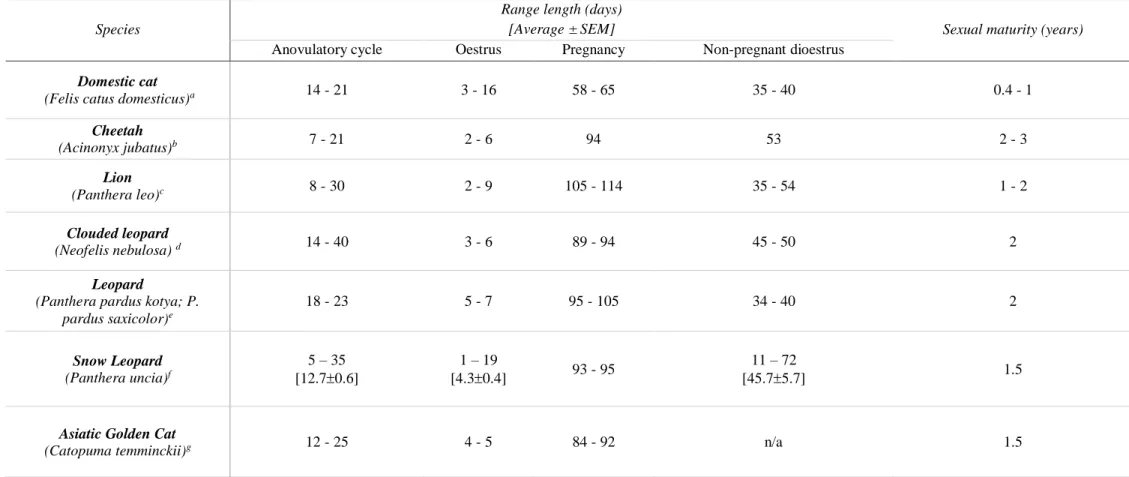
Table 1 summarizes some particularities of the physiology of domestic cat and the six species analysed in this study and Table 2 summarizes their reproductive cycles.
10
Table 1. Summary of reproductive cycles of studied female wild felid species (part 1): conservation status, geographic range, seasonality, type of ovulation and reproductive behaviour*.
Species IUCN Red List status
Geographic
Range Seasonality Type of ovulation Reproductive behaviour
1
Domestic cat
(Felis catus domesticus)a - Worldwide
Seasonal polyoestrous (with
day light increase) Mainly induced
Vocalise, roll, rub, high frequency of urination, lordosis with treading of the hind
feet.
Cheetah
(Acinonyx jubatus)b Vulnerable
Africa and central Asia
Non-seasonal
Polyoestrous Mainly induced
Vocalise, roll, rub, groomand high frequency of urination.
Lion
(Panthera leo)c Vulnerable Africa and Asia
Non-seasonal
Polyoestrous Mainly induced
Vocalise, roll, rub, groom, high frequency of urination, flirting run, lordosis and purr.
Clouded leopard
(Neofelis nebulosi) d Vulnerable Tropical Asia
Seasonal polyoestrous (with day light increase)
Spontaneous and induced
High frequency of urination, lordosis and prusten (chuff).
Leopard
(Panthera pardus kotya; P. pardus saxicolor)e
Endangered Africa and Asia
Seasonal or non-seasonal polyoestrous (depends on sub-species / geographical distribution) Spontaneous and induced
Vocalise, roll, rub, groomand high frequency of urination.
Snow Leopard (Panthera
uncia)f Vulnerable Central Asia
Seasonal polyoestrous
(with day light increase) Mainly induced
Vocalise, roll, rub, groomand high frequency of urination.
Asiatic Golden Cat
(Catopuma temminckii)g Near threatened Asia
Non-seasonal Polyoestrous
Vocalise, roll, rub, groomand high frequency of urination.
a Domestic cat - Brown et al., 2011; Maladain et al., 2011; Kanca et al., 2014. b Cheetah - Durant et al., 2015 (IUCN) ; Brown et al., 2011 ; Schulman et al., 2015. c Lion - Bauer
et al., 2016 (IUCN) ; Thongphakdee A, et al ; Brown et al., 2011; Schramm, Briggs and Reeves, 1994 (ovulation). d Clouded leopard - Grassman et al., 2016 (IUCN). e Leopard
- Stein et al., 2016 (IUCN) ; Paz et al., 2012 (ovulation). f Snow leopard - McCarthy et al., 2017 (IUCN); Reichert-Stewart et al., 2014. g Asiatic Golden Cat – McCarthy et al.,
2015 (IUCN); Olsen, 2012 (behaviour) ; personal communication Imke Lüders, 2019 (seasonality).
1Reproductive Behaviour – According to Stanton, Sullivan and Fazio, 2015 : Vocalise - cat produces sounds or calls, originating from the throat and mouth ; Roll - while lying
on the ground, cat rotates body from one side to another ; Rub - cat rubs any part or entire length of body against (modifier) ; Groom - cat cleans itself by licking, scratching, biting or chewing the fur on its body ; Lordosis - female cat raises hindquarters while lowering forequarters to the ground, presenting genitals to male, with tail often averted to one side ; Flirting run - female cat feigns running away from a breeding partner ; Purr – (type of vocalisation) low, continuous rhythmical tone produced during respiration while the cat’s mouth is closed ; Prusten (chuff) – (type of vocalisation) cat expels jets of air through the nose creating a low-intensity, soft, pulsed sound, described as being similar to the snorting of a horse.
Table 2. Summary of reproductive cycles of studied female wild cat species (part 2): characterization of some reproductive traits.
Species
Range length (days)
[Average ± SEM] Sexual maturity (years)
Anovulatory cycle Oestrus Pregnancy Non-pregnant dioestrus
Domestic cat
(Felis catus domesticus)a 14 - 21 3 - 16 58 - 65 35 - 40 0.4 - 1
Cheetah (Acinonyx jubatus)b 7 - 21 2 - 6 94 53 2 - 3 Lion (Panthera leo)c 8 - 30 2 - 9 105 - 114 35 - 54 1 - 2 Clouded leopard (Neofelis nebulosa) d 14 - 40 3 - 6 89 - 94 45 - 50 2 Leopard
(Panthera pardus kotya; P. pardus saxicolor)e 18 - 23 5 - 7 95 - 105 34 - 40 2 Snow Leopard (Panthera uncia)f 5 – 35 [12.70.6] 1 – 19 [4.30.4] 93 - 95 11 – 72 [45.75.7] 1.5
Asiatic Golden Cat
(Catopuma temminckii)g 12 - 25 4 - 5 84 - 92 n/a 1.5
a Domestic cat - Brown et al., 2011; Maladain et al., 2011; Kanca et al., 2014. b Cheetah - Brown et al., 2011 ; Schulman et al., 2015. c Lion - Thongphakdee A, et al. 2018 ;
Brown et al., 2011; Schmidt et al., 1979 (non pregnant dioestrus); Putman et al., 2015 (cycle). d Clouded leopard - Grassman et al., 2016 (IUCN). e Leopard - Stein et al., 2016
(IUCN) ; de Haas van Dorsser et al., 2007 (oestrus length, gestational period, sexual maturity, anovulatory cycle, non-pregnant luteal phase). f Snow leopard - Reichert-Stewart
12
5. ULTRASONOGRAPHY
Ultrasonography is a non-invasive, practical and relatively uncomplicated tool to access the reproductive status in both free-ranging and captive animals. Besides the detection of many reproductive pathologies, it can provide information on the size of the female reproductive organs, including the vagina, cervix, uterus and ovaries, and evidence of its functional structures, namely the number and size of follicles and CL. For this reason, the technology has been used in many domestic animals for breeding purposes. However, up to date, there are limited reports on the description of the normal reproductive tract of wild female felids, especially on the imaging of the ovaries (Schulman et al., 2015).
Technological advances in ultrasonography allowed, in recent years, to follow up follicular dynamics, identify the presence of follicles and CL (Malandain et at., 2011), assist the study of CL regression mechanisms (Gómez-Seco et al., 2017) and to confirm the efficiency of oestrus synchronization and superovulation protocols (Silva et al., 2017). This technique, therefore, becomes a potent tool in assisted reproduction, in that it allows better monitoring of the ovarian activity when performing artificial insemination and oocyte or embryo retrieval, IVF or ET. Ultrasound imaging is based on the creation of images using high-frequency sound waves, produced by the transducer into the tissues. Frequency varies from 1 to 30MHz (Lutz and Soldner, 2011). Higher frequencies produce an image with better resolution, which are more useful respecting the small size of reproductive organs in most felids, but do not penetrate so deep into the tissue. The principle of the technique is that sound waves, when meeting the tissue, are reflected or penetrate further. Because different organic tissues have different densities, the waves will reflect and penetrate in different amounts in each layer. The reflected wave is captured by the same transducer, transformed into an electric signal and displayed into an image on screen. The distance of each point is proportional to the time between sending the signal and receiving it and, as per its strength, the grey image created is shaded more black or white, namely hypoechoic and hyperechoic respectively. These differences respect to the echogenicity of tissues. High tissue densities (e.g. bone), reflect stronger waves and create white or hyperechoic images and low densities (e.g. liquid), create weak signals and darker or hypoechoic images. If the tissue does not reflect any waves, it is also called anechoic, which is the case of water and is represented black on the echography (Lutz and Soldner, 2011).
With the development of ultrasound technology, different modes have been created and improved. For reproductive assessment, the B-mode, or brightness modulation, is simple and easy to understand, producing a two-dimensional image that can be described as a slice through the tissue. There are also different transducers or probes designed for different purposes. The most used in reproduction are the linear probes with higher frequency waves and thus are better to visualise superficial tissues, while the convex transducers, with lower frequency, are better for deeper structures.
Due to the anatomic position of the reproductive organs in female felids, two approaches are considered: transrectal and transabdominal. In most studies in felids, only the transabdominal approach has been described, such as in lynx (Painer et al., 2014) and in lion and cheetah (Schulmann et al., 2015). However, the transrectal approach may give a better image quality, because of the proximity of the target organ to the thin rectal wall and better understanding of its anatomical position, allowing the visualisation of the full reproductive system from the vagina to the ovaries. Despite the transabdominal approach being a less invasive method, the transrectal ultrasonography has been shown to provide more information on the reproductive status (Wachter et al., 2011). The target structure, type of tissue and its anatomical position, type of examination and size of the animal are all factors that should be considered when choosing the approach.
The assessment of the reproductive cycle by ultrasonography is achieved by visualisation of major ovarian structures such as follicles and CL. Ovaries are oval, hyperechoic compared to the surroundings and with an irregular surface. Follicles appear spherical to oval shaped anechoic vesicles, while CL are iso- to hyperechoic irregular spherical structures with distinctive margins and non-smooth surfaces, sometimes presenting a hypoechoic or anechoic centre, showing homogenous fluid dark spaces, depending on its developmental stage (Chen et al., 2015). The vagina, cervix and uterus have not yet been described for the Felidae family. However, they may also be examined for a more accurate evaluation. The number, size and shape of ovarian structures, adding to the size, shape and position of the reproductive organs are characteristic of a particular stage in the cycle.
During prooestrus, the females present several numbers of follicles in one or both ovaries. In the domestic cat, during oestrus, 1 to 7 follicles are growing and maturing, with at least one achieving 3.5 mm of diameter (Malandain et al., 2011). The optimum size of the follicles for ovulation is an important measurement that should be taken into consideration when applying
14
ARTs. Malandain et al., (2011) suggested that if the cat is induced to ovulate on the first day of oestrus, there is a significantly lower number of oocytes retrieved and up to 83% of those oocytes degenerate. Moreover, in the absence of an LH peak, follicles start degenerating, gaining a more oval shape and reducing in size (follicular regression). Differences in the ovarian volume and uterine wall thickness in cheetahs during prooestrus or oestrus have been reported. It has been reported a significant increase in the ovarian volume and in the uterine wall thickness, which shows oedematous, more anechoic bands in cheetahs in prooestrus or oestrus when compared with a thinner, more echogenic uterine walls as observed in anoestrus (Wachter et al., 2011; Schulman et al., 2015). Irregularly shaped follicles that decreased in size may be seen during post-oestrus. After ovulation, the pre-ovulatory follicle is replaced by the forming CL. The walls of the vesicle start thickening, forming a corpus haemorrhagicum (CH) that develops into a full CL. The presence of these structures in the ovaries is indicative of a dioestrus. Because anoestrus is a period of ovarian inactivity, ovaries appear smaller in size, with no evident structures, and therefore are more difficult to visualise (Kirberger, Schulman and Hartman, 2011; Schulman et al., 2015).
With the use of ultrasonography for assisted reproduction, a more precise prediction of ovulation is possible. However, despite allowing the differentiation of cyclically active and inactive animals, in some species and individuals distinguishing a postovulatory CL is not always straightforward (Kirberger, Schulman and Hartman, 2011; Schulman et al., 2015).
6. VAGINAL CYTOLOGY
Because the vaginal epithelium is one of the target tissues for sex steroids, the vaginal cytology is used to assess the stage of the oestrous cycle and the presence of endocrine or reproductive pathologies, namely inflammatory or neoplastic diseases in domestic cats (Groppetti et al., 2012, Kanca et al., 2014). Changes in normal cell populations would reflect changes in the sexual cycle as well as any abnormalities due to either direct hormonal involvement or disease condition. Thus, vaginal cytology has become one of the simplest, more economically viable techniques to assess the reproductive cycle stage (Silva et al., 2017). In the breeding management of dogs (Canis lupus familiaris), it is a recurrent technique to identify the fertile period for natural breeding and timing of AI. However, in the domestic cat, this technique is not widely used because it is a polyestrous species. Furthermore, it is anecdotally thought that
there is a risk that the technique will induce ovulation, which defeats its purpose. Kanca et al. (2014) showed that swabbing for vaginal cytological examination does not seem to be a risk factor for unintended induction of ovulation.
To obtain a vaginal cytological specimen, a cotton swab moistened in a saline solution is inserted into the vagina, touching the walls, and rotated to capture shedding cells, which are then transferred into a clean slide with a rotation movement. The smear is stained and observed under the microscope (Mills, Valli and Lumsden, 1979; Shille, Lundström and Stabenfeldt, 1979; Johnston, Kustritz and Olson, 2001c).
It is possible to distinguish five different types of epithelial cells on the vaginal mucosa (Table 3). Epithelial cells located near the blood supply are little and healthy, becoming larger and with a more irregular shape when they move forward to the vaginal lumen due to the existence of oedema; also, its nuclei become progressively smaller until it disintegrates (Herron, 1997). From the least to the most differentiated, they are classified as basal, parabasal, intermediate and superficial cells. The nuclei of superficial cells may sometimes disappear as they disintegrate and thus this type may be divided in superficial nucleated and enucleated cells. Furthermore, superficial cells are also referred as cornified cells whereas basal, parabasal and intermediate cells are grouped in non-cornified cells (Johnston, Kustritz and Olson, 2001c). The same cell types can be found separately in a smear, in clusters when they are grouped next to each other or in piles, above each other. This arrangement is also important to identify the cycle stage (Mills, Valli and Lumsden, 1979; Shille, Lundström and Stabenfeldt, 1979).
16
Table 3. Classification and description of vaginal epithelial cells. Cycle stages as described elsewhere (Mills, Valli and Lumsden, 1979; Shille, Lundström and Stabenfeldt, 1979; Johnston, Kustritz and Olson, 2001c). Photographs: different phases of a mature African lion female, magnification of 400x and cut to scale.
Cell Description Cycle stage Microscopic appearance Basal Small, thick and round, with almost no
cytoplasm and a large spherical nucleus.
Mainly during anoestrus and dioestrus.
Parabasal Bigger than the previous, many times gaining an oval appearance. High
nucleus/cytoplasm ratio. They have smooth, rounded cytoplasmic and nuclear
membranes.
Mainly during anoestrus and dioestrus.
Intermediate Vary in size. With a growing cytoplasm, they have a round-to-oval shape, with smooth cytoplasmic and nuclear membranes gaining a slightly flattened appearance and smaller nucleus.
Mainly occur during prooestrus. May also occur during all other stages.
Superficial Nucleated
Flat and sometimes folded; the cytoplasmic membrane is rough and irregular, and the nucleus is small and oval to pyknotic with low nucleus/cytoplasm ratio.
Mainly during oestrus
Superficial Enucleated
Similar shape to superficial nucleated however they do not have an apparent nucleus.
Mainly during oestrus
Some other cells and artefacts might be observed in vaginal smears. Leucocytes or white blood cells, such as neutrophils may be seen in specific phases of the cycle but may also be a sign of vaginal or uterine disease. The consistency of their presence for cycle stage determination is nonetheless ambiguous. Some authors describe them as being a common feature in post-oestrus and anoestrus in the domestic cat (Toniollo et al., 1995) while others suggested them to be rare or inconstant (Shille et al., 1984; Paz et al., 2010). Neutrophils may originate from the uterine
capillaries, that pass into the vagina when oestrogen-dependent hyperaemia occurs, attracted by the presence of bacteria (Groppetti et al., 2012). Their role is to maintain the vaginal environment health, albeit their presence seems to be independent of bacterial proliferation (Sasaki, Nagata and Kobayashi, 2009). Erythrocytes, or red blood cells, are rarely seen during the normal cycle of the cat, contrarily to observations in dogs (Herron, 1997; Tonillo et al., 1995). Bacteria are present in the vagina during the oestrous cycle, being identified as very small cocci or bacilli, adherent or not, to epithelial cells. The flora composition may change regarding the cycle stage (Groppetti et al., 2012). In female domestic dogs, bacteria named Simonsiella spp. was sporadically identified during oestrus (Valle, Toledo and de Figueiredo, 2006). Mucus, with different organisation patterns, can also be distinguished in vaginal cytology, as well as different amounts of debris, composed mainly of dust; its presence usually depends on the collection technique and smear storage (Kanca et al., 2014).
Increased oestrogen concentrations determine cell proliferation and cornification, with thickening of the vaginal epithelial layers and consequent cell differentiation (Kanca et al., 2014). Because oestrogen starts increasing during prooestrus, this phase is characterised by an increasing percentage of intermediate and superficial nucleated cells, with a slightly dirty background and mucus. Neutrophils infiltrate the mucosa during this phase, preparing the vagina for mating, possibly responding to vaginal environment alterations, namely a pH decrease (Sasaki, Nagata and Kobayashi, 2009). During oestrus, the smear presents an accentuated reduction of debris, neutrophils disappear, and the proportion of superficial cells increases to more than 80% (Kanca et al., 2014). The clearing of the background in vaginal cytology has been proposed to be a very sensitive indicator of oestrogen activity in the cat (Shille et al., 1979). It is not possible to identify early or late oestrus as there are no apparent changes during this period (Kanca et al., 2014). After the follicular phase, neutrophils reappear in the vaginal cytology samples, along with the reappearance of intermediate and parabasal cells and reduction of superficial cells. Kanca et al. (2014), observed that the proportions of vaginal epithelial cells after oestrus change more rapidly when ovulation occurs, due to the rapid increase in progesterone levels, than when ovulation does not occur. These findings also suggest that when ovulation does not occur, oestrogens’ effects on the vaginal epithelium decrease extends for a longer period, possibly due to the slow degeneration of dominant follicles in the absence of CL. During dioestrus, the predominant type of cells is basal and parabasal. Debris and sometimes bacteria are also found in the background of the smear. It is not possible
18
to distinguish dioestrus from anoestrus vaginal smears (Kirberger, Schulman and Hartman, 2011; Schulman et al., 2015).
A single vaginal cytology allows to distinguish oestrus and dioestrus in domestic cats (Mills, Valli and Lumsden, 1979; Shille, Lundström and Stabenfeldt, 1979; Kanca et al., 2014), African lion (Kirberger, Schulman and Hartman, 2011) and cheetah (Schulman et al., 2015). The findings regarding post-oestrus and prooestrus are rather inconclusive due to its similarity. The same applies to dioestrus and anoestrus. However, if a continuous follow up of the female is possible, for example by correlating the findings with the monitoring of reproductive behaviour in captive animals, the assessment of the reproductive status by vaginal cytology can be more accurate. Continuous smear sample collection is another alternative to assess the reproductive cycle correctly. Still, in wild felids, despite the simple, non-invasive collection method, unless by rigorous training programmes, there is still the need to anesthetise the animal.
7. IDENTIFICATION OF THE FERTILE PERIOD
Techniques to identify the fertile (oestrus) period in non-domestic felids have been developed and studied, although this information is still limited, mostly due to the intractability of most wildlife species. Keeping in mind that distress could be one of the main causes of reproductive failure in captive animals (Terio, Marker and Munson, 2004), it is critical to value non-invasive techniques and minimise the handling of the animals. However, the accuracy of some non-invasive techniques remains poor and requires close monitoring over several days to months. Depending on the technique, for any sample collection, full anaesthesia is often required in potentially dangerous species.
Behavioural observation is a non-invasive, simple method to perform and is widely used in captive breeding programs. It is dependent on close monitoring over several days by a trained species-specific behaviour professional. However, besides a lack of current knowledge for feline behaviour in a number of species, several other factors make it difficult to read and interpret individuals: (i) variation of behaviour due to unsuitable environment, (ii) variations on the cycle length and behaviours between individuals and species, and (iii) some females may not show overt signs of heat, making it an unreliable method when used alone. Changes in natural habits and limited space may also influence alterations in reproductive behaviour of captive felids, as well as stress caused by visitors, handling and management (Silva et al., 2017).
In addition, some reproductive pathologies may influence behaviour, however they remain undetectable by observation only. Besides, some species also exhibit silent heat, not showing overt oestrus behaviours (Putman et al., 2015).
Although external signs of heat, such as vulvar swelling and discharge, are easily identified in some domesticated species, such as the domestic dog, in wild felines these changes are mild or inexistent or difficult to identify (Silva et al., 2017), similar to domestic cats. Thermography is another recently developed, non-invasive technique used to identify external genitalia changes, where temperatures of a surface are measured based on its infrared radiation emission. However, thermocameras that detect very small changes in temperature can be very expensive, as well as having maintenance costs (Metzar et al., 2014; Redaelli et al., 2014).
Another technique to identify the fertile period is to measure reproduction-associated hormones, which request the collection of biological samples from animals. Even though blood samples provide immediate and accurate information regarding the peripheral hormonal levels (Schwarzenberger and Brown, 2013), the collection method is invasive and requires physical and/or chemical contention of the animal, as well as a trained operator. Alternatively, knowing that circulating hormones are secreted into the saliva, deposited in the hair and excreted via faeces or urine (Heistermann, 2010), hormonal measurements can be obtained by collection of these samples. The discovery that, for felines nearly all gonadal metabolites are excreted in faeces within 1 to 2 days, has revolutionised the study of non-domestic feline reproductive endocrinology. Using this non-invasive approach, to date many research publications describe gonadal cycle patterns for approximately half of the known Felidae species (Andrews et al., 2018). Samples must be collected and processed and specialised facilities are usually necessary for the enzyme immunoassay (EIA) analysis used to measure hormones in faeces of this species, which may take up to a few weeks for the results. Therefore, the oestrus cycle can only be described retrospectively.
Other techniques for oestrous cycle determination, such as the ultrasonographic imaging of the ovaries and vaginal cytology, have been described in domestic species (Mills et al., 1979), as well as in the cheetah (Asa et al., 1992; Schulman et al., 2015). Ultrasonography can provide information not only about reproductive tract pathologies, but also about the size and shape of the ovaries and the presence of functional structures, such as corpora lutea or follicles. Vaginal cytology provides evidence of the hormonal changes during the different stages of the ovarian cycle, particularly regarding the stages of oestrogen dominance.
20
The technique chosen for reproductive assessment depends on each case, considering management arrangements and if close monitoring is possible. When studying the reproductive patterns, endocrinology is essential as it is at the base of reproductive physiology. However, when a direct assessment of the cycle stage is needed, more immediate results are an advantage. Both vaginal cytology and ultrasonography provide prompt information. The combination of more than one of these techniques for a correct reproductive assessment is usually more meaningful. Notwithstanding, more research is foreseen in this context, namely to associate the observation of external oestrus signs with the results from other techniques such as vaginal cytology, ultrasonography, hormone measurements or thermography (Silva et al., 2017).
Part II – Retrospective study of the association of ultrasonography
and vaginal cytology for oestrous cycle assessment
This dissertation intends to describe and compare two different techniques for accessing the reproductive cycle of non-domestic felids and to best predict the optimal time for the application of assisted reproductive technologies. In different stages of the reproductive cycle, variations in the ultrasonographic image of the female reproductive tract and variations on the vaginal mucosa described by vaginal cytology were assessed. The results of both techniques were related with the intention to describe the normal vaginal cell changes during different ovarian stages and to better determine oestrus in different non-domestic feline species. During the course of this research, the second question regarding the putative differences existing between the various species examined here, was raised.
1. MATERIAL AND METHODS
1.1. Animals
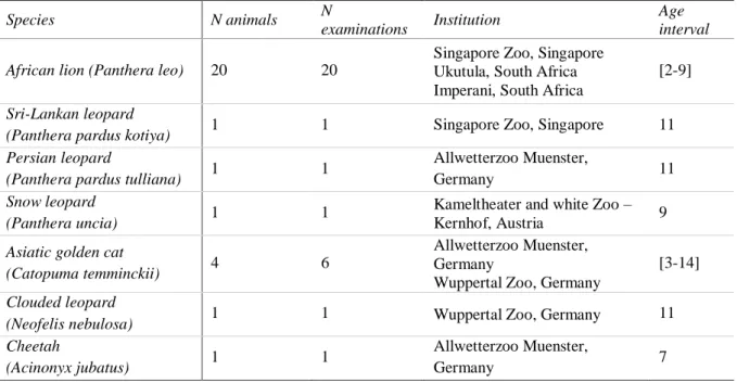
In this retrospective study, 29 animals from six different species of felids were examined over a period of three years, from February 2015 until June 2018, in a total of 31 examinations (Table 4). The enclosures were built following local legislations for each species, with an indoors shelter and outside area where animals could move freely. All the animals had access to water
ad libitum and were fed raw meat in quantities and frequency depending on the species. African
lions examined during this study were housed in groups of two to three females or in prides of one male and one to four females. The cheetah in this study was also housed together with another female. All other species were housed solitarily due to their nature. The average age of the 29 animals was six years, ranging from two to 14 years of age.
22
Table 4. Summary of examined animals, institutions where the examinations took place and age of animal at examination.
Species N animals N
examinations Institution
Age interval African lion (Panthera leo) 20 20
Singapore Zoo, Singapore Ukutula, South Africa Imperani, South Africa
[2-9]
Sri-Lankan leopard
(Panthera pardus kotiya) 1 1 Singapore Zoo, Singapore 11 Persian leopard
(Panthera pardus tulliana) 1 1
Allwetterzoo Muenster,
Germany 11
Snow leopard
(Panthera uncia) 1 1
Kameltheater and white Zoo – Kernhof, Austria 9
Asiatic golden cat
(Catopuma temminckii) 4 6
Allwetterzoo Muenster, Germany
Wuppertal Zoo, Germany
[3-14]
Clouded leopard
(Neofelis nebulosa) 1 1 Wuppertal Zoo, Germany 11 Cheetah
(Acinonyx jubatus) 1 1
Allwetterzoo Muenster,
Germany 7
1.2. Data collection
To obtain the data in the present study, in each felid a vaginal swab was taken and an ultrasonographic evaluation of the reproductive tract was performed under general anaesthesia.
1.2.1. Anaesthesia
During routine procedures for general health checks (n=15), due to reproductive failure (n=8) or preparation for AI (n=7), animals were anesthetised and a reproductive assessment was performed. In one case, the data was collected during a post mortem examination (n=1). The general anaesthesia was performed with a combination of medetomidine and ketamine, adding midazolam in some protocols.
1.2.2. Ultrasonography
For the ultrasound examination of the reproductive tract, a transrectal approach was the technique of choice. Firstly, an enema with 50-500ml temperate water was given with a lubricated rubber tube connected to a 100ml syringe (small felids) or a 3L container (large felids) to clean the rectum and provide better contact of the probe with the rectum wall. A 7-10 MHz endolinear probe (i739L-RS, scanner head size: 1.4 x 4.8 cm) mounted to a 50 cm long and 1.5cm of diameter PVC extension (to facilitate manipulation in larger species) was then inserted into the rectum. The probe, connected to a portable ultrasound machine (Logiq™