INTERVENTIONAL
Embolisation of prostatic arteries as treatment of moderate
to severe lower urinary symptoms (LUTS) secondary to benign
hyperplasia: results of short- and mid-term follow-up
Joao Martins Pisco&Hugo Rio Tinto&Luís Campos Pinheiro&Tiago Bilhim&Marisa Duarte&
Lúcia Fernandes&José Pereira&António G. Oliveira
Received: 9 August 2012 / Revised: 27 October 2012 / Accepted: 1 November 2012 # European Society of Radiology 2012
Abstract
Objectives To evaluate the short- and medium-term results of prostatic arterial embolisation (PAE) for benign prostatic hyperplasia (BPH).
Methods This was a prospective non-randomised study includ-ing 255 patients diagnosed with BPH and moderate to severe lower urinary tract symptoms after failure of medical treatment for at least 6 months. The patients underwent PAE between March 2009 and April 2012. Technical success is when selec-tive prostatic arterial embolisation is completed in at least one pelvic side. Clinical success was defined as improving symp-toms and quality of life. Evaluation was performed before PAE
and at 1, 3, 6 and every 6 months thereafter with the Interna-tional Prostate Symptom Score (IPSS), quality of life (QoL), International Index of Erectile Function (IIEF), uroflowmetry, prostatic specific antigen (PSA) and volume. Non-spherical polyvinyl alcohol particles were used.
Results PAE was technically successful in 250 patients (97.9 %). Mean follow-up, in 238 patients, was 10 months (range 1–36). Cumulative rates of clinical success were 81.9 %, 80.7 %, 77.9 %, 75.2 %, 72.0 %, 72.0 %, 72.0 % and 72.0 % at 1, 3, 6, 12, 18, 24, 30 and 36 months, respectively. There was one major complication.
Conclusions PAE is a procedure with good results for BPH patients with moderate to severe LUTS after failure of medical therapy.
Key Points
• Prostatic artery embolisation offers minimally invasive therapy for benign prostatic hyperplasia.
• Prostatic artery embolisation is a challenging procedure because of vascular anatomical variations.
• PAE is a promising new technique that has shown good results.
Keywords Benign prostatic hyperplasia . Therapeutic embolization . Prostatic diseases . Angiography . Catheterization
Abbreviations
BPH benign prostatic hyperplasia PAE prostatic artery embolisation
Introduction
Benign prostatic hyperplasia (BPH) has a prevalence of over 50 % in men over 60 years [1]. It is associated with lower
J. M. Pisco
:
H. Rio Tinto:
T. Bilhim:
M. Duarte:
L. FernandesInterventional Radiology, Saint Louis Hospital, Lisbon, Portugal
H. Rio Tinto
:
T. Bilhim:
L. FernandesRadiology Department, Hospital de São José, Lisbon, Portugal
H. Rio Tinto (*)
:
T. BilhimRadiology Department, Faculdade de Ciências Médicas, Universidade Nova de Lisboa, Lisbon, Portugal e-mail: [email protected]
L. Campos Pinheiro
Urology Department, Hospital de São José, Lisbon, Portugal L. Campos Pinheiro
Urology Department, Faculdade de Ciências Médicas, Universidade Nova de Lisboa, Lisbon, Portugal J. Pereira
Radiology Department, Hospital Santo António dos Capuchos, Lisbon, Portugal
J. Pereira
Saint Louis Hospital, Interventional Radiology, Lisbon, Portugal A. G. Oliveira
Biostatistics Department, Faculdade de Ciências Médicas, Universidade Nova de Lisboa, Lisbon, Portugal
urinary tract symptoms (LUTS) such as higher urinary fre-quency, urgency, leaking, hesitancy, interrupted and/or de-creased urinary stream, and in some patients sexual dysfunction, which may also be caused by medical therapy (ejaculation disorders and impotence) [2,3]. The indication for treatment depends on the severity and bother of urinary symptoms. LUTS severity is evaluated by the International Prostate Symptom Score (IPSS): mild LUTS (IPSS 1–7), moderate LUTS (8–19) and severe LUTS (20–35).
Medical therapy is usually the first-line treatment option and is indicated for patients with moderate lower urinary symptoms (patients with IPSS between 8 and 19) [4–6]. Medical therapies for BPH relief include alpha-blockers and 5-alpha-reductase inhibitors (5-ARI). Medical therapy is indicated for patients with moderate lower urinary symp-toms with no absolute surgical indications [6,7].
Minimally invasive treatments, including interstitial laser ablation, transurethral microwave treatment and transure-thral needle ablation, were originally conceived as an at-tempt to offer equivalent efficacy as operative therapy but without the burden and risk of operative morbidity [8].
Surgery is usually performed to improve symptoms and decrease progression of disease in patients who develop complications or who have inadequately controlled symp-toms while taking medical treatment.
Prostatectomy may be performed through the urethra (i.e. transurethral resection of the prostate, TURP) if the prostate is smaller than 60–80 cm3, or by open surgery if the prostate is larger. Both are associated with a significant complication rate. None of the minimally invasive treatments has proven superior to TURP from a cost/benefit standpoint, and TURP remains the standard effective treatment [9,10].
TURP is the most common surgical treatment for severe symptomatic BPH patients in whom medical therapy has failed. Although spinal anaesthesia is the most frequently used for TURP all patients should be suitable for general anaesthesia. Blood loss should be considered a frequent complication.
Although both medical and surgical treatment options for BPH are effective, they are associated with significant mor-bidity rates and some degree of sexual dysfunction. There-fore, there is the need for innovative technologies to continue to improve outcomes and minimise patient discom-fort and morbidity when managing BPH.
Prostatic arterial embolisation (PAE) for BPH has been shown to be safe and effective at inducing prostatic volume reduction in animals and humans [11–15]. Mauro [16] reported that BPH might be the next step after uterine artery embolisation (UAE) for fibroids.
Although the studies so far have been on few patients, short-term studies of BPH have shown that PAE is a safe and effective procedure, improves LUTS related to BPH and is associated with a decrease in prostate volume [14,15].
The purpose of this study was to retrospectively evaluate the short- and medium-term results of PAE in 251 patients with BPH with moderate to severe LUTS and failure of medical therapy for at least 6 months.
Materials and methods
From March 2009 to April 2012, 255 patients aged 45– 85 years (mean 65.5±7.4 years) who presented with a diag-nosis of BPH with moderate to severe LUTS refractory to medical treatment for at least 6 months were selected for PAE. This prospective study was approved by the hospital ethics committee and an informed consent form for PAE as an alternative treatment was signed by all participants. Efficacy variables of IPSS, quality of life-related symptoms (QoL), International Index of Erectile Function (IIEF), uroflowmetry (Qmax, peak urinary flow; PVR, post-void residual volume), prostatic specific antigen (PSA) and prostatic volume were assessed before PAE and at 1, 3, 6 and every 6 months after the procedure. The prostate volume was assessed by transrectal ultrasound (TRUS) and also measured by magnetic resonance (MR) before PAE and 6 months after PAE in the first 15 patients. The baseline data were obtained from the evaluation of these parameters (Table1). The inclusion criteria were male patients with age over 45 years and a diagnosis of BPH with moderate to severe LUTS (IPSS >18) and/or QoL at least 3, refractory to medical treatment for at least 6 months, and/or with Qmax inferior to 12 mL/s or with acute urinary retention and prostate larger than 40 cm3with sexual dysfunction or accepting the risk of developing sexual dysfunction after treatment.
Prostatic biopsy was performed in all cases of suspected prostatic malignancy due to a PSA level greater than 4 ng/mL or due to suspicious focal lesions detected on TRUS or MRI. Exclusion criteria were malignancy, advanced atheroscle-rosis and tortuosity of the iliac arteries, secondary renal insufficiency (due to prostatic obstruction), large bladder diverticula or stones, neurogenic bladder and detrusor failure.
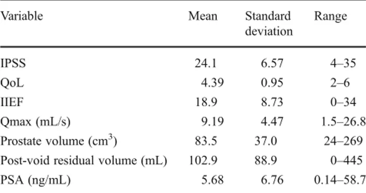
Table 1 Baseline values of efficacy parameters
Variable Mean Standard
deviation Range IPSS 24.1 6.57 4–35 QoL 4.39 0.95 2–6 IIEF 18.9 8.73 0–34 Qmax (mL/s) 9.19 4.47 1.5–26.8 Prostate volume (cm3) 83.5 37.0 24–269
Post-void residual volume (mL) 102.9 88.9 0–445
All patients were informed about the embolisation and other therapeutic options for their clinical situation including TURP, open surgery and laser treatment. The patients were allowed to choose freely between PAE, TURP/open surgery or minimally invasive treatment including laser surgery.
All the parameters mentioned above were evaluated in 238 patients at 1 month, 192 at 3 months 144 at 6 months, 89 at 12 months, 47 at 18 months, 21 at 24 months, 12 at 30 months and 8 at 36 months.
All patients were on medical therapy for BPH with per-sisting moderate to severe symptoms for more than 6 months. Eight patients had TURP years before and 32 patients had bladder catheters at the time, owing to acute urinary retention.
Pelvic magnetic resonance angiography (MRA) using a 1.5-T system (Philips, Eindhoven, the Netherlands) was
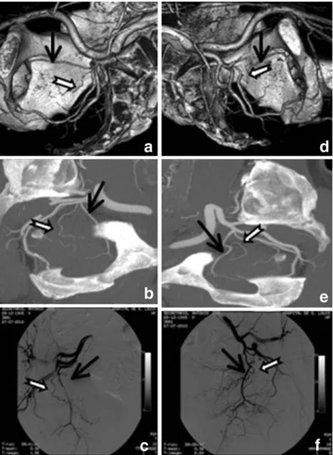
performed before PAE to evaluate the pelvic vessels for tor-tuosity and atherosclerotic changes of the iliac arteries in the first 15 patients. In the remaining patients pelvic CT angiog-raphy (CTA) was performed using a 16-row GE (R) Scanner (Fairfield, CT, USA). A specific CT angiography protocol was applied and post-processing using maximum intensity projec-tions (MIP) and volume rendering with 3D reconstrucprojec-tions were obtained [17,18]. The anatomy and the atherosclerotic involvement of the prostatic arteries could thereby be known in advance, before the procedure (Fig.1). We can also assess the degree of calcium and stenosis of prostatic origin, which also plays an important role in excluding some of the patients. CTA also reduces radiation during the procedure (digital subtraction angiography (DSA), or cone-beam CT eventually needed), the procedure time and contrast agent injection. It helps the team, giving them confidence, so that they do not get
Fig. 1 Computed tomography angiography (CTA) and digital subtraction angiography (DSA) of internal iliac arteries (IIA) to show the origin of prostatic ar-teries. a–c Right IIA. a Volume rendering with CT reconstruction of right IIA: right prostatic artery (open arrow) originates from the obturator (arrow). b Maximum intensity project (MIP) of the right IIA: prostatic artery (open arrow) originates from the obtu-rator artery (arrow). c DSA of the anterior trunk of the right IIA after selective catheterisation: confirms the origin of the pros-tatic (open arrow) artery from
the obturator (arrow). d–f Left
IIA. d Volume rendering with CT reconstruction of left IIA: left prostatic artery (open arrow) originates from the obturator (arrow). e MIP of left IIA: pros-tatic artery (open arrow) origi-nates from the obturator artery (arrow). f DSA of the anterior trunk of the left IIA after selec-tive catheterisation: confirms or-igin of the prostatic (open arrow) artery from the obturator (arrow)
lost with the anatomy during the procedure. It avoids catheter-isation of all other pelvic arteries and ultimately will reduce complications.
Embolisation technique
Patients started an acid-suppressing drug (omeprazole 20 mg, Bluepharma, once daily), an anti-inflammatory (nap-roxen 1,000 mg, naprosyn, Roche, twice daily) and an antibiotic (ciprofloxacin, 750 mg Jaba, twice daily) 2 days before the procedure and continued for 7 days following PAE. On the day of PAE, the patients had the same medi-cation at breakfast and at dinner 8 h after PAE. The patients were admitted to the hospital on the day of the procedure, 2 h before the intervention. During embolisation an anti-allergic (hydroxyzine 25 mg, Atarax) was given orally, an analgesic (metamizole 2 g i.v., Nolotil, Boehringer Ingel-heim) and an anti-inflammatory (ketorolac tromethamine 30 mg i.v., Toradol, Roche) were given intravenously.
The embolisation was planned in advance, on the bases of CT, particularly the volume rendering with CT recon-structions and the MIP. These examination results were available in the angiography suite at the time of the proce-dure (Fig.1).
Embolisation was performed under local anaesthesia us-ing a unilateral approach, mostly via the right femoral artery. For this purpose a 5-F RUC (Roberts Uterine Catheter, Cook Medical, Bloomington, USA) catheter or a cobra-shaped C2 catheter (Cook Medical, Bloomington, USA) was introduced into the right femoral artery in order to catheterise the left internal iliac artery (IIA) and its anterior
division. With the catheter at the initial segment of the IIA, DSA was obtained in the ipsilateral oblique view 35° and 10° cranio-caudal with 6 mL of iodine contract medium (Iopamiro 300, Iomeron, Bracco, Italy) at 3 mL/s, to visu-alise the prostatic arteries (Fig.2a). After identifying the left prostatic arteries, we obtained a road map with the catheter at the origin of the artery in which those arteries originate (Fig. 2b). Afterwards the prostatic vessels were selectively catheterised with a coaxial microcatheter (Progreat 2.7 or Progreat 2.4 with a GT microwire; Terumo, Tokyo, Japan) or 2.5 Cantata and Sagitta (Cook Medical) (Fig.2c). Anoth-er angiogram was pAnoth-erformed to confirm the position of the catheter in the ostium of the prostatic artery, and the prostate vascularisation in the same left anterior oblique (Fig. 2d). The microcatheter was then advanced distally into the prostatic artery before embolisation and an angiogram was obtained. Following that, another angio-gram of the prostatic artery in neutral projection was performed, in order to confirm that the catheter was in the prostatic artery by overlying it with the pubic bone (Figs. 3 and 4).
Upon finishing the embolisation of the left prostatic arteries, we removed the microcatheter and the Waltman loop was formed on the C2 or RCU and the right prostatic arteries were embolised in the same manner. After confirm-ing the position of the catheter in the ostium of the prostatic artery, we placed the microcatheter distally in the artery and the embolisation was carried out.
For embolisation 200-μm non-spherical polyvinyl alco-hol (PVA) particles were used (Cook Medical, USA) in the first 14 patients and 100 μm or 200 μm in the remaining
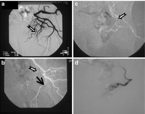
Fig. 2 Catheterisation of prostatic artery. a Digital subtraction angiography (DSA) of the left internal iliac artery (IIA). b Road map of the left pudenda artery (arrow) from which the prostatic artery (open arrow) originates. c Micro-guidewire (open arrow) placed in the prostatic artery. d Selec-tive angiogram of the prostatic artery through micro-catheter
patients. One millilitre of PVA particles was diluted in a solution of 50 mL saline and contrast medium in a 1:1 proportion and mixed with ketoprofene (50 mg) and cefa-zoline (100 mg). The particles were slowly injected through a 3-mL syringe under fluoroscopic control until we reached the end point. The end point of embolisation chosen was “near stasis” in the prostatic vessels with interruption of the arterial flow and prostatic gland opaci-fication (Fig.5).
Pain assessment was performed during and in the 6–8 h following PAE by visual analogue scale (VAS). Patients
were asked to rate their pain severity from 0 (sensation of no pain) to 10 (the worst pain).
Technical success was considered when selective prostat-ic arterial catheterisation and embolisation was achieved at least on one side of the pelvis.
IPSS, QoL, Qmax, IIEF, PVR, PSA and prostate volume were evaluated at 1, 3, 6 and every 6 months after the proce-dure. The prostate volume changes were evaluated by TRUS in all patients and in some patients also by MRI. All patients filled in the questionnaires by themselves without any exterior help (i.e. they were self-filled by the patients as recommended).
Clinical success was defined as improving symptoms (IPSS reduction at least 25 % of the total score and lower than 18 points) after PAE and improving of quality of life (reduc-tion of QoL of at least 1 point or equal to or below 3 points).
Statistical analysis
Response variables (IPSS, QoL, Qmax, PVR, PSA, prostate volume and IIEF) were analysed with random effects gen-eralized least-squares (GLS) regression with an AR (1) error structure. Prostate volume, Qmax and PSA were logarithmi-cally transformed to obtain a normal distribution. Observa-tion time was entered into the model as a categorical variable. No adjustment for multiplicity was done. Rates of clinical improvement over time were analysed using the Kaplan–Meier method to account for incomplete follow-up times. The Stata software release 12 (Stata Corp., College Station, TX, USA) was used for all analyses. Statistical differences were assumed at P<0.05.
Results
Prostatic artery embolisation was technically successful in 250 of the 255 (98 %) selected patients. In 5 patients (2 %) the procedure was impossible owing to tortuosity and ath-erosclerotic changes of the iliac arteries; surgery was per-formed in these cases. PAE was bilateral in 205 patients (82 %) and unilateral in 45 patients (18 %) also owing to the tortuosity and atherosclerotic changes of the iliac arteries.
The procedure time was between 20 and 185 min (mean 73 min) and the fluoroscopy time ranged between 7 and 64 min (mean 18 min). Mean follow-up time was 10 months (range 1–36 months).
The degree of pain ranged from 0 to 10 (mean 1.7); however 191/250 (76.4 %) patients did not feel any pain. Only one patient felt very severe pain (degree 9) during embolisation; the patient later developed a small area of bladder wall ischaemia.
Two hundred and twenty patients (88 %) were discharged from the hospital 3–8 h after the procedure and the remain-ing 30 patients (12 %) 18 h after, the next mornremain-ing.
Fig. 3 Digital subtraction angiography (DSA) of the right prostatic artery, oblique and neutral views. Prostate bed pacification. a DSA of the right prostatic artery, oblique view. b DSA of the right prostatic artery, AP view. c Prostate bed opacification of the right lobe (arrow), overlying the pubic bone
Follow-up data were available for 242 patients, who were observed for a mean of 10 months (range 1–36 months). Eight patients were lost to follow-up and 4 did not provide efficiency data. Efficacy data were available for 238 patients. Of these 238 patients, there was short-term clinical success at 1 month in 195 (81.9 %) and 43 (18.1 %) had clinical failures. Twenty four hours after PAE 102 patients (42.85 %) had already improved. All clinical failures had severe symptoms after PAE (IPSS persisted above 18 and QoL≥4). Clinical failure had no direct relationship with prostate volume reduction. Fifty-two of the 56 failures had complete follow-up data on prostate volume. In 23 of them there was no clinical success in spite of the significant (>15 %) prostate volume reduction. In the remaining 29 patients the prostate volume decreased less than 15 % in 16 and increased in 13 patients. On the other hand 12
patients had clinical success in spite of significant increase of prostate volume.
In 12 of the failures only unilateral embolisation could be performed and in 6 the embolisation was incomplete owing to advanced atherosclerosis that was not shown on CT angiog-raphy. In the remaining 25 patients with clinical failure the embolisation performed was bilateral and complete. PAE was successfully repeated and performed in the non-embolised prostatic artery in 4 of the 12 patients with unilateral emboli-sation. The procedure was repeated through another femoral approach, 2 weeks afterwards with clinical success.
Kaplan–Meier estimates of the cumulative rate of clinical success at other follow-up times were as follows: 80.7 % (95 % confidence interval (CI) 75–85.1 %) at 3 months, 77.9 % (95 % CI 71.9–82.7 %) at 6 months, 75.2 % (95 % CI 68.6–80.6 %) at 12 months, 72.0 % (95 % CI 64.1–
Fig. 4 Embolisation of the prostatic arteries, oblique and AP view. Digital subtraction angiography (DSA) of the right
prostatic artery (PA) (a–c).
Oblique view before embolisa-tion (a); AP view before embo-lisation (b); after emboembo-lisation (c). DSA of the left PA (d–f): oblique view before embolisa-tion (d); AP view before embolisation (e); DSA after embolisation (f)
78.5 %) at 18 months, 72.0 % (95 % CI 64.1–78.5 %) at 24 months, 72.0 % (95 % CI 64.1–78.5 %) at 30 months and 72.0 % (95 % CI 64.1–78.5 %) at 36 months (Table2).
A statistically significant improvement over time of all evaluated parameters was observed (Table3and Figs.6and7). Thirty-two patients were on urinary retention with blad-der drainage catheter. The bladblad-der catheters were removed between 5 and 60 days after the procedure in 30 of the patients who started to urinate successfully.
Ultrasound and MRI performed after PAE showed pros-tate volume changes and MRI showed ischaemia zones of the prostate (Figs.8,9).
Six of the 56 patients who were followed up to 18 months had failure with acute urinary retention and a bladder cath-eter was placed. Four of the patients improved after repeated PAE and the bladder catheter was removed. The remaining 2 patients had an open prostatectomy because of advanced atherosclerosis.
We considered IIEF improvement as any rise of IIEF score. In 199 patients with follow-up data on IIEF, the score
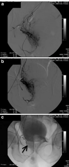
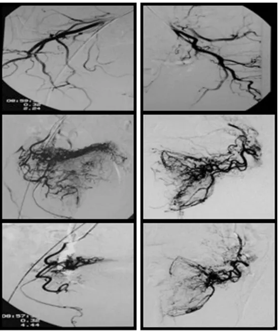
Fig. 5 Prostatic arteries embolisation. a Digital subtraction angiography (DSA) of the anterior division of right internal iliac artery (IIA). b DSA right prostatic artery (PA), before embolisation. c DSA right PA, after embolisation, fewer branches are shown. d DSA of the anterior division the left IIA. e DSA left PA, before embolisation. f DSA PA, after embolisation, fewer vessels are shown
Table 2 Cumulative probability of clinical improvement over time
Month Number at risk No. of failures % clinical improved 95 % confidence interval 1 238 43 81.9 76.4 86.3 3 192 3 80.7 75.0 85.1 6 144 5 77.9 71.9 82.7 12 89 3 75.2 68.6 80.6 18 47 2 72.0 64.1 78.5 24 21 0 72.0 64.1 78.5 30 12 0 72.0 64.1 78.5 36 8 0 72.0 64.1 78.5
improved in 96 (48.2 %), remained stable in 43 (21.6 %) and had no significant worsening in 60 (30.2 %). There were no cases of sexual impotence or retrograde ejaculation after PAE.
There was only one major complication: a bladder is-chaemia. A bladder mass was removed from that patient by simple surgery 1 month later and was confirmed by pathol-ogy to be necrosis and desquamation of the bladder wall. The patient suffered no further sequelae.
As an adverse event, 23/250 (9.2 %) patients experienced a burning sensation in the urethra and/or in the anus during the procedure. Nineteen of the 250 patients with urinary tract infections after embolisation (7.6 %) were treated with anti-biotics. Fourteen of the 19 patients with urinary tract infec-tion already had a urinary infecinfec-tion at the time of embolisation as proven by urine culture before the proce-dure. Transient haematuria occurred in 14/251 (5.6 %) patients, transient haematospermia in 10/250 (0.4 %), a small rectorrhagia in 6/250 (2.4 %) and balanitis in 4 (1.6 %) patients. All these minor complications disappeared sponta-neously without any treatment. Six patients had transient acute urinary retention after PAE. For relief, a temporary bladder catheter was placed at the time for a couple of hours.
Discussion
This prospective study shows that PAE performed in patients with BPH and moderate to severe LUTS refractory to medical therapy is a clinically successful procedure and may be an alternative to surgery. We thus present prostatic embolisation as an alternative to surgery. Therefore we accepted the same indications for PAE as those for surgery. In fact urinary retention and borderline urinary symptoms despite medical therapy were the main indications for patients selected for this trial. The European Association of Urology (EAU) guidelines
Table 3 Mean values over time of response variables
Variable Month No. pts. Mean 95 % confidence
interval Pa IPSS 0 238 24.0 23.3 24.9 <0.0001 1 236 12.2 11.4 13.1 3 224 11.0 10.2 11.8 6 167 11.5 10.4 12.5 12 101 10.4 9.1 11.7 18 58 10.1 8.4 11.9 24 25 9.0 6.4 11.6 30 14 8.1 4.8 11.5 36 9 9.1 4.1 14.1 QoL 0 238 4.40 4.28 4.52 <0.0001 1 236 2.48 2.32 2.64 3 221 2.23 2.08 2.38 6 167 2.27 2.08 2.47 12 102 1.96 1.72 2.20 18 54 1.83 1.50 2.16 24 25 1.76 1.36 2.16 30 13 1.85 1.43 2.26 36 9 1.67 1.12 2.21 Qmax (mL/s) 0 208 9.2 8.6 9.8 <0.0001 1 185 11.9 11.0 12.8 3 146 12.4 11.3 13.4 6 105 12.0 11.0 13.1 12 60 12.8 11.4 14.1 18 25 13.0 9.4 16.5 24 12 13.9 9.9 18.0 30 2 10.8 −75.6 97.2 PVR (mL) 0 210 102.9 90.8 114.9 <0.0001 1 175 65.6 54.1 77.0 3 134 59.2 49.4 69.1 6 99 62.8 50.0 75.5 12 58 51.7 37.1 66.4 18 23 75.4 46.5 104.3 24 13 91.9 24.2 159.6 30 3 95.3 −86.9 277.6 PV (mL) 0 238 83.5 78.8 88.2 <0.0001 1 183 66.8 62.6 71.0 3 147 68.3 63.2 73.3 6 111 66.6 60.8 72.4 12 63 69.9 61.8 78.0 18 29 72.0 57.5 86.5 24 14 90.9 62.6 119.1 30 4 72.0 13.7 130.3 PSA (ng/mL) 0 238 5.68 4.81 6.54 <0.0001 1 195 4.23 3.55 4.91 3 150 3.64 3.06 4.22 6 111 4.30 3.49 5.11 12 62 5.08 3.77 6.39 18 28 6.08 3.57 8.59 24 13 6.10 3.33 8.87 Table 3 (continued)
Variable Month No. pts. Mean 95% confidence
interval Pa 30 3 7.41 −3.91 18.7 IIEF 0 230 18.9 17.7 20.0 0.0002 1 197 20.6 19.5 21.7 3 152 20.9 19.7 22.1 6 110 20.5 18.2 21.9 12 65 20.1 18.2 22.0 18 25 20.4 17.1 23.6 24 12 18.7 12.2 25.2 30 3 20.0 −6.17 46.2 a
Random effects GLS regression. A significant P value is evidence that the mean value of an outcome variable changes over time
consider that although uroflowmetry does not allow one to make the diagnosis of obstruction, pressure flow studies are indicated before surgery only when Qmax is over 15 mL/s. Anyway we decided to be more strict and included patients with borderline urinary symptoms but with a Qmax lower than 12. For that reason we followed strictly the inclusion and exclusion criteria: IPSS should be more than 18, or the QoL at least 4, the Qmax under 12 mL/s or in acute urinary retention, the prostate volume larger than 40 cm3 and the patients should have been under refractory medical treatment for at least 6 months.
Most clinical changes occur in the first month after PAE. The clinical improvement is fast and 102 patients had im-proved in 24 h and another 93 had imim-proved by 1 month after the procedure. Therefore, at 1 month 195 had clinical improvement and 43 had clinical failure. Forty-three of the 56 clinical failures (76.8 %) occurred in the first month. Among those with clinical improvement by 3 months, only 20 (11 %) had not improved by 1 month. A retrospective review of the angiographic findings of the procedure in patients with clinical failures showed that in 12 patients
the embolisation was unilateral and in 6 it was incom-plete. However in the remaining 25/238 patients (10.5 %) both prostatic arteries were embolised. There-fore, to the best of our knowledge, there was no tech-nical reason for the clitech-nical failure. Because of this, the patients should be informed of the unpredictable results of PAE, even with complete embolisation of both pros-tatic arteries with a possible clinical failure rate of up to 25 % at 3 years.
Pre-procedural CT angiography is very important for evaluating the atherosclerotic changes of the iliac and pros-tatic arteries and the possible anastomoses of prospros-tatic ar-teries either to vesical or to rectal arar-teries, in order to avoid non-target embolisation. On the basis of CT angiography the patients have to be excluded if they have severe atheroscle-rotic changes or if the anatomy is not suitable. However, the CT angiograph may not show small plaques as occurred in 6 patients with incomplete embolisation, a finding that was not predictable before the procedure. There are no specific MR protocols to evaluate prostatic artery anatomy with the same information that CTA gives us. Alternatively,
Fig. 6 Change from baseline over time after PAE of IPSS, QoL, PVR and IIEF. Point estimates and 95 % confidence intervals. PAE prostatic artery embolisation, IPSS
International Prostate Symptoms Score, QoL quality of life, IIEF International Index of Erectile Function
Fig. 7 Percentage change from baseline over time after PAE of Qmax and prostate volume. Point estimates and 95 % confidence intervals. PAE prostatic artery embolisation, Qmax peak urinary flow, PV prostate volume
complementary cone-beam CT should be used in doubt-ful cases [19].
There was not a relationship between the reduction in prostatic volume and the clinical outcome (P00.12). Twenty-three of our patients with clinical failure had signifi-cant (>15 %) prostate volume reduction and 12 patients had clinical success in spite of a significant increase in prostate volume. Therefore the clinical success cannot be predicted on the basis of prostate volume reduction. Patients with the same prostate volume change may have different clinical outcomes. PAE is a safe and painless outpatient procedure with low morbidity in most cases. There was only one major
compli-cation—bladder wall ischaemia—that occurred early in our experience (patient 10). It manifested as an intraluminal volume of necrotic tissue that was removed by simple sur-gery without need for bladder reconstruction. To prevent complications a good knowledge of the prostatic anatomy is important in order to perform a superselective catheter-isation of prostatic arteries avoiding untargeted embolcatheter-isation of other arteries such as the vesical, rectal and dorsal artery of the penis [17,18].
PAE is usually a painless procedure; all patients were treated as outpatients and 220 patients (88 %) were dis-charged 3–8 h after the procedure.
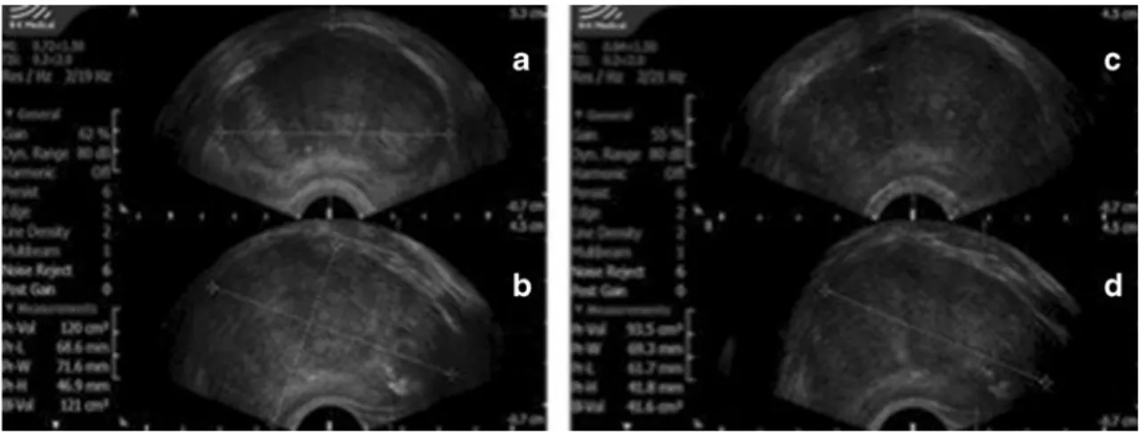
Fig. 8 Trans-rectal ultrasound. Prostate volume reduction after PAE. a, b Prostate volume before PAE (18 June 2009), 121 cm3. c, d Prostate
volume after PAE (20 August 2009), 93.5 cm3(i.e. 22.9 % reduction)
Fig. 9 MRI of the prostate before and after PAE. a MRI before PAE. b MRI 18 days after PAE. Lower intensity ones in both prostate lobes due to ischaemia
There were no cases of sexual dysfunction including impotence or retrograde ejaculation. The sexual function improved in 96 (48.2 %) of the patients and had no signif-icant differences in the remaining patients. The improve-ment in sexual function might be explained by the withdrawal of the prostatic medication that affects it and by the improvement in urinary symptoms and quality of life. The PAE compares favourably with surgery where retro-grade ejaculation occurs very frequently.
The main advantages of PAE are preservation of the sexual function with no cases of impotence or retrograde ejaculation, the minimally invasive nature of the procedure, the low morbidity, the possibility of stopping the daily BPH medication and the outpatient setting. In that way we believe that prostatic embolisation may be an alternative to surgery for treatment of moderately or severe LUTS secondary to BPH. In fact prostatic embolisation patients had an IPSS improvement of 62.5 % (from a mean IPSS of 15 before treatment to 9 at 36 months after PAE). In the same way Qmax increased 51 % (from 9.2 to 13.9 mL/s after PAE).
Although in this study the improvement of the mean uroflowmetry obtained by PAE patients is modest when compared to surgery patients (51 % for PAE and 125 % for TURP) the IPSS outcome after PAE is comparable to the one obtained by surgery (IPSS improvement of 62.5 % for PAE and IPSS improvement of 70.6 % for TURP). Future studies with longer follow-up will answer the question of longevity of the PAE outcome.
After PAE there is improvement in IPSS, Qmax and PVR. After 3 and 6 months there is a deterioration in those parameters in some patients. It is necessary to study the patients for longer periods of time in order to evaluate the durability of the results. Eight patients (3.4 %) followed up for 36 months showed continued improvement.
This study has some limitations and bias should also be considered. The variables used in this work are subjective although IPSS is a validated questionnaire. We only per-formed one uroflowmetry assessment and at least two would be helpful to define an average and reduce variability. How-ever the Qmax improvement is not as important as the IPSS improvement.
Prostate volume was measured by TRUS and it is always operator dependent. We tried to reduce bias by having the same operator measuring volumes in the same patient but sometimes that was not possible. Nevertheless, with all the limitations that they might have, they are the same param-eters used in almost any trial on BPH.
Pressure flow studies were not considered as part of the clinical evaluation in this trial because of their invasive nature. As in the majority of other trials, pressure flow studies were not included for the same reason.
We also tried to minimize some expected bias concerning the filling of the questionnaires. All patients filled the
questionnaires by themselves without any exterior help (i.e. they were self-filled by the patients as recommended).
Finally, this is a single-centre randomised and non-comparative study. Although the results are promising more studies are needed, especially multicentre randomised con-trolled trials with longer follow-up.
We conclude that PAE in selected BPH patients is a safe procedure with low morbidity, no sexual dysfunc-tion, and with good short- and medium-term follow-up and may be an alternative to surgery in moderately and severely symptomatic patients.
Conflict of interest There is no funding or potential conflicts of
interest for the authors regarding this article. The corresponding authors confirm that they have full access to all the data in this study and have final responsibility for the decision to submit for publication. All authors had full access to all of the data in the study and take responsibility for the integrity of the data and the accuracy of the data analysis.
References
1. Levy A, Samraj GP (2007) Benign prostatic hyperplasia: when to ‘watch and wait’, when and how to treat. Cleve Clin J Med 74:S15–S20 2. Garraway WM, Collins GN, Lee RJ (1991) High prevalence of benign prostatic hypertrophy in the community. Lancet 338:469–471 3. Michel MC, Mehlburger L, Bressel HU, Schumacher H, Schäfers RF, Goepel M (1998) Tamsulosin treatment of 19,365 patients with lower urinary tract symptoms: does co-morbidity alter tolerability? J Urol 160:784–791
4. McConnell JD, Bruskewitz R, Walsh P et al (1998) The effect of finasteride on the risk of acute urinary retention and the need for surgical treatment among men with benign prostatic hyperplasia. Finasteride Long-Term Efficacy and Safety Study Group. N Engl J
Med 338:557–563
5. Varkarakis J, Bartsch G, Horninger W (2004) Long-term morbidity and mortality of transurethral prostatectomy: a 10-year follow-up.
Prostate 58:248–251
6. Roehrborn CG, Rosen RC (2008) Medical therapy options for aging men with benign prostatic hyperplasia: focus on alfuzosin
10 mg once daily. Clin Interv Aging 3:511–524
7. Burnett AL, Wein AJ (2006) Benign prostatic hyperplasia in
primary care: what you need to know. J Urol 175:S19–S24
8. Baazeem A, Elhilali MM (2008) Surgical management of benign prostatic hyperplasia: current evidence. Nat Clin Pract Urol 5:540–549 9. Reich O, Gratzke C, Bachman A et al (2008) Morbidity, mortality and early outcome of transurethral resection of the prostate: a prospective multicenter evaluation of 10,654 patients. J Urol 180:246–249 10. Madersbacher S, Marberger M (1999) Is transurethral resection of
the prostate still justified? BJU Int 83:227–237
11. DeMeritt JS, Elmasri FF, Esposito MP, Rosenberg GS (2000) Relief of benign prostatic hyperplasia-related bladder outlet ob-struction after transarterial polyvinyl alcohol prostate
emboliza-tion. J Vasc Interv Radiol 11:767–770
12. Carnevale FC, Antunes AA, da Motta Leal Filho JM et al (2010) Prostatic artery embolization as a primary treatment for benign prostatic hyperplasia: preliminary results in two patients.
Cardio-vasc Intervent Radiol 33:355–361
13. Pisco JM, Pinheiro LC, Bilhim T, Duarte M, Mendes JR, Oliveira AG (2011) Prostatic arterial embolization to treat benign prostatic
14. Sun F, Sánchez FM, Crisóstomo V et al (2008) Benign prostatic hyperplasia: transcatheter arterial embolization as potential
treat-ment—preliminary study in pigs. Radiology 246:783–789
15. Jeon GS, Won JH, Lee BM et al (2009) The effect of transarterial prostate embolization in hormone-induced benign prostatic hyper-plasia in dogs: a pilot study. J Vasc Interv Radiol 20:384–390 16. Mauro MA (2008) Can hyperplastic prostate follow uterine
fib-roids and be managed with transcatheter arterial embolization? Radiology 246:657–658
17. Bilhim T, Casal D, Furtado A, Pais D, O’Neill JE, Pisco JM (2011)
Branching patterns of the male internal iliac artery: imaging
find-ings. Surg Radiol Anat 33:151–159
18. Bilhim T, Pisco JM, Furtado A et al (2011) Prostatic arterial supply: demonstration by multirow detector angio CT and catheter angiography. Eur Radiol 21:1119–1126
19. Sapoval M, Daroles J, Rio Tinto H et al (2012) C-arm cone-beam CT in BPH: techniques and considerations for the safe treatment of benign prostatic hyperplasia. Endovascular Today April:61–63