Ana Regina Silva
Cerqueira
PPA e a sua fosforilação na sinalização STAT3
induzida por Gα
oAPP and APP phosphorylation in Gα
o-induced
Ana Regina Silva
Cerqueira
PPA e a sua fosforilação na sinalização STAT3
induzida por Gα
oAPP and APP phosphorylation in Gα
o-induced
STAT3 signaling
Dissertação apresentada à Universidade de Aveiro para cumprimento dos requisitos necessários à obtenção do grau de Mestre em Biomedicina Molecular, realizada sob a orientação científica da Professora Doutora Sandra Vieira, Professora Auxiliar Convidada da Secção Autónoma de Ciências da Saúde da Universidade de Aveiro.
Este trabalho contou com o apoio do Centro de Biologia Celular (CBC) da Universidade de Aveiro, e é financiado por fundos FEDER através do Programa Operacional Factores de Competitividade – COMPETE e por Fundos nacionais da FCT – Fundação para a Ciência e a Tecnologia no âmbito dos projectos PTDC/QUI-BIQ/101317/2008, PTDC/SAL-NMC/111980/2009 e PEst-OE/SAU/UI0482/2011.
Dedicada ao meu avô, Rui Silva.
o júri
presidente Odete Abreu Beirão da Cruz e Silva
Prof. Auxiliar Com Agregação, Secção Autónoma de Ciências da Saúde, Universidade de Aveiro
Sandra Isabel Moreira Pinto Vieira
Prof. Auxiliar Convidada, Secção Autónoma de Ciências da Saúde, Universidade de Aveiro
Ana Sofia Duarte
agradecimentos À minha orientadora, Sandra Vieira, agradeço a motivação, sabedoria, inspiração e dedicação que contribuíram para uma melhor aprendizagem. À professora Odete da Cruz e Silva por me ter dado a oportunidade de realizar esta etapa no laboratório de neurociências do Centro de Biologia Celular. Aos meus pais e à minha irmã pelo apoio incondicional, confiança, afecto, compreensão, dedicação e incentivo, que tornaram a concretização desta etapa possível.
À linda família que tenho a sorte de ter e por, apesar das adversidades face ao trabalho, me proporcionarem o melhor ambiente familiar.
Ao Bruno, à Pati, à Lili e à Ana Cristina por me darem o prazer de desfrutar diariamente de uma boa e verdadeira amizade.
Ao João, à Carina, Daniela, Juliana, Ana Silva e Catarina por demonstrarem que a distância é um mero pormenor, quando o sentimento é verdadeiro. À Ana, Vanessa, Liliana, ao Tiago e Diogo por serem a prova que o tempo voa, mas que a amizade se pode manter.
Ao Ivo, Marcelo e Rafael pela boa disposição e momentos de descontração, que tornam não só os jantares como, principalmente, a companhia insubstituíveis.
A todos os meus colegas do CBC, particularmente aos meus colegas do laboratório de Neurociências. Ao Roberto e à Joana Rocha, pelo companheirismo, altruísmo e respeito que foram fundamentais para a realização deste trabalho.
À FCT pelo financiamento dos projetos PTDC/QUI-BIQ/101317/2008 e PTDC/SAL-NMC/111980/2009.
palavras-chave Proteína precursora de amilóide de Alzheimer (PPA); fosforilação da PPA na S655; sinalização da STAT3; proteína Gαo; células SH-SY5Y
resumo A proteína precursora de amilóide de Alzheimer (PPA) é uma proteína cerebral fundamental, envolvida em mecanismos celulares como a adesão e migração celular e neuritogénese. Como fosfoproteína, a PPA apresenta oito resíduos fosforiláveis no seu domínio citoplasmático. Um desses resíduos, Serina 655 (S655), localiza-se no motivo funcional 653YTSI656, e a sua fosforilação altera o tráfego e o processamento da PPA, podendo ainda mediar a ligação desta a outras proteínas através de um hydrophobic pocket localizado imediatamente a jusante ao motivo YTSI (657His-Lys676). Nomeadamente, a proteína Gαo, uma
subunidade α de proteínas G heterotrimérica, liga-se à PPA neste local. A Gαo
está envolvida em várias cascatas de sinalização e é o membro da família Gi/o
mais abundante no cérebro. Quando ativada, a Gαo consegue induzir fatores
de crescimento relacionados com a via JAK2/STAT3 via Rap, provavelmente através da interação e sequestro de proteínas RapGAP. Adicionalmente, a Gαo
foi sugerida como um transdutor da PPA, funcionando esta como um guanidine
exchange factor (GEF) para a Gαo. No entanto, ainda não foi atribuído nenhum
papel funcional a esta interação.
O presente trabalho teve como principal objetivo determinar o papel da PPA na cascata de sinalização da STAT3 induzida por Gαo, pela análise dos efeitos da
co-transfecção de PPA-Gαo nos níveis de STAT3 e STAT3 fosforilada através
de Western blot. Para isto, células humanas de neuroblastoma SH-SY5Y são co-transfectadas por 6h e 24h com PPA-GFP cDNAs (Wt ou fosfomutantes S655: S655A, SA, e S655E, SE) e Gαo ou GαoCA (um mutante Q205L que
mimetiza um estado constitutivamente activo da Gαo). Adicionalmente, foi
estudado como é que os metabolismo da PPA e da Gαo afetam-se um ao
outro, através da quantificação dos níveis de Gαo, PPA e PPAs secretada por
Western blot.
Em geral, os resultados obtidos mostram que as co-expressões de PPA-GFPs com Gαo/GαoCA levam a um decréscimo da fosforilação da STAT3 induzida
por Gαo. Isto parece ocorrer através da indução de um efeito retro-inibitório nas
vias Gαo-STAT3 após a ativação inicial da Gαo e, enquanto este efeito é mais
pronunciado para o mutante que reproduz desfosforilação do resíduo S655 da PPA, a fosforilação neste mesmo resíduo parece retardar este efeito. Em concordância, a PPA invariavelmente diminui os níveis de Gαo e a Gαo
também diminui os níveis de PPA, potencialmente envolvendo um mecanismo de inibição da ativação da STAT3 induzida pela Gαo através da PPA por
keywords Alzheimer’s Amyloid Precursor Protein (APP); APP phosphorylation at S655; STAT3 signalling; Gαo protein; SH-SY5Y cells
abstract The transmembranar Alzheimer’s Amyloid Precursor Protein (APP), an important brain protein, is potentially involved in cellular mechanisms such as cell adhesion, migration and neuritogenesis. As a phosphoprotein, APP has eigth phosphorylatable residues in its cytoplasmic domain. One of these residues, S655, lies within the APP 653YTSI656 sorting motif, and its phosphorylation alters APP trafficking and processing, and may potentially mediate its binding to other proteins by means of a hydrophobic pocket localized immediately downstream the YTSI motif (657His-Lys676). This is the pocket to which Gαo, an alpha subunit of heterotrimeric G proteins, binds to.
Gαo mediates various signaling pathways and is the most brain-enriched
member of the Gαi/o family. When activated, Gαo can induce the growth
factors-related JAK2/STAT3 pathway via Rap, probably through interaction and sequestration of RapGAP proteins. Gαo has been suggested to be a transducer
of APP, with APP acting as a guanidine exchange factor (GEF) for Gαo.
Nonetheless, no functional role has been attributed to this interaction.
In the work here described, the role of APP in Gαo-induced STAT3 signaling
was assayed, by analysing the effects of APP-Gαo co-transfection in STAT3
and phospho-STAT3 levels by Western blot means. For this, human SH-SY5Y neuroblastoma cells were co-transfected for 6h and 24h, with APP-GFP fusion cDNAs (Wt or S655 phosphomutants: dephosphomimicking S655A and phosphomimicking S655E) and Gαo or GαoCA (a Q205L mutant mimicking
constitutively active Gαo). Additionally, the effects of APP and Gαo in each other
metabolism were also evaluated, by quantifying the levels of Gαo, APP and
medium secreted sAPP by Western blot analysis.
In general, the results obtained show that APP-GFPs co-expression with Gαo/GαoCA leads to a decrease in Gαo-induced STAT3 phosphorylation. This
appears to occur via a retro-inhibition effect on the Gαo-STAT3 pathways
following an initial Gαo activation and, while is more pronounced for the S655
dephosphomimicking mutant, phosphorylation at S655 appears to delay this effect. Accordingly, APP invariably decreases Gαo half-life, and Gαo also
decreases APP levels, with APP/Gαo lysosomal co-degradation being a
Index
Abbreviations ... 3
1. Introduction ... 7
1.1. Alzheimer’s Disease ... 7
1.1.1. Alzheimer’s Disease pathology ... 7
1.1.2. Hallmarks of AD ... 9
1.1.3. Risk factors ... 11
1.2. The β-Amyloid Precursor Protein (APP) ... 14
1.2.1. APP gene family, isoforms and domains ... 15
1.2.2. APP metabolism: trafficking and processing ... 18
1.2.3. Phosphorylation of APP ... 23
1.3. APP as a potential mediator of G proteins signaling ... 25
1.3.1. G proteins ... 25
1.3.1.1. Gαo Protein: genetics, expression pattern and function ... 29
1.3.1.2. Modulation of Gαo activity ... 30
1.3.1.3. Gαo-mediated signaling transduction ... 31
1.3.1.4. APP-Gαo binding and Alzheimer’s disease ... 32
1.3.2. STAT3 signaling ... 33
1.3.2.1. STAT proteins ... 33
1.3.2.2. STAT3 function ... 35
1.3.2.3. APP and Gαo in STAT3 signaling ... 36
2. Aims of this thesis ... 39
2.1. General Aims... 39
2.2. Specific Aims ... 39
3. Materials and Methods ... 41
3.1. Culture and maintenance of the SH-SY5Y cell line ... 41
3.2. Wt and S655 Phosphomutants APP-GFP cDNAs ... 41
3.3. Wild-type and constitutively active G-protein alpha o – Gαo and GαoCA (Q205L) cDNAs ... 41
3.4. SH-SY5Y cells transfection with APP-GFP and Gαo or GαoCA cDNAs ... 42
3.4.1. Transfection by the Polyplus method ... 42
3.4.2. Transfection using the TurboFectTM reagent ... 43
3.5. Cell collection and quantification of protein content (BCA) ... 44
3.6. Antibodies ... 45
3.7. Western Blot Assays ... 46
3.8. GFP-trap® pull-down assays ... 47
3.9. Ponceau S staining of proteins bands ... 48
3.10. Fluorescence Microscopy ... 49
3.11. Data analysis ... 49
4. Results ... 51
4.1. Optimization of APP/Gαo cDNAs transfection ... 51
4.2. Pilot experiment – time periods of Gαo transfection ... 52
4.3. APP effects on Gαo–induced STAT3 signaling ... 54
4.3.1. APP and Gαo effects on STAT3 activation levels ... 54
4.3.2. Effect of APP overexpression in Gαo levels ... 59
4.3.3. Effect of Gαo overexpression in APP levels ... 61
4.4. Visual analysis of cellular APP/Gαo levels ... 64
4.5. Analysis of APP-Gαo binding ... 66
5. Discussion ... 67
6. Conclusion ... 72
7. References ... 75
Abbreviations
AD Alzheimer’s disease
ADP Adenosine diphosphate
AGS Activator of protein signaling
AICD APP intracellular C-terminal domain
AMP Adenosine monophosphate
APLP Amyloid precursor-like proteins
APLP Amyloid precursor-like protein
APOE Apolipoprotein E gene
ApoE Apolipoprotein E protein
APP β-amyloid precursor protein
APPL Amyloid precursor protein-like
APs Amyloid plaques
Aβ Amyloid β-peptide
BACE1 β-site APP-cleaving enzyme
BSA Bovine serum albumin
Ca2+ Calcium
CaMKII Calmodulin-dependent protein kinase II
cAMP Cyclic adenosine monophosphate
CB1 Cannabinoid 1 receptor
Cdc2 Cyclin-dependent protein kinase 2
Cdk5 Cyclin-dependent kinase 5
cDNA Complementary deoxyribonucleic acid
CNS Central nervous system
CNTF Ciliary neurotrophic factor
CTF Carboxy-terminal fragment
CuBD Copper-binding domain
DAPI 4',6-diamidino-2-phenylindole
ECL Enhanced chemiluminescence
ER Endoplasmic reticulum
FAD Familial Alzheimer’s disease
G protein GTP-bindign protein
Gαi inhibitory G-protein alpha
GAIP Gα-interating protein
GAP-43 Growth cone-associated protein
GAPs GTPase-activating proteins
Gβ G-protein beta-gamma subunits
GDP Guanosine diphosphate
GEFs Guanine nucleotide exchanger factors
GFP Green fluorescent protein
Gi inhibitory G-protein
Go Heterotrimeric 'other' G protein
gp130 Glycoprotein 130
GPCRs G protein-coupled receptors
GPR G protein regulatory
Gq G protein q
GRIN G protein-regulated inducer of neurite outgrowth
GSK-3β Glycogen synthase kinase 3β
GTP Guanosine triphosphate
Gz G-protein z
Gαo G-protein alpha-subunit o
GαoCA constitutively active G-protein alpha o
JAK Janus Kinase
JNK c-Jun N-terminal kinase
K+ Potassium
KPI Kunitz-type protease inhibitor
LD Linker domain
LIF Leukemia inhibitory factor
MAPK Mitogen-activated protein kinase
ND N-domain
NFTs Neurofibrillary tangles
NT non-transfected cells
PARP poly ADP ribose polymerase
PI3K Phosphadylinositol-3-kinase
PKC Protein kinase C
PM Plasma membrane
PPCs Proprotein convertases
PS1 and PS2 Presenilin 1 and 2 protein
pSTAT3 STAT3 phosphorylated
PTX Pertussis toxin
Rac Rho family of GTPase subfamily
Rap Ras-related protein or a small GTPase protein Rap-GAP Rap GTPase activating protein
Ras Small G protein superfamily
RGS Regulators of G protein signaling
S/SerXXX Serine amino acid residue at position XXX S655A APP Serine 655 mutated at alanine
S655E APP Serine 655 mutated at glutamate SA APP/SA Dephosphorylated S655 APP mutant
sAPP Secreted APP
SE APP/SE Phosphorylated S655 APP mutant
SEM Standard Error of he mean
SH2 Src homology 2 domain
Src Non-receptor protein tyrosine kinase
STAT Signal transducer and activator of transcription STAT3 Signal transducer and activator of transcription 3 TACE Tumor necrosis factor-α converting enzyme
TGN Trans-Golgi network
Thr Threonine amino acid residue at position XXX Tip60 Tat interactive protein, 60kDa
TK Tyrosine-Kinase
Tyr Tyrosine amino acid residue at position XXX
VPS Vacuolar protein sorting
WB Western Blot
1. Introduction
1.1. Alzheimer’s Disease
1.1.1. Alzheimer’s Disease pathology
Alzheimer’s disease (AD), a disorder of unknown etiology, is the most common form of dementia in the elderly. AD is a chronic disorder, apparently preceded by a clinically silent period of several years or even decades, which slowly and irreversibly destroys neurons and causes serious cognitive disability (Arendt, 2001; Bojarski, Herms et al., 2008).
Since it was presented for the first time in 1906 by Alois Alzheimer, this neurodegenerative disease is affecting an increasing number of individuals. In 2006, approximately 26.6 million people worldwide suffered from AD, and because of the growing life expectancy, the global prevalence of Alzheimer’s can be predicted to be more than 100 million in 2050 (Selkoe, 2004; Brookmeyer, Johnson et al., 2007).
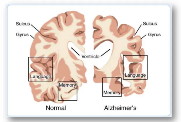
AD cases have been described according to lesions type, the type of onset, the cause (genetic or sporadic) and associated lesions (e.g. vascular lesions). Clinically, in the beginning, AD is characterized by a mild cognitive impairment and deficits in short-term and spatial memory, which can be confused with the normal changes of aging. The symptoms become more severe with disease progression and not only destroy a person’s memory, but the patient also presents disturbances in language use, perception, ability to learn necessary skills, reason, solve problems, think abstractly, make judgment, communicate and carry out daily activities. In some patients, AD can even lead to personality and behavioural changes (Marotta, Majocha et al., 1992; Arakawa, Kita et al., 2008; Hooijmans and Kiliaan, 2008). In Figure 1 is depicted a comparison between a normal and an AD brain, through a brain cross-section, highlighting some of the neuropathological alterations in AD brains, including brain shrinkage.
The clinical symptoms of AD result from neuropathological lesions, including deposition of amyloid plaques (APs) and neurofibrillary tangles (NFTs), in characteristic brain regions, together with downstream reactive processes such as inflammation and oxidative stress, leading to loss of effective neural network connectivity, synaptic degeneration and progressive neuronal cell death (Bojarski, Herms et al., 2008; Duyckaerts, Delatour et al., 2009; Hampel, Shen et al., 2010; Lin and Luo, 2011). Of note, some reports indicate that AD could be a disease where not only neurons are affected, but also peripheral cells such as fibroblasts, lymphocytes and platelets (Bojarski, Herms et al., 2008).
The neuropathological and biochemical features of AD develop the disorder and delay the diagnosis, since the symptoms appear later on (Lemere and Masliah, 2010). Although there are currently accepted clinical guidelines for the probable diagnosis of AD, the definitive diagnosis of AD happens at autopsy with the postmortem examination of the brain, which reveals large quantities of NFTs and APs within the parenchyma (Marotta,
Fig. 1: This image represents a cross-section of the brain as seen from the front. On the left the cross-section
represents a normal brain and the one on the right represents a brain with Alzheimer's disease. Reproduced from American health assistance foundation (January 2012), http://www.ahaf.org/alzheimers/about/understanding/brain-with-alzheimers.html
Majocha et al., 1992). In Figure 2, we can observe AP and NFT in a microscopic analysis of the AD brain.
Several symptomatic treatments are in use for AD, but they are incapable of reversing the pathology of disease, namely, the widely used acetylcholinesterase inhibitors, which reduce inflammation and alleviate the symptoms. Recently, new therapeutic drugs are being developed, including vaccines aiming to destroy AP plaques before or after being aggregated.
1.1.2. Hallmarks of AD
The two major pathological hallmarks of AD that appear to be more correlated with the clinical symptoms are the extracellular amyloid plaques (also called senile plaques), and the intracellular neurofibrillary tangles.
The APs are aggregated, insoluble, dense cores of 5-10 nm amyloid fibrils with surrounding reactive cells, dystrophic neurites, lysosomes, abnormal mitochondria, astrocytic processes and activated microglia (Fig. 3). The main proteinaceous component of APs is the neurotoxic β-amyloid (Aβ) peptide, produced by proteolytic cleavages of a type I transmembranar glycoprotein named β-amyloid precursor protein, APP (Selkoe, 1999; Duyckaerts, Delatour et al., 2009; Hampel, Shen et al., 2010; Lemere and Masliah, 2010). Amyloid-containing senile plaques are a prominent feature of specific AD brain
Fig. 2: Histopathological analysis of an AD brain tissue, post-mortem. In this tissue cut we can observe the
neurofibrillary tangles and amyloid plaques. Reproduced from Experimental genetics group (January 2012), http://med.kuleuven.be/legtegg/AD.html
regions, affect limbic and cortical structures, like cerebral cortex, amygdaloid nucleus, corpus striatum, diencephalon and hippocampus, but they also have been described in the cerebellum (Marotta, Majocha et al., 1992; Lukiw and Bazan, 2000; Merlo, Spampinato et al., 2010). The mechanisms responsible for the excessive accumulation of β-amyloid in the AD brain are unknown, but a few genetic, molecular biology and protein chemistry hypotheses have been proposed to explain the disease process (Marotta, Majocha et al., 1992).
The neurofibrillary tangles (Fig. 3) derive from the intracellular accumulation of paired helical filaments resulted from hyperphosphorylation of a microtubule-associated protein known as Tau, causing it to aggregate into an insoluble form. In AD, this abnormal Tau protein induces the microtubule structures to collapse inside neuronal cell bodies, axons and dendrites (Hooijmans and Kiliaan, 2008; Duyckaerts, Delatour et al., 2009; Merlo, Spampinato et al., 2010). The progression of Tau pathology is stepwise and stereotyped from the entorhinal cortex, through the hippocampus, to the neocortex (Marotta, Majocha et al., 1992).
Fig. 3: Representation of nerve cells within AD brains. The tissue with AD presents a much smaller number of nerve
cells and synapses than a healthy brain; APs are abnormal deposits of protein fragments between nerve cells; the dead and dying nerve cells contains neurofibrillary tangles. Reproduced from health news (January 2012), http://www.healthinformer.net/alzheimer-disease-plaques-seen-with-conventional-mri-in-animal-model-for-the-first-time.html
Although controversies still exist on the real causes of AD and on the precise relationship between Aβ, Tau and AD pathogenesis, the ability to determine both proteins levels in living people could markedly improve AD diagnosis and treatment, and these proteins still considered the most possible targets for new therapies (Merlo, Spampinato et al., 2010).
The synaptic loss in AD is difficult to evaluate, however it is an early pathological hallmark of AD that is age dependent and largely independent of Aβ. More than Aβ deposits, this hallmark could be the neuropathological alteration that is more correlated with neuronal damage, cognitive decline and memory impairment (Hooijmans and Kiliaan, 2008; Duyckaerts, Delatour et al., 2009). Furthermore, synaptic dysfunction and failure of brain connectivity appears to predate neuronal loss (Coleman, Federoff et al., 2004; Hooijmans and Kiliaan, 2008). Studies indicate that the neurodegenerative process initiates in the temporal lobe structures, like entorhinal cortex, followed by synapse disfunction and loss in the hippocampus (Coleman, Federoff et al., 2004; Duyckaerts, Delatour et al., 2009).
The plaques and tangles formation accompany this process, however research demonstrate that synaptic loss and memory impairment precede APs in the limbic system (Mucke, Masliah et al., 2000), and AD has been associated with synaptic disruption also in specific cortical areas (Duyckaerts, Delatour et al., 2009).
These evidences demonstrate that synaptic failures of AD begin prior to neuronal loss, plaques and tangles development, and also prior to clinical detection of disease. The explicit recognition that AD starts long before the appearance of the traditional ways of detecting the illness will highlight the need of the development of early diagnosis (Coleman, Federoff et al., 2004).
1.1.3. Risk factors
As mentioned above, the cause or causes of AD are not yet identified and there is no cure, so efforts are needed for preventing the development of the dementia. Many theories have been proposed, like the “amyloid cascade hypothesis”. The amyloid deposition is seen as the primary pathway leading to neurodegeneration and AD. Although this hypothesis is one of the most persistent, there are yet controversy in some results, such as: the conflicting results about the neurotoxicity of deposited Aβ in vivo (Bishop and
Robinson, 2002); the fact that Aβ accumulation is not the earliest hallmark and is not always correlated to neuronal loss (Mucke, Masliah et al., 2000); the fact that many non-demented persons show large amounts of AD neuropathologicals events (Hooijmans and Kiliaan, 2008).
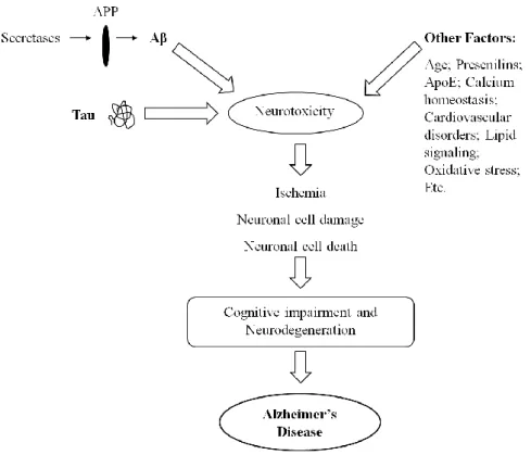
So, it becomes necessary to study the AD risk factors and underlying theories to explain the etiology of this illness. Some researchers suggest various theories and risk factors that play an important role in AD pathogenesis, like deregulation of calcium homeostasis (Bojarski, Herms et al., 2008), cardiovascular disorders (Hooijmans and Kiliaan, 2008), abnormal activation of lipid signaling (Lukiw and Bazan, 2000), induction of oxidative stress, and others.
AD risk factors can be subdivided in sporadic and familial AD. About 95% of all AD cases appear to be of random, idiopathic or sporadic origin (Hooijmans and Kiliaan, 2008). These sporadic cases involve the most significant risk factor – age – in concert with genetic and environmental risk factors. Some researchers suggest that a combination of lifestyle, genetic, and amyloid related factors, which enhance each other contribution in the onset and course of AD, will probably be at the etiology of the disease instead of being only one mechanism (Hooijmans and Kiliaan, 2008). In the group of risk factors for sporadic AD, the environmental factors are associated with a combination of life events, such as early-life childhood and adolescent environment, psychosocial and mental inactivity, loss of motivation and mental stress, a lower level of education and occupation, improper diet, higher age, exposure to neurotoxic factors, brain injury, and vascular disease (Arendt, 2001). The best-known genetic risk factor in late-onset sporadic AD is the apolipoprotein E (APOE) gene. This gene is located on chromosome 19 that encode the apolipoprotein E (ApoE) protein (Ling, Morgan et al., 2003). ApoE is polymorphic with three isoforms, ApoE2, ApoE3 and ApoE4, which translate into three alleles of the gene and differ from each other at two amino acids. ApoE plays an important role in cholesterol transport in the central nervous system and in AD development, which relates to its ability to interact with Aβ (Ling, Morgan et al., 2003; Hooijmans and Kiliaan, 2008; O'Brien and Wong, 2011). The mechanism by which ApoE4 predisposes individuals to AD is not clear. However, pathologically the evidence is that ApoE4 accelerates brain Aβ deposition, APs formation and loss of neuronal function by stimulating APP processing and reducing Aβ clearance (Ling, Morgan et al., 2003; Hooijmans and Kiliaan, 2008).
Familial AD is the second most important risk factor group. About 1% of AD cases occur due to this group of risk factors (Chiba, Nishimoto et al., 2007). This early onset AD develops because of missense mutations in the genes coding for the amyloid precursor protein (APP) on chromosome 21, presenilin 1 (PSEN1) on chromosome 14, and presenilin 2 (PSEN2) on chromosome 1, all causing abnormal processing of APP to Aβ and production of Aβ-related neurotoxic peptides (Lukiw and Bazan, 2000; Hooijmans and Kiliaan, 2008).
As mentioned before, APP is the precursor of Aβ and these proteins are implicated in the pathogenesis of both sporadic and familial AD. The APP gene localizes in the same chromosome that causes Down’s syndrome (trisomy 21), chromosome 21. The patients with this syndrome reveal the classical neuropathological features of AD, like APP overexpression, which results in brain Aβ accumulation, and formation of neurofibrillary tangles (Hooijmans and Kiliaan, 2008; O'Brien and Wong, 2011). This finding led to a specific search for families with early-onset AD with genetic linkage to chromosome 21 and resulted in the identification of several missense mutations in APP, associated to familial AD (Hooijmans and Kiliaan, 2008). In familial AD, mutations of APP gene cause a change in amino acids adjacent to the BACE1 (enzyme essential for the generation of Aβ) cleavage site, or occur around the -secretase cleavage site, and result in alteration of APP proteolytic processing to Aβ, leading to increased production of total Aβ or to production of a more toxic Aβ peptide (Chiba, Nishimoto et al., 2007; Cole and Vassar, 2007; O'Brien and Wong, 2011).
Presenilins proteins (PS1 and PS2) are integral components of a multiprotein protease complex, called -secretase, which is responsible for the cleavage of APP and other important proteins such as Notch, an essential cell development protein, similar to APP in size and intracellular localization (Ling, Morgan et al., 2003; Bojarski, Herms et al., 2008). Presenilins are involved in a range of physiological and biological processes (Ling, Morgan et al., 2003). The familial AD mutations in PSEN1 and PSEN2 show that the efficiency of presenilin-mediated Notch cleavage is reduced, in contrast to the effects on APP processing (Ling, Morgan et al., 2003). Indeed, mutations in presenilins genes lead to an increased production of the more toxic Aβ42, a form of Aβ peptide that is particularly prone to precipitate and aggregate, relative to Aβ40 (Ling, Morgan et al., 2003; O'Brien and Wong, 2011).
The Figure 4 summarizes the various AD risk factors above mentioned.
1.2. The β-Amyloid Precursor Protein (APP)
As mentioned previously, Amyloid Precursor Protein (APP) is the precursor of the major protein component of senile plaques in Alzheimer’s disease, Aβ. APP is an integral type-I transmembranar glycoprotein that suffers cleavages, by protease activity, in different fragments including the Aβ.
APP is ubiquitously expressed, as e.g. in endothelial cells, smooth muscle cells and all peripheral cells; in the central nervous system (CNS) it is abundantly expressed in neurons. Other brain cells that also express APP and release variable amounts of Aβ are glial cells, such as astrocytes (Perez, Zheng et al., 1997; King and Scott Turner, 2004).
Despite years of intense investigation, the biological functions of normal APP are still far from clear. It is known that APP is important for normal CNS function. APP resembles a cell surface signaling receptor, and has been involved in memory regulation,
Fig. 4: Various risk factors involved in neurotoxicity and AD onset. Proposed sequence of events in the development
cell-cell interaction, cell adhesion, cell survival, cell growth, neurite outgrowth, and protease inhibition (King and Scott Turner, 2004). APP has also been implied in pro- and anti-apoptotic functions. (De Strooper and Annaert, 2000).
1.2.1. APP gene family, isoforms and domains
In humans, the APP gene is located on the chromosome 21q21, and contains 19 exons, of which exons 7, 8 and 15 can be alternatively spliced (De Strooper and Annaert, 2000; da Cruz e Silva and da Cruz e Silva, 2003; Ling, Morgan et al., 2003). APP belongs to a family of related proteins that includes the amyloid precursor-like proteins (APLP1 and APLP2) in mammals and amyloid precursor protein-like (APPL) in Drosophila, Caenorhabditis elegans, and Xenopus (De Strooper and Annaert, 2000; O'Brien and Wong, 2011). All these are homologous type I transmembranar proteins with overlapping expression in brain, similarly metabolism, and that exhibit some functional redundancy (King and Scott Turner, 2004). Nonetheless, only APP cleavage gives rise to the Aβ peptide, which is derived from the domain encoded by parts of exons 16 and 17 (da Cruz e Silva and da Cruz e Silva, 2003; Ling, Morgan et al., 2003).
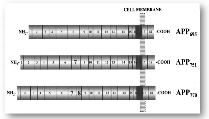
Alternative splicing of the APP transcript generates at least 8 isoforms, ranging from 365 to 770 amino acids (da Cruz e Silva and da Cruz e Silva, 2003; King and Scott Turner, 2004; O'Brien and Wong, 2011). The three most common isoforms, however, predominate in a cell-type specific manner, and differ only in the size of their extracellular sequence: APP695, APP751 and APP770 (Fig. 5). APP695 is expressed predominantly in the
CNS neurons, and the APP751 and APP770 are more expressed in non-neuronal cells, but are
also found in brain glial cells (Selkoe, 2001; King and Scott Turner, 2004; O'Brien and Wong, 2011). All of these transcripts encode multidomain proteins with a single membrane-spanning region. The APP751 and APP770 differ from the APP695 in the
expression of exon 7, which encodes a Kunitz serine protease inhibitor (KPI) domain (Selkoe, 2001).
Studies revealed that the different APP isoforms respond differently under various experimental conditions, like Aβ addition. All these three isoforms contain exons 16 and 17, but APP695 responds more readily than the other isoforms to the accumulation of Aβ
(da Cruz e Silva and da Cruz e Silva, 2003).
The complete crystal structure of APP and the ligands or receptors that interact with the large APP ectodomain are not yet much understood (De Strooper and Annaert, 2000). However, a number of distinct, large independently-folding structural domains have been identified in the APP sequence. APP possesses three general domains: a large glycosylated extracellular component, a single membrane-spanning region and a short intracellular cytoplasmic domain (Hampel, Shen et al., 2010) (Fig. 6).
Fig. 5: Schematic representation of the three major APP isoforms found in mammalian tissues. The number and
vertical lines delineate the corresponding exons. The most abundant neuronal isoform is APP695 (exons 1-6, 9-18) that
comprises 695 amino acids. As illustrated, APP751 (exons 1-7, 9-18) and APP770 (exons 1-18) are alternatively spliced
isoforms that differ from APP695 in the expression of exons 7 and 8. The sequences encoded by the APP gene exons are
indicated approximately to scale. The Aβ domain it’s represented in this figure as the solid gray region, whose sequence is divided between exons 16 and 17. Reproduced from da Cruz e Silva and da Cruz e Silva (2003)
The large extracellular region can be subdivided into several functional domains (Fig. 6). At the N-terminal is a cysteine-rich region consisting in a growth factor domain (GFD) and a copper-binding domain (CuBD) that interact tightly together. The GFD binds heparin and can stimulate neurite outgrowth (Kong, Miles et al., 2008). The CuBD of APP may regulate proteolytic processing or act as a metallotransporter. The copper binding to this domain affects the dimerization state of APP leading to reduction in Aβ production, whereas copper binding to the Aβ generates toxic species (Bossy-Wetzel, Schwarzenbacher et al., 2004; Kong, Miles et al., 2008). Studies revealed that the cysteine-rich region also contains a zinc-binding domain. This APP metal-binding site was assumed to play mainly a structural role, modifying its conformation and interfering with APP binding to constituents of the extracellular matrix (De Strooper and Annaert, 2000).
The copper-binding domain is followed by an acidic domain, which links the cystein-rich region to a Kunitz-type protease inhibitor (KPI) domain and an OX2 domain. The KPI and OX2 domains can be spliced out, to produce three main isoforms: APP770,
APP751 and APP695. The longer isoforms APP751 and APP770 contain a 56-amino acid KPI
domain, located in the middle of the APP ectodomain, which inhibits serine proteases. The OX2 domain is only present in APP770 (King and Scott Turner, 2004). Following these
domains there is a glycosylated domain referred to as E2 (sometimes called the D6a, like in Figure 6) and an unstructured region, D6b, which precedes the transmembranar domain. The E2 domain possesses the RERMS motif that appears to have putative growth-promoting properties and also has a heparin binding site (De Strooper and Annaert, 2000; Kong, Miles et al., 2008).
Another specific domain that implicates a role for APP as a cell surface receptor is theC-terminal cytoplasmic tail. This region contains several consensus motifs that regulate
Fig. 6: Schematic representation of functional domains arrangement of APP, highlighting some important regions.
The N-terminal growth factor domain (GFD) is followed by copper-binding domain (CuBD), an acidic-rich region, Kunitz-type protease inhibitor (KPI) and OX2 domains that occur in some APP isoforms, a couple of glycosylated domains (D6a, sometimes called the E2 domain, followed by an unstructured domain, D6b), a transmembrane region (TM) and a cytoplasmic tail. The location of the Aβ region, a major component of Alzheimer’s disease plaques, is shown in red. Reproduced from Kong, Miles et al. (2008)
its trafficking and suggest a role in signal transduction, through interaction with several proteins (De Strooper and Annaert, 2000; Evin and Weidemann, 2002). The APP cytoplasmic domain has been shown to bind to the Fe65 protein and G proteins, for example. Some APP hereditary mutations that are linked with AD can cause constitutive activation of Go, a member of the heteromeric G protein family that serve as signal
transducers of various cell surface receptors (Kong, Miles et al., 2008).
1.2.2. APP metabolism: trafficking and processing
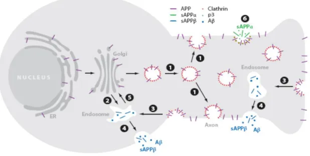
As described above, APP is produced in large quantities in neurons and is metabolized very rapidly. APP traffic (Fig. 7) is tightly regulated and along it APP can be cleaved by specific proteases. APP follows the constitutive secretory pathway, being N-glycosylated in the endoplasmic reticulum (ER) (immature APP) and further O-glycosylated (APP maturation) in the Golgi, where it is highly abundant (Thinakaran and Koo, 2008; Vieira, Rebelo et al., 2010). After sorting in the ER and Golgi, APP is delivered to the axon, where it is transported by fast axonal transport to presynaptic terminals (O'Brien and Wong, 2011). APP can be packaged into secretory vesicles in the Trans-Golgi network (TGN) and delivered to the plasma membrane (PM). On the cell surface, APP may be proteolytically processed or suffer internalization, via its YENPTY motif, being delivered into the endocytic pathway (endosomes). Then, APP is either transport to lysosomes, where it suffers lysosomal degradation, or recycled by transport vesicles to the TGN or to the cell surface (Vieira, Rebelo et al., 2010).
Crucial steps in APP metabolism occur at the cell surface and in the TGN. From the TGN, APP is transported via a clathrin coat complex, which mediate two steps: the transport directly to an endosomal compartment, and its reinternalization from the cell surface into the endocytic pathway, connecting the cell surface to the endosome (O'Brien and Wong, 2011). APP contains the NPXY (asparagiNe-Proline-any-tYrosine) amino acid motif (YENPTY domain), a conserved sorting signal that regulates the targeting of proteins for clathrin pit localization and their transport via clathrin-associated vesicles from the cell surface to the endosome (Evin and Weidemann, 2002; Small and Gandy, 2006; O'Brien and Wong, 2011).
Another complex that is implicated in APP transport is the retromer complex. The retromer is a multi-subunit complex that mediates the retrograde transport of several transmembranar proteins from endosomes to the TGN (Vieira, Rebelo et al., 2010). This complex consists of two sorting nexin subunits and a cargo-recognition trimer, vacuolar protein sorting (VPS) 26, VPS29, VPS35 (Small and Gandy, 2006). Several findings indicate that a dysfunctional retromer complex and/or one of its sorting receptor components, sorLA (a type-1 transmembrane molecule), can be related to late onset AD pathology (Vieira, Rebelo et al., 2010). Indeed, the TGN retrieval pathway, involving sorLA and the retromer, has been inversely correlated with Aβ production. Further studies revealed that phosphorylation of APP in the Ser655 residue enhances APP binding affinity for sorting proteins, such as the retromer-related VPS35 protein (at the core of the retromer) (Vieira, Rebelo et al., 2010). Ser655 phosphorylation was found to enhance both the protein exit from the Golgi, and its recycling back to the TGN from endosomes, increasing its cleavage to sAPPα during this trafficking cycle.
Fig. 7: Schematic representation of APP trafficking in neurons. Newly synthesized APP (purple) is transported from
the Golgi down the axon to the cell surface (1) or into a cell body endosomal compartment (2), with clathrin-associated vesicles (red) mediating both steps. In the cell surface, some APP is cleaved by α-secretase (6), generating the sAPPα fragment (green), and some is re-internalized into endosomes (3), where Aβ and sAPPβ are generated (blue). Following proteolysis, the endosome recycles to the cell surface (4), releasing Aβ and sAPPβ. Transport from the endosomes to the Golgi prior to APP can also occur, mediated by the retromer (5). Reproduced from O'Brien and Wong (2011)
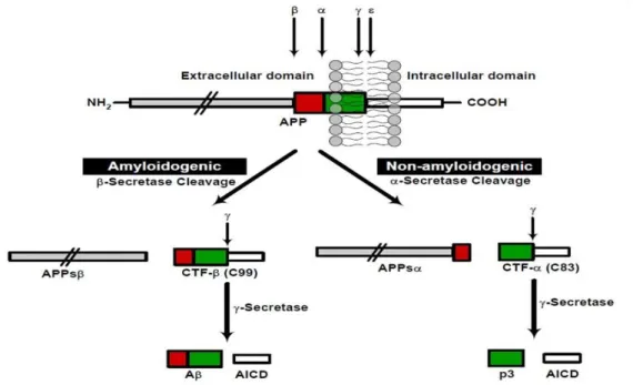
In what concerns cleavage, there are mainly two pathways for APP proteolytic processing: the amyloidogenic and non-amyloidogenic, distinguished by the mutually exclusive action of two different enzymes in the first cleavage event (Fig. 8).
In the non-amyloidogenic pathway, cleavage of APP by α-secretase produces a large soluble extracellular N-terminal fragment, sAPPα, which has neuroprotective properties, and a C83 membrane-bound C-terminal fragment. This cleavage divides the Aβ domain and precludes the formation of intact Aβ. So, stimulating α-cleavage of APP leads to a significant decrease in Aβ generation (Evin and Weidemann, 2002; Ling, Morgan et al., 2003). This activation is a relatively major and ubiquitous pathway of APP metabolism in most cells. Membrane-bound disintegrin and metalloproteinases including ADAM17 (also called TACE), ADAM10, ADAM9 and MDC-9 are proteins that have been identified as having α-secretase-like activity (Evin and Weidemann, 2002; Ling, Morgan et al., 2003). The constitutive α-secretase activity occurs primarily at the cell surface, while the
Fig. 8: Schematic diagram of APP proteolytic processing. APP can undergo proteolytic processing via two pathways:
amyloidogenic and non-amyloidogenic. Cleavage of APP by β-secretase occurs at the beginning of the Aβ domain and generates a shorter soluble N-terminus, APPsβ, as well as an amyloidogenic C-terminal fragment (CTF-β, C99). Alternatively, α-secretase cleavage, within the Aβ domain, generates the large soluble N-terminal, APPsα, and a non-amyloidogenic C-terminal fragment (CTF-α, C83). Further proteolysis of these fragments by -secretase results in generation of either the Aβ (amyloidogenic pathway) or p3-fragment (non-amyloidogenic pathway) and a cytoplasmic APP intracellular C-terminal domain (AICD). Reproduced from Kumar and Walter (2011)
regulated activity is predominantly located within the Golgi apparatus (Ling, Morgan et al., 2003; Small and Gandy, 2006). There are several factors that can increase this pathway, by mechanisms involving the formation and release of secretory vesicles from the TGN, and thus enhancing APP (and possibly α-secretase) trafficking to the cell surface, as for example the activation of protein kinase C by phorbol esters (Evin and Weidemann, 2002; Ling, Morgan et al., 2003; da Cruz e Silva, Rebelo et al., 2009).
The alternative, amyloidogenic pathway is characterized by the cleavage of APP by β-secretase at its β-cleavage site, producing the large soluble sAPPβ peptide and the carboxy-terminal fragment (CTF) C99. This pathway constitutes the first step in the formation of Aβ and is particularly enriched in neurons (Evin and Weidemann, 2002). Two novel transmembrane aspartyl proteases homologues, β-site APP-cleavage enzymes BACE1 and BACE2, were identified to cleave APP at the β-secretase sites. The major neuronal β-secretase that governs the first enzymatic step in this APP processing is BACE1. This protease appears to be produced as a pro-enzyme predominantly within the nuclear envelope and the ER (Ling, Morgan et al., 2003). The prodomain in BACE1 does not support activity but appears to facilitate correct folding of the active protease domain. This domain is cleaved by proprotein convertases (PPCs) immediately before trafficking through the Golgi (Ling, Morgan et al., 2003). Although the interaction of APP with BACE1 can occur in the ER and on the cell surface, evidence suggests that active BACE1 predominantly localizes in the TGN and endosomes, consistent with the amyloidogenic cleavage of wild-type APP during endocytic/recycling steps (Evin and Weidemann, 2002; Ling, Morgan et al., 2003; Small and Gandy, 2006). The precise role of BACE2 in APP processing remains unclear. BACE2 shows similar substrate specificity, cleaving APP at the β-secretase site, but it also shows a distinct cellular localization pattern and intracellular protease specificity in the cleavage of APP that differentially affects the generation of Aβ (Ling, Morgan et al., 2003). Indeed, BACE2 can also have an α-secretase activity, cleaving APP in the middle of the Aβ domain, between Phe19 and Phe20 (Ling, Morgan et al., 2003).
The second enzymatic step is determined by cleavage of the membrane-bound C-terminal APP fragments (CTFs) of each pathway by -secretase. The C83 and C99, fragments resulting from α-secretase and β-secretase cleavage, respectively, remain anchored in the membrane and may become degraded or can be further cleaved by
-secretase, leading to the release and secretion of non-pathogenic p3 peptide and Aβ (Evin and Weidemann, 2002). In addition, in both pathways, -secretase cleavage generates a cytoplasmic APP intracellular C-terminal domain (AICD). -secretase is a multimeric complex composed of four essential transmembrane proteins: presenilin 1 or 2 (PS1 or PS2, respectively), nicastrin, APH-1 and PEN-2 (Small and Gandy, 2006; Thinakaran and Koo, 2008; O'Brien and Wong, 2011). This complex is essential for the sequential intramembranous proteolysis of a variety of transmembrane proteins, like APP, Notch and Cadherin. APH-1 aids the formation of a precomplex, which interacts with PS1 or PS2 while Pen-2 enters the complex to facilitate the proteolytic cleavage of PS1 or PS2, which are critical to the γ-secretase complex (O'Brien and Wong, 2011). The ectodomain of nicastrin recognizes and binds to the previously cleaved transmembrane proteins (O'Brien and Wong, 2011).
Several data indicates the presence of -secretase complex and its enzymatic activity in multiple compartments, including the ER, Golgi, TGN, endosomes and plasma membrane, but studies in cell lines have showed that APP cleavages occur mainly in the TGN and endosomes (Thinakaran and Koo, 2008). The -secretase complex cleaves at multiple sites within the APP transmembrane domain. The cleavage of C83, CTF resulting of α-secretase, leads to AICD and p3 fragments production. The processing of C99, consequent CTF of the amyloidogenic via, results in different Aβ forms and AICD. The AICD, free from the membrane, has been suggested to function as a transcription factor, but genes regulated by AICD have not been unambiguously identified (Hampel, Shen et al., 2010). The release of the cytoplasmic tail of APP by -cleavage may function in gene expression, with AICD being targeted to the nucleus, where it forms a multimeric complex with the nuclear adaptor protein Fe65 and the histone acetyltransferase Tip60, potentially stimulating the transcription of various genes, including APP itself (Ling, Morgan et al., 2003).
Noteworthy, APP metabolism may be regulated by direct APP phosphorylation, and the aberrant production of Aβ associated with AD may result from deregulated, abnormal APP phosphorylation (da Cruz e Silva and da Cruz e Silva, 2003).
1.2.3. Phosphorylation of APP
The direct phosphorylation of APP might be important in APP-mediated roles such as neuronal differentiation, possible by altering APP traffic, by regulating APP proteolytic cleavage to its physiological fragments and/or the binding of APP to specific signal transducers. APP is a phosphoprotein with several well defined phosphorylated residues at its extracellular and intracellular portions (Fig. 9).
Only one phosphorylatable domain was identified in the extracellular ectodomain, and the residues shown to be phosphorylated are Ser198 and Ser206 (da Cruz e Silva, Fardilha et al., 2004). This phosphorylation appears to occur in two distinct cellular locations: in a post-Golgi secretory compartment and at the cell surface, by ectoprotein kinases (da Cruz e Silva, Fardilha et al., 2004). Particularly, phosphorylation of APP cytoplasmic tail comprises three crucial motifs, 653YTSI656, 667VTPEER672, and
682
YENPTY687, which have been shown to regulate the interaction of APP with some of its binding proteins (da Cruz e Silva and da Cruz e Silva, 2003; Lee, Kao et al., 2003; da Cruz e Silva, Fardilha et al., 2004).
Fig. 9: Schematic representation of the APP phosphorylatable residues. The top diagram represents APP and
indicates the two phosphorylated Serine residues present in its ectodomain. The bottom diagram shows the complete amino acid sequence of the cytoplasmic tail, and the three phosphorylatable functional domains. Serine, threonine and tyrosine putative phosphorylation sites are shown enlarged (numbering is relative to APP695). The relative localization of
The 653YTSI656 (amino acid numbering according human APP695 isoform), is a
typical YXXI sorting signal, responsible for mediating APP basolateral sorting. This motif contains Thr654 and Ser655, two consensus residues for phosphorylation known to be phosphorylated in vitro and in vivo. The possible putative kinases involved in their phosphorylation are protein kinase C (PKC) and calmodulin-dependent protein kinase II (CaMKII) (Isohara, Horiuchi et al., 1999; da Cruz e Silva and da Cruz e Silva, 2003; Schettini, Govoni et al., 2010). Ser655 was also reported to be phosphorylated by APP kinase I (da Cruz e Silva and da Cruz e Silva, 2003). Phosphorylation of Ser655 was observed to occur predominantly in mature rather than immature APP isoforms (da Cruz e Silva, Fardilha et al., 2004). As described above, various studies revealed that phosphorylation within this sorting motif modulates APP trafficking. Mimicking phosphorylation at the Ser655 residue was reported to enhance APP secretory traffic, e.g. by increasing APP binding to the retromer complex and the subsequent retromer-mediated APP retrieval to the TGN (Vieira, Rebelo et al., 2010). S655 phosphorylation, potentially via PKC, also increases sAPPα production by the alpha-secretase pathway (da Cruz e Silva, Rebelo et al., 2009; Vieira, Rebelo et al., 2009).
The 667VTPEER672 motif contains Thr668. This residue can be phosphorylated by neuronal cyclin-dependent kinase 5 (Cdk5), cyclin-dependent protein kinase 2 (Cdc2), glycogen synthase kinase 3β (GSK-3β), c-Jun-N-terminal kinase 3 and stress-activated protein kinase 1b (da Cruz e Silva and da Cruz e Silva, 2003; da Cruz e Silva, Fardilha et al., 2004; Schettini, Govoni et al., 2010). Phosphorylation at Thr668 might be involved in a neuron-specific aspect of APP metabolism and/or function, because this phosphorylation in mature APP occurs only in the brain, being detected in neurites (da Cruz e Silva, Fardilha et al., 2004). Phosphorylation of this domain appears to be an important factor in the control of interactions of APP with other proteins. However, there are contradictory results for the role of APP phosphorylation at Thr668 in Aβ production, in Fe65 binding and in nuclear translocation of AICD or AICD/Fe65 complex (Schettini, Govoni et al., 2010).
Tyrosine phosphorylation of APP may functionally link APP processing and neurotrophic signaling to intracellular pathways associated with cellular differentiation and survival. This phosphorylation occurs in residues of the amino acid sequence
682
YENPTY687, encompassing an NPXY signaling motif, which is a typical internalization signal for membrane-associated receptor proteins (da Cruz e Silva and da Cruz e Silva,
2003). This motif is present in many Tyrosine-Kinase (TK) receptors and non-receptors TKs; it is generally phosphorylated and represents the docking site for several interacting APP-binding proteins, involved in cell signaling and gene transcription (Russo, Venezia et al., 2005; Schettini, Govoni et al., 2010). The phosphorylation of APP occurs in Tyr682 and Tyr687, although phosphorylation at this last residue is still somehow controverse (da Cruz e Silva and da Cruz e Silva, 2003; da Cruz e Silva, Fardilha et al., 2004; Rebelo, Vieira et al., 2007). In cell-culture studies, Tyr682 can be phosphorylated by the overexpression of the nerve growth factor receptor TrkA, by a constitutively active form of the tyrosine kinase Abl or by the Src kinase (da Cruz e Silva, Fardilha et al., 2004; Russo, Venezia et al., 2005; Schettini, Govoni et al., 2010). In some studies using HEK293 cells, it was reported that the phosphorylation of APP at Tyr687 is important for its processing by α- and - secretases, increasing CTF-α and AICD generation (Takahashi, Niidome et al., 2008). Of note, it is possible that APP may only become Tyr682 phosphorylated after full-length APP cleavage by the -secretase complex (da Cruz e Silva, Fardilha et al., 2004).
1.3. APP as a potential mediator of G proteins signaling
1.3.1. G proteins
G proteins, also called guanine nucleotide-binding proteins, are a family of heterotrimeric proteins that have a crucial role as molecular switches in signal transduction pathways mediated by G protein-coupled receptors (GPCRs). They are involved in transmitting extracellular messages from hormones, neurotransmitters, chemokines and other signaling factors that interact with GPCRs and G proteins to activate many intracellular signaling pathways (Hewavitharana and Wedegaertner, 2012).
The heterotrimeric G proteins are signal-transducing protein complexes composed of α, β and subunits, located on the cytoplasmic side of the plasma membrane (Knust, 2001). The Gα subunit contains the nucleotide binding site and GTPase activity, along with sites for binding receptors, effectors and Gβ, and a helical domain whose function is not clear (Neer, 1995). This subunit determines G protein diversity. The β and subunits form a dimer, a single complex that only dissociates when it is denatured and is, therefore, a functional monomer (Neer, 1995). Today it is known that both α and β subunits positively
regulate effectors, acting as signaling molecules by activating other second messengers or by gating ion channels directly (Neer, 1995).
Different types of G proteins share a common mechanism. They are activated in response to a conformation change in the GPCR receptor. The GPCRs constitute a large and the most versatile protein family of transmembrane receptors in the mammalian genome (Gudermann, Schoneberg et al., 1997; Wettschureck and Offermanns, 2005). The diversity of this superfamily is a result of the large number of members, their ability to form different dimer combinations and their ability to respond to several stimuli, as well as by the large amount of intracellular signaling pathways they activate. Signaling by GPCRs is not just limited to second messenger molecules but also includes transcription factors and molecules that affect the cytoskeleton. Despite their structural and functional diversity, all GPCRs share a similar molecular architecture (Knust, 2001). They consist of an extracellular N-terminus, an intracellular C-terminus, and seven transmembrane domains in between, linked by alternating intracellular and extracellular loops (Gudermann, Schoneberg et al., 1997).
In the inactive state, the Gα subunit has GDP in its binding site. When a chemical or physical signal stimulates the receptor, the receptor becomes activated and changes its conformation. The Gα subunit responds with a conformational change that decreases GDP affinity, so that GDP comes off the binding site (Fig. 10). Once GTP is bound, the Gα assumes its activated conformation and dissociates both from the receptor and from Gβ (Knust, 2001). Then, both free Gα-GTP and Gβ can interact with downstream effectors proteins and diverse downstream signaling cascades, while the receptor is able to interact with other G proteins and amplify signal transduction (Knust, 2001). In addition to activation of second messenger molecules, Gα subunits can also modulate the activity of transcription factors, thereby regulating gene expression (Ram and Iyengar, 2001). The activated state lasts until the GTP is hydrolyzed to GDP by the inherent GTPase activity of Gα, allowing re-association with Gβ. G protein becomes inactive and returns to the receptor, starting a new cycle (Wettschureck and Offermanns, 2005).
The GTPase cycle of G protein activation and deactivation is subject to regulation by many protein factors that either influences the rate of guanine nucleotide exchange or the rate of hydrolysis of bound GTP.
The family of regulators of G protein signaling (RGS), for example, has been found to play a role in desensitization. These proteins are GTPase-activating proteins (GAPs) that are involved in mechanisms that accelerate the rate of GTP hydrolysis and thereby negatively regulate G protein signaling (Diverse-Pierluissi, Fischer et al., 1999; Hewavitharana and Wedegaertner, 2012). RGS proteins block G protein function by accelerating α subunit GTPase activity, physically blocking the binding to G protein effectors and/or altering the level of free β subunits available to their downstream effectors, therefore allowing for rapid modulation of G protein-mediated signaling (Wettschureck and Offermanns, 2005; Blazer, Roman et al., 2010).
By contrast, guanine nucleotide exchanger factors (GEFs) promote the activation of G proteins by increasing the rate of GDP dissociation and GTP association (Hewavitharana and Wedegaertner, 2012). The ligand-bound GPCRs are widely recognized as guanine exchange factors for heterotrimeric G proteins.
The functional versatility of the G protein mediated signaling pathways is based on its modular architecture and on the fact that there are numerous subtypes of G proteins. In
Fig. 10: Activation and deactivation of G proteins by guanine nucleotide exchange and GTP hydrolysis. A ligand
binds to the receptor and is activated to become a catalyzer, enhancing the rate of detachment of bound GDP from the α-subunit of the G protein. The rapidly exchange of GDP to GTP in the α-α-subunit site is rapidly processed (1) and causes activation of both α- and β-subunits, enabling them in turn to activate specific effector enzymes. The interaction between receptor and G protein is transient, allowing the receptor to catalyze guanine nucleotide exchange on a succession of G protein molecules. The system returns to the resting state following hydrolysis of the bound GTP, and promotes deactivation of G protein (2). Reproduced from Gomperts, Kramer et al. (2009)
humans, there are genes encoding 16 Gα, 5 Gβ and 12 G subunits (Hewavitharana and Wedegaertner, 2012). The α-subunits increase the diversity, define the basic properties of a heterotrimeric G protein, and can be divided into four families: Gi/o; Gs; Gq and G12/13
(Wettschureck and Offermanns, 2005; Shan, Chen et al., 2006; Hewavitharana and Wedegaertner, 2012). Each family consists of various members that often show very specific expression patterns. Members within each family have structural and functional homologies (Wettschureck and Offermanns, 2005; Hewavitharana and Wedegaertner, 2012).
The G proteins of the Gi/o family are widely expressed. The main members of these
types of G proteins are Gi proteins (Gi1, Gi2 and Gi3) that inhibit various types of adenylyl
cyclases and thus lower the levels of the second messenger cyclic AMP (cAMP). The function of members of the Gi/o family is affected by pertussis toxin (PTX), which is able
to ADP-ribosylate the Gi/o α-subunit, and, subsequently, uncouples Go and Gi from their
receptors, leading to the disruption of their signaling. A less widely distributed member of this family is Gz, which is not affected by PTX, is expressed in various tissues (nervous
system and platelets) and was recently shown to interact specifically with various other proteins including Rap1GAP and certain RGS proteins. Other α-subunits that belong to the Gi/o family, like gustducin and transducin are involved in specific sensory functions, taste
and visual, respectively. The Go member is particularly abundant in the nervous system
and will be discussed later on, with its action being greatly mediated by its β-complex. Indeed, activation of Gi/o is believed to be the major coupling mechanism that results in the
activation of β-mediated signaling processes.
The ubiquitously expressed G protein Gs stimulates adenylyl cyclase, resulting in
increases intracellular levels of cAMP (Wettschureck and Offermanns, 2005).
The Gq/G11 family of G proteins binds to and activates members of phospholipase
C β-isoform family (Wettschureck and Offermanns, 2005). Interestingly, there is no obvious difference between the abilities of both G protein α-subunits to regulate β-isoforms of phospholipase C. The α-subunits of Gq and G11 are almost ubiquitously
expressed and are involved in various biological signaling pathways (Wettschureck and Offermanns, 2005).
In the G12/13 family, the two members G12 and G13 appear to be expressed
leading to the activation of various downstream effectors including other G proteins (Hewavitharana and Wedegaertner, 2012). Of note, the analysis of cellular signaling processes regulated by these G proteins has been difficult since there are no specific inhibitors available. The G12 and G13 Gα subunits were described to bind to and regulate
cell adhesion proteins, and interact directly with the cytoplasmic domain of some members of the Cadherin family of cell surface adhesion molecules (Wettschureck and Offermanns, 2005; Hewavitharana and Wedegaertner, 2012).
The much less understood G protein β and subunits, but with a very important role in the regulation of various effectors, are tightly associated and can be regarded as one functional unit. The different subunits could produce several different combinations, but not all possible pairs are formed (Neer, 1995). The β subunits show very high sequence identity and exhibit a more or less ubiquitous expression pattern, whereas subunits are considerably more diverse in sequence and tissue expression (Gudermann, Schoneberg et al., 1997; Hurowitz, Melnyk et al., 2000). This suggests that through their diversity these specific subunits couple selectively to effectors, although the molecular determinants of Gβ-effector coupling are not well known.
1.3.1.1. Gαo Protein: genetics, expression pattern and function
The Gαo protein was accidently discovered during the purification of Gαi from
bovine brain. Gαo was designed as the ‘other’ GTP-binding protein to differentiate from
Gαs, Gαi and transducin. PTX-mediated ADP ribosylation and electrophoresis samples
detected an additional Gα subunit with molecular weight of 39 kDa and revealed a highly Gαo expression in brain tissue (Jiang and Bajpayee, 2009; Bromberg, Lyengar et al., 2011).
In contrast to the well characterized pathways transduced by other G-proteins, only recently some mechanism details of Gαo signaling were elucidated. Nonetheless, the
identification of direct effectors of Gαo and the determination of Gαo-induced cellular
responses are still far from known.
The cDNAs encoding Gαo have been cloned and identified in several species, such
as human, rat, mouse, bovine, Drosophila. Gαo has a highly degree of similarity among the
species, suggesting that Gαo-mediated signaling are important to receive, integrate and
Analysis of genomic cDNA clones encoding Gαo revealed that the human Gαo gene
localizes on chromosome 16q13 and contains 11 exons. Both Gαo isoforms, Gαo1 (GoAα)
and Gαo2 (GoBα), are generated via alternative splicing and are identical in the first
two-thirds of the amino acid sequence. The two forms differ by only 20 amino acids in human cells. Gαo1 and Gαo2 share the first 6 exons that encode 241 N-terminal amino acids, and
each isoform has unique exons 7, 8 and 9. Different sets of exons suggest that each isoform may contribute to distinctive biological functions in the body and, especially, in the human brain (Jiang and Bajpayee, 2009).
The Gαo protein has a less ubiquitous expression pattern than Gαi or Gαs. The
protein is expressed widely in the CNS and was estimated to compose 0.5%-1% of membrane proteins in brain (Jiang and Bajpayee, 2009; Bromberg, Lyengar et al., 2011). However, the regional distribution of Gαo protein in all brain is not homogeneous. At the
anatomic level, high concentrations of Gαo have been detected in the frontal cortex,
cerebellum, hypothalamus, hippocampus and substantia nigra, being principally found on the cytoplasmic face of the plasma membrane coating the cell body and the neurites, particularly at ‘cell-to-cell’ contacts. It was also determined that Gαo is one of the most
abundant proteins in the neuronal growth cones, structure at the tip of the growing neurite that is generated during neuronal differentiation. In addition, Gαo expression has also been
observed in heart tissue, pituitary gland and pancreatic islets (Jiang and Bajpayee, 2009). The Gαo protein has been received considerable attention aiming to determine its
physiological role in the body, mainly due to its high expression in CNS. Gαo has been
associated to cell survival, neuronal development, neuronal adhesion, migration and some diseases as Alzheimer’s and Parkinson’s. Studies with Gαo knockout mice have reported
several neurological deficiencies, including poor motor coordination, hyperactive and abnormal behaviour, as well as hyperalgesia, when subjected to the lack of this protein (Jiang and Bajpayee, 2009). Another study has revealed that Gαo activation can induce
neurite outgrowth in PC12 cells through inhibition of PKC and modulation of intracellular calcium levels (Strittmatter, Fishman et al., 1994).
1.3.1.2. Modulation of Gαo activity
Many protein factors are involved in signal transduction through G proteins to regulate the strength, duration, efficiency and specificity of signaling. Gαo is a highly
effective molecular signaling transducer and many GPCRs have been identified to couple to Gαo protein, such as the cannabinoid 1 (CB1) receptor, muscarinic cholinergic receptors,
α2-adrenergic receptors, and as previously mentioned, these are widely recognized as GEFs
for heterotrimeric G proteins. However, several non-receptor proteins have also shown to affect the Gαo subunit G protein activity, including the growth cone-associated protein with
molecular weight of 43 kDa (GAP-43), APP, Presenilin 1 and the activator of protein signaling (AGS) (Jiang and Bajpayee, 2009).
GAP-43, a protein highly expressed in the growth cones of developing and regenerating neurons, has the ability to enhance Gαo binding to GTP in the distal tips of
growth cones (acting as a GEF), suggesting that Gαo protein can have a neurite outgrowth
role. Interestingly, PTX does not alter the Gαo activation by GAP-43 like in the others
GEFs. AGS is another GEF involved in regulating Gαo-mediated signaling, enhancing
GTPS binding to both Gi and Go proteins. APP and Presenilin 1 have also been
hypothesized to act as a GEF for Gαo protein (Jiang and Bajpayee, 2009).
Other protein factors have also been identified, but these act as decreasers of the guanine nucleotide exchange rate. A RGS protein identified was the Gα-interacting protein (GAIP) that is responsible to selectively accelerate the deactivation of Gαo protein. This
RGS protein is not only responsible for deactivation of Gαo protein signaling, but also
regulates the duration and specificity of receptor-stimulated Gαo protein signaling (Jiang
and Bajpayee, 2009). Other studies revealed the existence of G protein regulatory (GPR) or GoLoco motifs, which bind to Gαo and stabilize it in the GDP-bound conformation while
simultaneously competing with Gβ for Gα binding (Siderovski, Diverse-Pierluissi et al., 1999; Jiang and Bajpayee, 2009).
1.3.1.3. Gαo-mediated signaling transduction
The signaling transduction mechanism of Gαo protein and its intracellular effectors
has received widely attention, in an attempt to identify the ligands that might stimulate the signal, the downstream signaling pathways subsequently activated, and the cellular functional output of the signaling cascades.
Several studies have shown that Gαo is required to activate phospholipase C