UNIVERSIDADE DE LISBOA
FACULDADE DE FARMÁCIA
DEPARTAMENTO DE MICROBIOLOGIA
Molecular Epidemiology and Evolution of
HIV-1 in Portugal and Portuguese Speaking African
Countries
Inês Isabel Fernandes Bártolo
DOUTORAMENTO EM FARMÁCIA
MICROBIOLOGIA
UNIVERSIDADE DE LISBOA
FACULDADE DE FARMÁCIA
DEPARTAMENTO DE MICROBIOLOGIA
Molecular Epidemiology and Evolution of
HIV-1 in Portugal and Portuguese Speaking African
Countries
Inês Isabel Fernandes Bártolo
Dissertação Orientada pelo Prof. Doutor Nuno Taveira e co-orientada pelos
Prof. Doutora Patrícia Cavaco Silva e Prof. Doutor José Moniz Pereira
DOUTORAMENTO EM FARMÁCIA
MICROBIOLOGIA
Todas as afirmações efectuadas no presente documento são
da exclusiva responsabilidade da sua autora, não cabendo
qualquer responsabilidade à Faculdade de Farmácia de Lisboa
pelos conteúdos nele apresentados.
Inês Bártolo teve o apoio financeiro da Fundação para a Ciência e Tecnologia através de
uma bolsa de doutoramento (SFRH/BD/30343/2006).
“Be a virus, see the world”
Gary Larson
i
Os meus agradecimentos dirigem-se a todos que de alguma forma contribuíram para a conclusão desta tese.
Ao Prof. Doutor Nuno Taveira, meu orientador, agradeço-lhe a sua orientação científica e a sua amizade durante estes 13 anos, sem as quais esta tese não teria sido efectuada.
À Prof. Doutora Patrícia Cavaco Silva, minha co-orientadora, agradeço-lhe a sua orientação científica.
Ao Professor Doutor José Moniz-Pereira, Coordenador do Departamento de Microbiologia e Imunologia da Faculdade de Farmácia de Lisboa, e meu co-orientador, agradeço-lhe por me ter recebido para estágio de licenciatura no longínquo ano de 1998 e me ter permitido durante todos estes anos o desenvolvimento de trabalho científico.
A todos os médicos, que se prontificaram a fazer a recolha das amostras, vai o meu agradecimento.
À Fundação GlaxoSmithKline das Ciências da Saúde, à Fundação Calouste Gulbenkian e à Fundação para a Ciência e Tecnologia agradeço o apoio financeiro para a realização dos trabalhos práticos.
A todos os meus colegas de laboratório, aos que cá estão e aos que por cá passaram, agradeço-vos os bons momentos de convívio.
A
ii
À Cheila agradeço-lhe a sua preciosa ajuda e apoio nos bons e nos maus momentos. És uma óptima amiga!
Ao Pedro, por todo apoio quer no trabalho de bancada quer na parte informática, e principalmente pela sua contagiosa boa disposição!
À Lena agradeço-lho o constante incentivo e amizade. Já lá vão 13 anos!
À minha sogra pelos inúmeros, vou chegar mais tarde, os miúdos podem jantar aí, por favor dê-lhes banho, o meu muito, muito obrigada!
Aos meus irmãos e cunhados por todo o apoio e força, muito obrigada!
Aos meus pais que estão sempre disponíveis e que sem eles a minha vida seria um caos! Obrigada!
Aos meus filhos, que são a luz da minha vida. Quando estou cansada, desmotivada ou triste, basta o vosso sorriso e tudo volta de novo a fazer sentido!!!
Por fim, a ti Jorge. MUITO OBRIGADA. Nada do que eu possa escrever descreve o que eu sinto, digo apenas, sem ti a minha vida não tinha sentido!!!
iii
The research desccribed in this thesis was performed from March of 2007 to December of 2010 under the supervision of Prof. Doutor Nuno Taveira and the co-supervision of Prof. Doutora Patrícia Cavaco Silva and Prof. Doutor José Moniz Pereira.
The studies described in this thesis were performed at the Unidade de Retrovírus e Infecções Associadas – Centro de Patogénese Molecular da Faculdade de Farmácia de Lisboa. The results obtained were published in manuscripts already published or in preparation:
Bártolo I, Camacho R, Barroso H, Bezerra V e Taveira N. Rapid clinical progression to AIDS and death in a persistently seronegative HIV-1 infected heterosexual young man. AIDS.
2009;23(17):2359-2362;
Bártolo I, Casanovas J, Bastos R, Rocha C, Abecasis AB, Folgosa E, Mondlane J, Manuel R e Taveira
N. HIV genetic diversity and transmitted drug resistance in health care settings in Maputo,
Mozambique. J Acquir Immune Defic Syndr. 2009;51(3):323-31;
Bártolo I, Rocha C, Bartolomeu J, Gama A, Fonseca M, Mendes A, Epalanga M, CristinaF, ThammS, Cavaco Silva P, Taveira N. Antiretroviral drug resistance surveillance among treatment-naive
HIV-1-infected individuals in Angola: evidence for low level of transmitted drug resistance.
Antimicrobial Agents and Chemotherapy. 2009;53(7):3156-3158. Epub 2009 May 11;
P
iv
Bártolo I, Rocha C, Casanovas J, Bastos R, Abecasis AB, Folgosa E, Mondlane J, Manuel R e Taveira
N. HIV genetic diversity and transmitted drug resistance in 2002-2004 in Maputo, the capital
city of Mozambique. Reviews in Antiviral Therapy. 2009;1:34-35;
Bártolo I, Rocha C, Bartolomeu J, Gama A, Marcelino R, Fonseca M, Mendes A, Epalanga M, Cavaco
Silva P, Taveira N. Highly divergent subtypes and new recombinant forms prevail in the HIV/AIDS
epidemic in Angola: New insights into the origins of the AIDS pandemic. Infect Genet Evol.
2009;9:672-682. Epub 2008 May 9;
Bártolo I, Abecasis AB, Borrego P, Barroso H, McCutchan F, Camacho R, Taveira N. Origin and epidemiologic history of HIV-1 CRF14_BG. (Manuscript accepted for publication in PLoS ONE);
Oliveira V, Bártolo I, Borrego P, Rocha C, Valadas E, Barreto J, Almeida E, Antunes F, Taveira N.
Genetic diversity and drug resistance profiles in HIV-1 and HIV-2 infected patients from Cape Verde Islands. (Manuscript submitted to AIDS Research and Human Retroviruses).
Other Publications
Published manuscripts:
Borrego P, Barroso H, Bártolo I, Família C, Quintas A e Taveira N. Evolutionary and structural
features of the C2, V3 and C3 envelope regions underlying the differences in HIV-1 and HIV-2 biology and infection. PLoS ONE. 2011;6(1):e14548.
Submitted manuscripts:
SantosA, Clemente S, Bártolo I, Palladino C, Cavaco Silva P, FrancoV, Epalanga M, PintoR, Taveira N. Evaluation of the diagnostic performance of the rapid test VIKIA HIV1/2 in a highly complex
HIV-1 epidemic. Diagnostic Microbiology and Infectious Diseases. 2011; (In press);
Borrego P, Calado R, Marcelino JM, Bártolo I, Família C, Rocha C, Cavaco-Silva P, Doroana M, Antunes F, Maltez F, Caixas U, Barroso H, Taveira N. Baseline susceptibility of primary Human
Preface
v
Book chapters:
Taveira N, Borrego P, Bártolo I. Biologia Molecular de VIH - Manual sobre SIDA - 3ª Edição. Editor Francisco Antunes. Permanyer Portugal. 2008; 27-50;
Rocha C, Bártolo I, Barroso H, e Taveira N. New insights into the relationship between HIV-2
evolution and disease progression - VIII HIV/AIDS Virtual Congress – Current research insights into
HIV/AIDS and related diseases. SIDAnet, Associação Lusófona. 2008; 251-262;
Rocha C, Barroso H, Bártolo I, Marcelino J, Rosado L e Taveira N. Evolução molecular do gene env
do HIV-2 e colapso do sistema imunitário em doentes infectados por via vertical - VII HIV/AIDS
Virtual Congress – O VIH na Criança e no Idoso. SIDAnet, Associação Lusófona. 2007; 251-262;
Palma C, Pinheiro C, Lobão D, Silva J, Barroso H, Bártolo I, Carvalho P, Matoso P, Rocha C, Bezerra V. HIV Medicine 2006, Versão Portuguesa. Flying Publisher. 2007.
vii
A infecção VIH/SIDA é um grave problema de saúde pública, particularmente na África subsaariana, onde vivem 67% dos infectados. O diagnóstico, a terapêutica e a prevenção da infecção VIH/SIDA dependem do conhecimento aprofundado da sua epidemiologia bem como dos agentes causadores desta infecção. A diversidade genética de VIH-1 pode ter impacto a vários níveis, nomeadamente, no diagnóstico, transmissão, progressão da doença e emergência de resistências aos fármacos antirretrovirais (revisto no Capítulo 1). Com a recente introdução da terapêutica antirretroviral combinada (TARc) nos países em vias de desenvolvimento, o prognóstico dos doentes que têm acesso a estes fármacos melhorou substancialmente, com uma marcada diminuição das taxas de mortalidade e morbilidade. No entanto, há preocupações relativas à baixa eficácia da TARc nestes países, o que pode levar à rápida e descontrolada emergência de variantes resistentes aos fármacos antirretrovirais e à sua subsequente transmissão. Há vários aspectos que podem levar à baixa eficácia da TARc, nomeadamente a monitorização limitada da carga viral nos doentes em terapêutica, o uso não regulado de fármacos antirretrovirais comprados no mercado negro e a falta de sistemas de vigilância de resistências.
Os estudos de epidemiologia molecular e de resistência aos fármacos antirretrovirais na infecção VIH-1 descritos nesta dissertação foram realizados em Angola, Moçambique, Cabo Verde e Portugal. Estes países têm fortes laços históricos, sociais, culturais e económicos. Contudo, a infecção VIH/SIDA afecta estes países de forma muito diferente. Moçambique é um dos países da África subsaariana com maior taxa de prevalência da infecção VIH-1 (11,5%), tendo províncias com prevalências superiores a 25%. Angola tem uma prevalência moderada de 6,5%, sendo Luanda a
R
viii
província que apresenta a prevalência mais elevada do país (13,6%). Cabo Verde apresenta uma taxa de prevalência muito baixa para um país Africano, inferior a 1%. Em Portugal a prevalência da infecção VIH/SIDA sendo baixa (0,6%) é, no entanto, uma das mais elevadas da Europa Ocidental. Em Angola, Moçambique e Cabo Verde, tal como na maioria dos países Africanos, a transmissão da infecção faz-se sobretudo por via heterossexual enquanto em Portugal os toxicodependentes por via endovenosa correspondem a cerca de 40% dos casos de infecção. Em Portugal, os subtipos B e G e a forma recombinante CRF14_BG dominam a epidemia de infecção por VIH-1 mas pouco se sabe sobre a sua origem e história epidemiológica. Relativamente a Angola, Moçambique e Cabo Verde, há pouca ou nenhuma informação sobre os subtipos virais, sobre a sua origem e história epidemiológica e sobre o seu potencial impacto no diagnóstico, progressão da doença e susceptibilidade e resistência aos fármacos antirretrovirais. Pouco se sabe também sobre a natureza e a prevalência das mutações de resistência (primárias e secundárias). Deste modo, os objectivos desta dissertação foram: 1) caracterizar a diversidade genética de VIH-1 em Portugal, Angola, Moçambique e Cabo Verde, e investigar a origem e história epidemiológica deste vírus nestes países; 2) analisar o impacto da diversidade genética no diagnóstico, progressão para a doença e susceptibilidade à terapêutica antirretroviral; e 3) determinar a prevalência das resistências transmitidas em doentes não tratados dos três países Africanos.
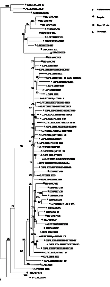
Foram colhidas amostras de plasma de doentes VIH-1 tratados e não tratados, entre 1993 e 2007, de Angola, Moçambique e Cabo Verde (Capítulos 2-5). Para subtipagem e/ou análise de mutações de resistência, foram obtidas sequências dos genes gag (p17), pol (PR and RT) e/ou env (C2C3) utilizando para amplificação um protocolo de PCR desenvolvido no nosso laboratório. Os genótipos virais foram determinados por análise filogenética. As mutações de resistência foram determinadas recorrendo ao Stanford Genotypic Resistance Interpretation Algorithm. As mutações associadas com resistência transmitida foram seleccionadas de listas publicadas recentemente. Para definir se uma mutação era um polimorfismo natural de um determinado subtipo, as frequências de todos os polimorfismos encontrados nas sequências de doentes tratados e não tratados, foram comparadas com sequências de todo o mundo do mesmo subtipo e/ou de subtipo B. Nos doentes não tratados infectados com vírus resistentes, o perfil mutacional foi analisado para se poder indicar o regime antirretroviral de 1ª linha mais efectivo. Nos doentes tratados, sempre que possível correlacionou-se a precorrelacionou-sença de mutações de resistência com parâmetros imunológicos e virológicos e com o regime antirretroviral em uso. Usaram-se métodos filogenéticos para determinar a origem dos vírus resistentes e para determinar se os doentes eram epidemiologicamente relacionados. Na investigação de um caso de infecção VIH-1 seronegativa fez-se uma caracterização inicial de vírus recombinantes CRF14_BG com base em sequências clonais parciais dos genes gag e env (Capítulo 6). Para a caracterização completa destes CRF14_BGs Portugueses, obtiveram-se sequências genómicas completas a partir de amostras de três doentes, um jovem adulto com infecção seronegativa infectado por via heterossexual e duas crianças infectadas por via vertical, em 1997 (Capítulo 7). A reconstrução da história evolutiva desta CRF foi realizada através de molecular clock analysis.
Resumo
ix
Métodos genéticos e filogenéticos foram usados para determinar o tropismo de um número significativo de isolados CRF14_BG de doentes Portugueses e investigar a selecção positiva na região V3.
Em Angola encontraram-se praticamente todas as formas genéticas de VIH-1 do grupo M excepto o subtipo B (Capítulo 2). Quarenta e sete por cento dos doentes estava infectado com vírus recombinantes, dos quais 36% eram de 2ª geração. Encontraram-se 58 perfis de recombinação diferentes. Encontraram-se ainda numerosos isolados não tipáveis e dois novos grupos de sequências dentro da radiação do subtipo A que podem constituir novos sub-subtipos A. Os dados epidemiológicos disponíveis e a elevada divergência genética intra-subtipo dos isolados Angolanos são consistentes com uma epidemia antiga neste país. Pensamos que a guerra colonial com os Portugueses (1961-1974) terá sido a principal causa da disseminação de VIH-1 em Angola. A epidemia de VIH/SIDA em Portugal (Capítulo 7) e Cabo Verde (Capítulo 5) é causada por uma grande proporção de subtipos não-B e recombinantes, alguns fortemente relacionados com os isolados Angolanos. Deste modo, a grande migração de pessoas durante a guerra colonial com Angola pode também ter promovido a disseminação de subtipos não-B de VIH-1 para Portugal e Cabo Verde, possivelmente em meados dos anos 60, e daí para o resto do mundo. Tendo isto em conta, é provável que Angola tenha sido um dos epicentros da actual epidemia de subtipos não-B em todo o mundo.
Em Maputo, Moçambique, a maior parte (81%) dos doentes estava infectada com vírus de subtipo C de origem diversa (Capítulo 4), contudo, também se detectaram quase todos os outros subtipos. De relevância particular neste contexto, foi a identificação de um cluster de isolados CRF37_cpx que terão sido importados recentemente para Moçambique. Em geral, os nossos resultados indicam que a epidemia VIH-1 em Moçambique (Maputo) está a evoluir rapidamente em complexidade genética devido à recente introdução de todos os principais subtipos e formas recombinantes. No futuro será importante determinar de que modo esta alteração da diversidade viral terá impacto na epidemia de VIH/SIDA em Moçambique.
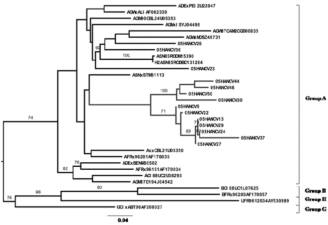
Em Cabo Verde caracterizou-se pela primeira vez as formas genéticas de VIH-1 e de VIH-2 em circulação (Capítulo 5). O subtipo G de VIH-1 foi o subtipo mais prevalente (48%). Encontraram-se ainda outros subtipos bem como variantes não tipáveis, sozinhos ou em formas recombinantes. Os isolados de subtipo G de Cabo Verde são extremamente divergentes em relação aos isolados de referência e na sua maioria parecem ter origem em Portugal e/ou Angola. Isto não é surpreendente visto Cabo Verde ter fortes relações históricas, sociais e económicas com estes dois países. Encontrou-se um recombinante C(pol)/G(env) ainda não descrito que importa agora sequenciar a nível genómico para determinar se poderá vir a definir uma nova CRF_CG. Todos os isolados VIH-2 pertenciam ao grupo A, um grupo que é também endémico em Portugal e nos países da África Ocidental relacionados com Cabo Verde. Encontraram-se dois isolados VIH-2 que partilhavam um ancestral comum com o histórico VIH-2ROD, o primeiro VIH-2 a ser sequenciado em 1987. Estes
x
resultados sugerem que a origem do VIH-2 ROD é Cabo Verde e que este país poderá ser ter sido um dos epicentros da actual epidemia de VIH-2.
Não se encontraram mutações de resistência major aos inibidores da protease (IPs) na PR dos isolados de VIH-1 presentes em Angola, Moçambique e Cabo Verde (Capítulos 3, 4, 5), o que é consistente com o facto dos regimes de primeira linha utilizados nestes países não inclurem esta classe de fármacos. No entanto, na PR dos isolados não-B foram identificados um grande número de polimorfismos naturais em posições que no subtipo B são consideradas mutações minor de resistência aos IPs (exs. L10I/V, V11I e T74P). Adicionalmente, foram encontrados novos polimorfismos ainda não descritos nas bases de dados de sequências de PR referentes a doentes não tratados. Estes resultados confirmam que os vírus que circulam nestes países são extremamente divergentes e sugerem que estes vírus poderão ter uma barreira genética de resistência diminuída para alguns IPs.
Na RT dos isolados VIH-1 de Angola, Moçambique e Cabo Verde também se encontraram numerosos polimorfismos que poderão ter impacto na susceptibilidade aos inibidores da RT (Capítulos 3, 4, 5). Adicionalmente, foram encontradas mutações de resistência aos INRTs (M41L, D67N, M184V, L210W, T215F/Y, K219Q) e INNRTs (K103N) nalguns doentes não tratados de Angola (2 doentes, 2%), Moçambique (4, 6%) e Cabo Verde (3, 12%). Nenhum destes isolados com mutações de resistência foi totalmente sensível aos regimes antirretrovirais de primeira linha usados nestes países. A baixa prevalência de resistência transmitida encontrada em Angola é consistente com a baixa disponibilidade de antirretrovirais no período de estudo (1993-2001). Pelo contrário, as taxas de resistência transmitida encontradas em Moçambique e Cabo Verde foram inesperadas dado que os fármacos antirretrovirais só começaram a estar disponíveis nestes países por volta de 2004. Os nossos resultados sugerem que a maior parte dos isolados resistentes terão sido importados de países onde a terapêutica está disponível há mais tempo. No entanto, o uso não monitorizado de antiretrovirais poderá também ser responsável por estas taxas relativamente elevadas de transmissão de vírus resistentes.
Em Portugal estudámos um caso raro de infecção seronegativa por VIH-1 com rápida progressão para SIDA e morte (Capítulo 6). O doente estava infectado com um vírus recombinante B(env)/G(gag) semelhante à CRF14_BG encontrada originalmente em Espanha em toxicodependentes. A análise filogenética indicou que o doente tinha sido infectado pela sua parceira sexual, uma toxicodependente, e que havia uma evolução genética muito restrita do vírus. A análise genética do tropismo indicou que o doente foi infectado selectivamente com uma variante CCR5. Estes dados demonstraram que na ausência de pressão selectiva por anticorpos os vírus CCR5 podem provocar SIDA e morte. No global, os resultados sugeriram uma infecção massiva com uma estirpe tipo CRF14_BG extremamente agressiva e/ou a presença de um problema imunológico não identificado no doente que impediu a formação de anticorpos específicos contra VIH-1.
Por sequenciação e análise filogenética do genoma completo de três doentes não relacionados, infectados com recombinantes B/G, fizemos a primeira caracterização molecular e evolutiva dos
Resumo
xi
vírus CRF14_BG em circulação em Portugal (Capítulo 7). Os nossos resultados sugerem que a CRF14_BG terá emergido em Portugal em 1992 (1989-1996) após o início da epidemia, tendo-se disseminado para Espanha no fim dos anos 90, provavelmente como consequência da migração de toxicodependentes, e depois para o resto da Europa. Verificámos que a grande maioria dos isolados CRF14_BG usa o co-receptor CXCR4 e estão associados à rápida depleção das células CD4 e progressão para SIDA. Finalmente, verificámos que só um aminoácido na V3 loop dos CRF14_BG está sobre pressão selectiva enquanto que no subtipo B há quatro aminoácidos sobre pressão selectiva. No conjunto, os resultados sugerem que os vírus CRF14_BG são particularmente patogénicos, que esta característica está em geral associada ao uso do co-receptor CXCR4, e que o uso preferencial deste co-receptor pode resultar de uma evolução convergente determinada pela fuga eficiente à resposta humoral neutralizante.
xiii
The aims of this thesis were to better characterize HIV-1 diversity in Portugal, Angola, Mozambique and Cape Verde and to investigate the origin and epidemiological history of HIV-1 in these countries. The impact of these issues in diagnosis, disease progression and susceptibility to ARV therapy was also investigated. Finally, the nature, dynamics and prevalence of transmitted drug resistance (TDR) was determined in untreated HIV-1 infected patients.
In Angola, practically all HIV-1 genetic forms were found, including almost all subtypes, untypable (U) strains, CRFs and URFs. Recombinants (first and second generation) were present in 47.1% of the patients. HIV/AIDS epidemic in Angola probably started in 1961, the major cause being the independence war, subsequently spreading to Portugal. In Maputo, 81% of the patients were infected with subtype C viruses. Subtype G, U and recombinants such as CRF37_cpx, were also present. The results suggest that HIV-1 epidemic in Mozambique is evolving rapidly in genetic complexity. In Cape Verde, where HIV-1 and HIV-2 co-circulate, subtype G is the prevailed subtype. Subtypes B, C, F1, U, CRF02_AG and other recombinant strains were also found. HIV-2 isolates belonged to group A, some being closely related to the original ROD isolate. In all three countries numerous new polymorphisms were identified in the RT and PR of HIV-1 viruses. Mutations conferring resistance to the NRTIs or NNRTIs were found in isolates from 2 (2%) patients from Angola, 4 (6%) from Mozambique and 3 (12%) from Cape Verde. None of the isolates containing TDR mutations would be fully sensitive to the standard first-line therapeutic regimens used in these
A
xiv
countries. Close surveillance in treated and untreated populations will be crucial to prevent further transmission of drug resistant strains and maximize the efficacy of ARV therapy.
In Portugal, investigation of a seronegative case infection with rapid progression to AIDS and death revealed that the patient was infected with a CRF14_BG-like R5-tropic strain selectively transmitted by his seropositive sexual partner. The results suggest a massive infection with a highly aggressive CRF14_BG like strain and/or the presence of an unidentified immunological problem that prevented the formation of HIV-1-specific antibodies. Near full-length genomic sequences obtained from three unrelated patients enabled the first molecular and phylogenomic characterization of CRF14_BG from Portugal; all sequences were strongly related with CRF14_BG Spanish isolates. The mean date of origin of CRF14_BG was estimated to be 1992. We propose that CRF14_BG emerged in Portugal in the early 1990s, spread to Spain in late 1990s as a consequence of IDUs migration and then to the rest of Europe. Most CRF14_BG strains were predicted to use CXCR4 and were associated with rapid CD4 depletion and disease progression. Finally, we provide evidence suggesting that the X4 tropism of CRF14_BG may have resulted from convergent evolution of the V3 loop possibly driven by an effective escape from neutralizing antibody response.
xv
3TC lamivudine ABC abacavir
AIDS acquired immunodeficiency syndrome APV amprenavir
ART antiretroviral ATV atazanavir AZT zidovudine
bDNA branched-chain DNA signal amplification CA coreceptor antagonist
cART combined antiretroviral therapy CCR5 C-C chemokine receptor type 5 CD4bs CD4 binding site
cpx complex
CRF circulant recombinant form CXCR4 C-X-C chemokine receptor type 4 DBS dried blood spots
ddI didanosine d4T stavudine DDC zalcitabine
DRC Democratic Republic of Congo DRM drug resistance mutation
A
xvi DRV darunavir dS synonymous substitution EFV efavirenz EI entry inhibitor ETR etravirine fAPV fosamprenavir
FDA Food and Drug Administration FI fusion inhibitor
FTC emtricitabine
HAART highly active antiretroviral therapy HIV-1 human immunodeficiency virus type 1 HIV-2 human immunodeficiency virus type 2 HLA human leukocyte antigen
HR1 heptad repeat 1 HR2 heptad repeat 2
HTLV human T cell lymphotrophic virus IDU intravenous drug user
IDR immunodominant region
IDV indinavir
II integrase inhibitor INT integrase
LPV lopinavir
M major or main HIV-1 group mAb monoclonal antibody MVC maraviroc
MSM men who have sex with men MTCT mother-to-child transmission N non-M non-OHIV-1 group
NAb neutralizing antibody
NAAT nucleic acid amplification testing
NASBA nucleic acid sequence-based amplification NFV nelfinavir
NNI nearest-neighbor interchange
NNRTI non-nucleoside reverse transcriptase inhibitor NRTI nucleos(t)ide reverse transcriptase inhibitor NVP nevirapine
O outlier HIV-1 group P P HIV-1 group
Abbreviations
xvii
PCR polymerase chain reaction
PENTA Pediatric European Network for Treatment of AIDS PI protease inhibitor
PR protease RAL raltegravir
qRT-PCR quantitative real time polymerase chain reaction RT reverse transcriptase
RT-PCR reverse transcriptase polymerase chain reaction (RT) PCR real time polymerase chain reaction
RTV ritonavir
sdNVP single dose nevirapine SQV saquinavir
SIV simian immunodeficiency virus
T20 enfurvitide TARc tenofovir TDF tenofovir
TDR transmitted drug resistance TPV tipranavir
U untypable or unclassified URF unique recombinant form USA United States of America WHO World Health Organization
xix Agradecimentos/Acknowledgements i Preface iii Resumo vii Abstract xiii Abbreviations xv
Table of Contents xix
Chapter 1: General Introduction 1
Acquired Immunodeficiency Syndrome 3
Genetic Diversity of HIV-1 3
Origin and Classification of HIV 4
Geographic Distribution 7
Antiretroviral Therapy and Resistance 13
Antiretroviral Drugs 13
Nucleos(t)ide Reverse Transcriptase Inhibitors (NRTIs) 13 Non Nucleoside Reverse Transcriptase Inhibitors (NNRTIs) 13
Protease Inhibitors (PIs) 14
Entry Inhibitors (EIs) 15
T
xx
Integrase Inhibitor (II) 15
Antiretroviral Regimens 15
Resistance to Antiretroviral Drugs 15
Transmitted Drug Resistance 16
Impact of HIV-1 Genetic Diversity 17
Transmission 17
Disease Progression 19
Diagnosis and Disease Management 20
Vaccination 26
Response to Antiretroviral Therapy 29 Resistance to Antiretroviral Therapy 31 Performance of Genotypic and Phenotypic Drug Resistance Assays 34 Aim of the Studies and Work Plan 39
Chapter 2: Highly divergent subtypes and new recombinant forms prevail in the HIV/AIDS epidemic in Angola: new insights into the origins of the AIDS pandemic
43
Abstract 45
Introduction 47
Materials and Methods 48
Patients 48
Viral RNA Extraction, PCR Amplification and Sequencing 48 Phylogenetic and Recombination Analysis 49
Statistical Analysis 50
GenBank Accession Numbers 51
Results 51
Discussion 59
Acknowledgments 61
Chapter 3: Antiretroviral drug resistance surveillance among treatment-naive HIV-1-infected individuals in Angola: evidence for low level of transmitted drug resistance
63
Abstract 65
Short-form paper 67
Supplemental Material 71
Chapter 4: HIV genetic diversity and transmitted drug resistance in health care settings in Maputo, Mozambique
79
Abstract 81
Table of Contents
xxi
Methods 84
Population 84
Viral RNA Extraction, Polymerase Chain Reaction Amplification, and Sequencing 84 Phylogenetic and Recombination Analysis 85 Origin of Subtype G and CRF37_cpx 85 Resistance Mutation Analysis 85
Statistical Analysis 85
GenBank Accession Numbers 86
Results 86
Study Population 86
HIV-1 Genetic Diversity 86
Drug Resistance Mutations and Other Polymorphisms 87
Discussion 94
Chapter 5: Genetic diversity and drug resistance profiles in HIV-1 and HIV-2 infected patients from Cape Verde Islands
99
Abstract 101
Introduction 103
Materials and Methods 104
Population 104
Viral RNA Extraction, PCR Amplification and Sequencing 104 Phylogenetic and Recombination Analysis 106 Resistance Mutation Analysis 106
Statistical Analysis 106
Results 106
Discussion 116
Acknowledgments 118
GenBank Accession Numbers 119
Chapter 6: Rapid clinical progression to AIDS and death in a persistently seronegative HIV-1 infected heterosexual young man
121
Abstract 123
Research Letter 125
Acknowledgments 130
Chapter 7: Origin and epidemiologic history of HIV-1 CRF14_BG 131
Abstract 133
xxii
Results 136
Molecular Epidemiology of Partial and Near Full-Length HIV-1 Sequences
136 CRF14_BG Was Originated in Portugal in Early 1990s 136 Most CRF14_BG Isolates Use CXCR4 141 Positive Selection Might Explain the Evolution to CXCR4 Usage in
CRF14_BG Isolates 141
Discussion 141
Methods 144
Sample Collection and Sequencing 144 Subtyping of HIV-1 Sequences 144 Dating the Origin of CRF14_BG 145 Co-receptor Usage, Selective Pressure and Divergence Rates
Estimation 145
Statistical Analysis 146
GenBank Accession Numbers 146
Author Contributions 146
Chapter 8: General Discussion and Conclusions 147
Appendix 155
G
G
e
e
n
n
e
e
r
r
a
a
l
l
I
I
n
n
t
t
r
r
o
o
d
d
u
u
c
c
t
t
i
i
o
o
n
n
C
General Introduction
3
Acquired Immunodeficiency Syndrome
Acquired immunodeficiency syndrome (AIDS) is a complex disease of the human immune system caused by the human immunodeficiency virus type 1 (HIV-1) and type 2 (HIV-2) [1]. AIDS was first recognized as a disease in the United States in 1981 after the observation of a high incidence of rare opportunistic infections in homosexual men caused by a general immune deficiency [2]. HIV-1 was isolated from this population in 1983 [3, 4]; HIV-2, which has a slightly different genomic structure [5], was isolated from West African AIDS patients in 1985 [6]. HIV-1 is responsible for the global pandemic while HIV-2, that appears to be less pathogenic [9, 10], is mainly restricted to West Africa [7] and countries with historical links to that area, like Portugal [8].
The overall growth of the global AIDS epidemic appears to have stabilized [11]. The annual number of new HIV infections has been steadily declining since the late 1990s and there are fewer AIDS-related deaths due to the significant scale up of antiretroviral (ARV) therapy over the past few years [11]. By the end of 2009 an estimated 33.3 million adults and children were living with HIV/AIDS worldwide. More than 67% (22.5 millions) of these individuals resided in Sub-Saharan Africa where the average prevalence of HIV infection among adults was 5.0%. In 2009, there were an estimated 2.6 million people who became newly infected with HIV and 1.8 million AIDS-related deaths among adults and children [11].
The introduction of highly active antiretroviral therapy (HAART), in developed countries in 1996 and in developing countries in 2004, has caused a significant decrease in the mortality and morbidity rates associated with HIV-1 infection and, the prognosis for patients who have access to these drugs has also improved significantly [12-16].
Genetic Diversity of HIV-1
HIV-1 is characterized by a very high rate of molecular evolution which allows the virus to diversify into numerous genetic forms, escape the human immune system and develop drug resistant variants [17]. HIV-1 can diversify by at least two ways: by mutation (substitutions, deletions, and insertions) occurring during viral DNA synthesis by the viral reverse transcriptase (RT) due to its lack of DNA proofreading activity; and by recombination, which is due to the RT strand-transfer activity and occurs in cells infected simultaneously with two or more virus strains [18-22].
The mutation rate of HIV-1 is 2.4x10⁻⁵ nucleotide substitution per nucleotide per cell infection [21]
and recombination occurs at an estimated rate of at least 2.8 crossovers per genome per cycle [22]. The effective recombination rate, i.e., the product of super-infection and crossovers, is 1.4x10⁻⁵
recombinations per site and generation [23]. In nature, the high genetic diversity of HIV is enhanced by a high level of virus production (108 to 1010 virions per day) [24-26], a large number of viral
replication cycles (107 to 108 cycles per day) [24, 25], and a large pool of infected individuals.
Within its host HIV exists as a pool of closely related genetic variants, known collectively as quasispecies [27].
4
Origin and Classification of HIV
HIV-1 and HIV-2 are distinguished on the basis of their genome organizations and phylogenetic (i.e., evolutionary) relationships with other primate lentiviruses. The two human viruses are related to different simian immunodeficiency viruses (SIVs) and therefore have different evolutionary origins [17]. HIV-1 is more related to SIV isolated from chimpanzees (SIVcpz) [28-31] and from gorillas (SIVgor) [32] and HIV-2 is more closely related to SIVsm, which is found with high prevalence in sooty mangabey monkeys [33].
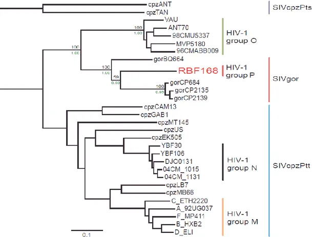
HIV entered the human population as a result of zoonotic, or cross-species, transmission [31, 34, 35]. Phylogenetic analysis shows that there have been many cross-species transmissions to humans; for instance in HIV-2 this number might be at least eight [34], whereas the independent origin of HIV-1 groups M (major or main), N (non-M, non-O) and O (outlier) could be explained by three jumps from chimpanzees to humans [36, 37]. Recently a new human immunodeficiency virus closely related to SIVs from gorillas, SIVgor, was identified [32]. This new HIV-1 variant is distinct from the three established groups of HIV-1, namely M, N, and O and was designated HIV-1 group P (Fig. 1.1).
Figure 1.1 - Evolutionary relationship between the HIV-1 different groups. The unique isolate of HIV-1 group P is shown in red letters. Adapted from Plantier et al., Nat Med 2009 [32].
General Introduction
5
HIV-1 groups N and P seem to be restricted to Cameroon and have few documented cases [32, 37-39]. Group O viruses have been identified in persons with epidemiological links to Central Africa, mainly Cameroon and some neighboring countries [40].
The earliest documented cases of HIV-1 group M infection, dating from 1959 and 1960, were identified in patients from the Democratic Republic of Congo (DRC) (former Zaire) [41, 42]. Phylogenetic analyses put the 1959 sequence closest to the ancestral node of subtype D and the 1960 sequence closest to subtype A [42]. These data showed that diversification of HIV-1 in West-Central Africa took place long before the recognition of the AIDS pandemic. Using different methods of molecular clock analysis it was estimated that group M was originated in the 1930s (1915-1941) [42-45]. For HIV-2, the date of the most recent common ancestor of group A strains was estimated to be 1940 (1924-1956), and that of B strains was estimated to be 1945 (1931-1959) [46].
Since its emergence, HIV-1 group M has diversified into nine subtypes, A, B, C, D, F, G, H, J, and K, seven sub-subtypes (A1-A5 and F1-F2), multiple circulating recombinant forms (CRFs) and countless unique recombinant forms (UFRs) [47-51]. Subtypes are genetically equidistant from one another, with the intersubtype nucleotide distances ranging from 11 to 17% in the gag gene and from 18 to 22% in the env gene. Intrasubtype distances range from 5 to 7% in the gag gene and 11 to 14% in the env gene. Intersub-subtype diversity ranges from 10 to 12% in the gag gene and from 16 to 17% in the env gene. CRFs are recombinant forms that have become epidemic [52-54]. Like subtypes, CRFs are defined by significant clustering in phylogenetic analysis and, additionally, by an identical recombinant structure. At present, 49 CRFs were recognized of which 37 are first generation recombinants (Table 1.1) and 12 are second generation recombinants (Table 1.2) [54]. Second generation CRFs result from recombination between more than one first generation CRF or from recombination between CRFs and different subtypes (Table 1.2) [53, 55-67]. CRFs are described by their specific structure and by the subtypes they contain; when more than three subtypes are present, the designation „„cpx‟‟ (complex) is used [53]. URFs are recombinant forms that were identified in a single individual and therefore have a unique subtype composition and recombinant structure. The URFs represent a large and heterogeneous group; they usually reflect the mixture of subtypes in the population where they are found, and some may be generated within individuals with a dual HIV-1 infection [68, 69].
For HIV-2 there are eight recognized groups (A-H). HIV-2 groups A and B have infected a substantial number of people in West Africa, while groups C to H have each been identified only in single individuals [70-73].
6
T
TAABBLLEE11..11
First Generation HIV-1 Circulant Recombinant Forms
Name Subtypes Reference
CRF01_AE A, E [74] CRF02_AG A, G [75] CRF03_AB A, B [76] CRF04_cpx A, G, H, K, U [77] CRF05_DF D, F [78] CRF06_cpx A, G, J, K [79] CRF07_BC B', C [80] CRF08_BC B', C [81] CRF09_cpx A, G, U [81, 82] CRF10_CD C, D [83] CRF12_BF B, F [84] CRF14_BG B, G [85] CRF16_A2D A2, D [86] CRF17_BF B, F [84] CRF18_cpx A1, F, G, H, K, U [87] CRF19_cpx A1, D, G [88] CRF20_BG B, G [89] CRF21_A2D A2, D [90] CRF23_BG B, G [89] CRF24_BG B, G [89] CRF25_cpx A, G, U [91] CRF26_AU A, U [51] CRF27_cpx A, E, G, H, J, K, U [92] CRF28_BF B, F [93, 94] CRF29_BF B, F [93, 94] CRF31_BC B, C [95] CRF35_AD A, D [96] CRF38_BF B, F1 [97] CRF39_BF B, F1 [98] CRF40_BF B, F [98] CRF41_CD C, D *
General Introduction
7
T
TAABBLLEE11..11 (continued)
Name Subtypes Reference
CRF44_BF B, F1 [99]
CRF45_cpx A, K, U [100]
CRF46_BF B, F1 [101]
CRF47_BF B, F1 [102]
CRF49_cpx A1, C, J, K, U [103]
* Not yet published.
T
TAABBLLEE11..22
Second Generation HIV-1 Circulant Recombinant Forms
Name Subtypes Reference
CRF11_cpx A, CRF01, G, J [59] CRF13_cpx A, CRF01, G, J, U [65] CRF15_01B CRF01, B [55] CRF22_01A1 CRF01, A1 [104] CRF30_0206 CRF02, CRF06 [58] CRF32_06A1 CRF06, A1 [56] CRF33_01B CRF01, B [62] CRF34_01B CRF01, B [63] CRF36_cpx A, G, CRF01, CRF02 [61] CRF37_cpx A, G, CRF01, CRF02, U [60] CRF43_02G CRF02, G [66] CRF48_01B CRF01, B [57]
Geographic Distribution
In Table 1.3 the main geographic distribution of HIV-1 subtypes and recombinants are listed, along with an estimate of relative global prevalences. Five HIV-1 strains dominate the global epidemic: subtypes A, B, and C, along with CRF01_AE and CRF02_AG, with subtype C accounting for almost 50% of all HIV-1 infections worldwide (Table 1.3) [52, 105-107]. Molecular epidemiological studies show that, with the exception of sub-Saharan Africa where almost all subtypes, CRFs, and several URFs have been detected, there is a specific geographic distribution pattern for HIV-1 subtypes
8
(Table 1.3) [52, 105-107]. Subtype A is prevalent in Central and Eastern Africa (Kenya, Uganda, Tanzania, and Rwanda) [108-110], Iran [111], Eastern Europe [112, 113], and Central Asia [114, 115]. In all these cases, sub-subtype A1 is more prevalent, whereas sub-subtypes A2-A5 are primarily found in Africa and rarely in Europe.
Subtype B is the most disseminated variant. It predominates in developed countries, such as United States of America (USA) and Canada [116-119], in Brazil [95, 120, 121], countries of Western and Central Europe [122-130] and Australia [131, 132], and is also common in several countries of Southeast Asia [133, 134], northern Africa [135] and the Middle East [136], and among South African and Russian homosexual men [52, 105-107, 137]. The global spread of subtype B is considered a major event in the history of HIV/AIDS because it marks the point when the virus first entered the large, wealthy and highly mobile populations of the Western world. Gilbert and collaborators estimated that the HIV-1 subtype B entered in Haiti from Africa around 1966 (1962-1970) [138]. This timescale corresponds well with a period when many Haitians returned to their home country from the DRC, after the latter‟s independence from Belgium and subsequent political crises. The migration of HIV-1 subtype B from Haiti to the United States and beyond is dated to the 1966–1972 period (1969) [138], suggesting that HIV-1 was circulating cryptically in the USA for approximately 12 years before the initial recognition of AIDS in 1981 [2]. The introduction of HIV-1 subtype B into Europe occurred mainly through homosexual contacts or needle sharing in or from the USA [139-141], or through heterosexual contacts with individuals from Central Africa [142-144].
Subtype C is the overwhelming prevailing strain in Southern Africa [145-148], in India and neighbor countries [149, 150] and in the southern region of Brazil [121]. Subtype D strains are found mainly in East Africa, and to a lesser extent in West Africa [78, 108, 110, 151]. Subtype F predominates in Central Africa [152, 153], South America [154, 155] and Eastern Europe [156, 157]. Subtype G viruses are prevalent in Central and Western Africa [158, 159], as well as in Portugal [122, 160-162] and Spain [124, 163]. Subtypes H and J were described in Central Africa [164-166] and in Angola [167, 168]. Subtype K was identified in DRC and Cameroon [169].
Some CRFs have high impact in local AIDS epidemics, such as CRF01_AE in Southeast Asia [170, 171] and CRF02_AG in Western and Central Africa [172-174]. CRF01_AE represents a putative subtype A/E recombinant that is spreading epidemically in Asia, but originated from Central Africa [74, 170]. No “pure” full-length genome has been found for subtype E. CRF02_AG [175] is a subtype A/G recombinant form that is highly prevalent in West and Central Africa [75], but has also been reported in Taiwan [176]. CRF03_AB represents a subtype A/B recombinant that was first found in Kaliningrad, and is circulating in Russian and Ukrainian cities, primarily in injecting drug users (IDUs) [76, 177]. Other CRFs linked to this transmission group (IDUs) are: CRF07_BC and CRF08_BC in China [80, 81, 178-180], CRF14_BG in Portugal and Spain [85, 161, 181], CRF32_06A1 in Estonia [56], CRF33_01B [62] and CRF4801_B in Malaysia [57], CRF15_01B [55] and CRF34_01B in Thailand [63] and CRF35_AD in Afghanistan [96].
General Introduction
9
The first B/G recombinant form – CRF14_BG - was described in Spanish and Portuguese IDUs [85, 161, 181]. This CRF was first isolated in 2002 from IDUs in Galiza, Spain [85]. So far, only 10 CRF14_BG isolates have been characterized by full-genome sequencing. These were obtained from Spanish (5/10, 50%), and German (1, 10%) IDUs, and from Portuguese patients, one IDU, one patient infected by heterosexual transmission and two children infected by perinatal transmission [85, 182, 183].
Until 2007, several sub-genomic sequences related to CRF14_BG were reported in Germany (1), Italy (2), United Kingdom (2), Estonia (15), Spain (38) and Portugal (50) suggesting that this CRF spread efficiently throughout Europe [56, 161, 162, 181, 182, 184-195]. The other B/G recombinant forms identified so far, CFR20_BG, CRF23_BG and CRF24_BG, were described in Cuban patients, [89, 196]. Two complex recombinants with an African origin, CRF18_cpx [87] and CRF19_cpx [88], were also first described in Cuba. The pronounced genetic complexity of HIV-1 in Cuba is not unexpected, considering that large numbers of Cuban military and civilian personnel had been stationed in the 1960s and 1970s in several African countries [197], and when they returned home probably brought with them different genetic forms of HIV-1 present in those countries [167, 168, 198].
In South America (Argentina, Cuba, Bolivia, Brazil, Chile, and Uruguay) the epidemic is a mixture of subtype B and B/F recombinants [84, 93, 97-99, 101, 199, 200] with a small proportion of infections by subtype C [121]. CRF42_BF described in Luxembourg and CRF47_BF described in Spanish patients, were the first B/F1 CRFs to be identified outside South America [84, 93, 97-99, 101, 102, 200, 201]. The 21 clinical isolates of CRF42_BF form a cluster clearly distinct from previously described South American CRF_BF strains [201]. The subtype F segments from CRF47_BF are distinct from South American subtype F1 sequences [102].
CRF11_cpx was the first second generation CRF described in 2000 in patients from Cameroon [202]. This CRF circulates in Cameroon, Central African Republic, Gabon, DRC, and Angola although its exact prevalence rate remains to be determined [67, 168, 203-207]. Second generation CRFs are becoming common in complex epidemics with multiple subtypes and recombinant forms. At present they have been detected in Africa, East Asia, Thailand, Malaysia, Estonia and Saudi Arabia (Table 1.3).
HIV-1 group O seems to be endemic in Cameroon and neighboring countries in West-Central Africa and represents only about 1-5% of HIV-1 positive samples in this region [208, 209]. Elsewhere in the world, group O viruses have been identified mainly from people with epidemiological links to the referred Central African countries [40]. HIV-1 groups N and P circulate exclusively in Cameroon [32, 38, 210, 211]. In some regions of the world, little information is available about HIV diversity, particularly in North Africa, the Middle East, and parts of Central Asia.
HIV-1 Genetic Forms Main Geographic Localizations Global Prevalence (%)
Reference
Subtypes A Central and Eastern Africa (Kenya, Uganda, Tanzania, and Rwanda), Iran, Eastern Europe and Central Asia 12.3 [108-115] B
America, Western and Central Europe, Australia, and is also common in several countries of Southeast Asia, Northern Africa
and Middle East 10.4
[52, 95, 105-107, 116-136, 212]
C Southern Africa, India and neighbor countries and Southern region of Brazil 49.9 [121, 145-150]
D East and West Africa 2.5 [78, 108, 110, 151]
F Central Africa, South America and Eastern Europe 0.6 [152-157]
G Central and Western Africa, Portugal and Spain 6.3 [122, 124, 158-163]
H Central Africa and Angola 0.17 [164, 165, 167, 168]
J Central Africa and Angola 0.14 [164-168]
K DRC and Cameroon 0.04 [169]
1st Generation CRF01_AE Southeast Asia 4.7 [74, 170, 171]
CRFs CRF02_AG West and Central Africa and Taiwan 6.7 [75, 172-174, 176]
CRF03_AB Former Soviet Republics 0.1 [76, 177]
CRF04_cpx Cyprus and Greece 0.003 [77, 213, 214]
CRF05_DF Central Africa 0.0001 [78]
CRF06_cpx West Africa 0.005 [215, 216]
CRF07_BC China 0.005 [80, 178, 179]
T
TAABBLLEE11..33 (continued)
HIV-1 Genetic Forms Main Geographic Localizations Global Prevalence
(%)
Reference
1st Generation CRF09_cpx West Africa and Angola 0.0003 [81, 82]
CRFs CRF10_CD East Africa (Tanzania, Madagascar) 0.0007 [83, 217, 218]
CRF12_BF Argentina, and Uruguay 0.001 [84, 97]
CRF14_BG Portugal and Spain 0.0006 [122, 161, 181]
CRF16_A2D Kenya, South Korea, and Argentina 0.00005 [86, 219]
CRF17_BF Argentina, Uruguay, and Bolivia 0.00002 [84]
CRF18_ cpx Cuba 0.0003 [87, 220] CRF19_ cpx Cuba 0.0003 [88, 220] CRF20_BG Cuba 0.00007 [89, 196] CRF21_A2D Kenya <0.00001 [86, 90] CRF23_BG Cuba 0.00005 [89, 196] CRF24_BG Cuba 0.00009 [89, 196]
CRF25_cpx Cameroon, DRC, and Saudi Arabia 0.00003 [66, 91, 207]
CRF26_AU DRC 0.00004 [51, 207]
CRF27_cpx DRC 0.00003 [92]
CRF28_BF Brazil <0.00001 [93, 94]
CRF29_BF Brazil 0.00001 [93, 94]
CRF31_BC Brazil 0.00006 [95]
CRF35_AD Afghanistan, and Iran 0.0001 [96, 221]
CRF38_BF Uruguay <0.00001 [97]
HIV-1 Genetic Forms Main Geographic Localizations Global Prevalence (%) Reference 1st Generation CRF40_BF Brazil 0.00002 [98] CRFs CRF41_CD * * * CRF42_BF Luxembourg 0.00006 [200, 201] CRF44_BF Chile 0.00004 [99]
CRF45_cpx Cameroon, Gabon, and DRC 0.00006 [100]
CRF46_BF Brazil 0.00003 [101]
CRF47_BF Spain 0.00003 [102]
CRF49_cpx Gambia <0.00001 [103]
2nd Generation CRF11_cpx Central Africa (Cameroon, Central African Republic, Gabon, and DRC) 0.003 [59, 203-207]
CRFs CRF13_cpx Central and Southern Africa (Cameroon, Angola and DRC) 0.0007 [65, 168, 207, 222]
CRF15_01B Thailand 0.00006 [55, 64] CRF22_01A1 Cameroon 0.0001 [67] CRF30_0206 Nigeria <0.00001 [58] CRF32_06A1 Estonia <0.00001 [56] CRF33_01B Malaysia 0.0001 [62, 134] CRF34_01B Thailand 0.00002 [63] CRF36_cpx Cameroon 0.00005 [61]
CRF37_cpx Cameroon, Mozambique and DRC 0.00008 [60, 148, 207]
CRF43_02G Saudi Arabia 0.00003 [66]
CRF48_01B Malaysia <0.00001 [57]
1The global prevalence of each form is expressed as a percentage of the total number of HIV-1 isolates identified worldwide. Isolates from HIV-1 groups O and N
General Introduction
13
Antiretroviral Therapy and Resistance
Since the advent of HAART in developed countries in 1996, and in developing countries in 2004 we have seen an improved prognosis of HIV-1 infected patients, with a significant decrease in morbidity and mortality rates associated with this infection [12-16]. The currently available treatment provides better quality of life to HIV/AIDS patients, minimizing viral replication, promoting partial recovery of the immune system and, thus, delaying or halting disease progression [223].
Antiretroviral Drugs
In March of 1987, zidovudine (AZT) was the first HIV-1 drug approved by the USA Food and Drug Administration (FDA) for AIDS treatment. Between 1991 and 2008, 25 additional drugs were approved [224]. These drugs fall into five categories, according to the viral protein target (Table 1.4). Currently, the standard treatment is a combination of drugs, named HAART. It consists of two nucleos(t)ide reverse transcriptase inhibitors (NRTIs) and a non-nucleoside reverse transcriptase inhibitor (NNRTI) or a protease inhibitor (PI) [225]. Due to high costs, more recent ARV drug classes, such as entry inhibitors (EIs) and integrase inhibitors (IIs), are primarily used for salvage therapy in cases of multiple resistances to the previous classes [225].
Nucleos(t)ide Reverse Transcriptase Inhibitors (NRTIs)
The target of NRTIs is the RT. There are eight NRTIs (Table 1.4). NRTIs are phosphorylated by cellular enzymes and converted into their active NRTI triphosphate form. Once activated, they compete with the natural nucleoside triphosphates for binding the RT polymerase active site, and after their incorporation into the primer strand, act as terminator of DNA synthesis due to the lack of a 3‟-hydroxyl group [226]. NtRTIs (tenofovir) should be clearly distinguished from the NRTIs as they are nucleotide analogues (not nucleoside analogues), which means that they only need two (not three) phosphorylation steps to be converted into their active form [224].
Non Nucleoside Reverse Transcriptase Inhibitors (NNRTIs)
There are at present four NNRTIs licensed for clinical use in the treatment of HIV infections (Table 1.4). NNRTIs are designed to bind to an RT hydrophobic pocket (binding site), located in the palm domain of the p66 subunit at a close distance from the active (catalytic) site of HIV-1 RT, modifying its structure allosterically and impairing the polymerase domain catalytic site, thereby exerting a non-competitive inhibition [227].
New strategies to inhibit RT enzymatic activities and to overcome viral resistance are under investigation: a new NNRTI, rilpivirine (TMC278) is currently in Phase III clinical trials [228].
14
T
TAABBLLEE11..44
Antiretroviral Drugs in Clinical Use
Drug Class Drug Release Year
Nucleos(t)ide reverse transcriptase inhibitors (NRTIs) Zidovudine (AZT) 1987 Didanosine (ddI) 1991 Zalcitabine (ddC) 1992 Stavudine (d4T) 1994 Lamivudine (3TC) 1995 Abacavir (ABC) 1998 Tenofovir (TDF) 2001 Emtricitabine (FTC) 2003 Non-nucleoside reverse transcriptase
inhibitors (NNRTIs)
Nevirapine (NVP) 1996
Delavirdine (DLV) 1997
Efavirenz (EFV) 1998
Etravirine (ETR) 2008
Protease inhibitors (PIs)* Saquinavir (SQV) 1995
Indinavir (IDV) 1996 Nelfinavir (NFV) 1997 Amprenavir (APV) 1999 Lopinavir (LPV) 2000 Atazanavir (ATV) 2003 Fosamprenavir (fAPV) 2003 Tipranavir (TPV) 2005 Darunavir (DRV) 2006
Entry inhibitors (EIs)
Fusion inhibitor (FI) Enfuvirtide (T-20) 2003 Coreceptor antagonist (CA) Maraviroc (MVC) 2007 Integrase inhibitor (II) Raltegravir (RAL) 2007 * All PIs are boosted with Ritonavir (RTV) with exception of ATV.
Protease Inhibitors (PIs)
There are ten PIs presently available for the treatment of HIV infections (Table 1.4). PIs are mimetics of viral peptides and bind to the active site of the protease (PR) enzyme, preventing viral maturation in a late step of the virus life cycle, [229].
General Introduction
15
Entry Inhibitors (EIs)
There are three crucial steps for entry of HIV into the CD4+ T cells: binding of HIV to the CD4 receptor, binding to coreceptors (CCR5 and/or CXCR4), and fusion of virus and cell. There are only two EIs currently available for the treatment of HIV infection, enfuvirtide (T20) – a fusion inhibitor, and maraviroc (MVC) – a CCR5 antagonist [230, 231] (Table 1.4). T20 is a polypeptide of 36 amino acids that is homologous to the heptad repeat 2 (HR2) region of the viral glycoprotein gp41. T20 interacts with HR1 region of gp41 and as a consequence of this interaction, fusion of the virus particle with the outer cell membrane is blocked [232]. MVC interacts with the coreceptor CCR5 used by R5-tropic HIV strains to enter the target cells. MVC inhibits the binding of gp120 to CCR5 by changing its shape such that gp120 can no longer recognize it [233].
Integrase Inhibitor (II)
Raltegravir (RAL) is the only integrase inhibitor available (Table 1.4); it binds to the viral integrase (INT) and prevents the integration of the viral double-stranded cDNA into the host cellular genome during the early steps of the virus cycle [234].
Antiretroviral Regimens
According to UNAIDS, in the end of 2009, there were 15 million of people that needed ARV therapy in low- and middle-income countries [11]: only 36% of those were receiving it. The ARV regimens recommended in these countries are the following [235]: first line regimens - one NNRTI + two NRTIs, one of which should be AZT or TDF; second line regimens - one PI RTV-boosted (ATV/RTV or LPV/RTV) + two NRTIs, one of which should be AZT or TDF.
By the end of 2008, only 2% of the adults in ARV treatment, in low- and middle-income countries, were receiving second-line regimens [236].
In developed countries the recommended ARV regimens should be [225]: first line regimens - two NRTIs (TDF+FTC or ABC+3TC) + one NNRTI (EFV); two NRTIs (TDF+FTC or ABC+3TC) + one PI (ATV/RTV); second line regimens - two NRTIs (TDF+FTC or ABC+3TC) + one PI (DRV/RTV). As an alternative, the third drug in an ARV regimen could be, LPV/RTV, fAPV/RTV or MVC.
Resistance to Antiretroviral Drugs
Management of HIV infection has greatly improved because of the development of new treatment protocols, involving the combination of highly potent drugs [12-16]. However, combination therapy is not effective in all patients and drug resistance is the inevitable consequence of incomplete suppression of HIV replication. Severe side effects, nonadherence to ARV regimens, pharmacokinetic interactions and lack of potency of drugs, are some factors that may contribute to the emergence of resistant viruses [237-240].
16
Generally, there are two main processes leading to resistance related to treatment failure: resistant viruses may preexist at low frequencies in drug naive patients and are rapidly selected in the presence of ARV drugs (primary or transmitted drug resistance), or, resistant viruses are absent at the start of therapy but are generated by residual viral replication during therapy (secondary or acquired drug resistance) [241].
Currently, drug resistance mutations (DRM) have been described for all drugs in clinical use (Appendix Figure 1) [242]. In the polymerase domain of RT, 15 positions were correlated with loss of drug susceptibility to NRTI, and 14 positions to NNRTI (Appendix Figure 1). In the PR region, 35 positions interfere with PI susceptibility (Appendix Figure 1). Seven positions in gp41 are associated with FIs resistance and four positions in the viral integrase are related to integrase inhibitor resistance (Appendix Figure 1) [242].
There are two types of DRMs, major or primary mutations and minor or secondary mutations, also called compensatory or accessory. Major mutations normally are close to the catalytic site of an enzyme and a single mutation can lead to loss of susceptibility to one or more ARV drugs. Sometimes, one mutation alone can lead to cross resistance to all ARVs within a given drug class [242]. The mutated enzyme is less fit than the wild-type [243, 244]. Minor DRMs could help the mutated enzyme to recover fitness (increase replicative capacity) [245, 246]. However, two or more minor DRMs can lead to loss of drug susceptibility [242]. Some mutations considered major to one ARV can be considered minor DRMs to other drugs [242].
Transmitted Drug Resistance
Transmitted drug resistance (TDR) is a major public health problem, especially in resource limited settings as it can determine rapid loss of effectivity of first line ARV regimens at the population level. Drug naive individuals that acquired a virus with DRMs begin ARV therapy with a lower genetic barrier to resistance, a higher risk of virologic failure and a higher risk of developing resistance even to those drugs in their regimen that were originally fully active [247, 248]. The recent initiation of ARV treatment programs in resourced limited countries [236] highlights the importance of studying this problem. A list of DRMs for surveillance of TDR is available since 2009 (Table 1.5) [249].
In Europe and North America, surveillance studies have reported prevalences of TDR ranging from 8% to 15% in newly diagnosed individuals [250, 251]. In Europe, contrary to USA, TDR prevalence seems to be stabilizing [250-253]. In most Sub-Saharan [254-259] and South and South-East Asia countries [260, 261] TDR is still residual (<5%). This low prevalence is consistent with the very recent introduction of ARVs in these regions [236].
Recently three pathways were proposed to describe the evolution of resistant viruses after transmission to a new host [262]. In the first pathway, the mutation reverts to wild-type when the replicative capacity of the virus is severely diminished; in the second pathway there is a replacement of mutations by atypical amino acids that also improve viral replication capacity; in the third pathway the mutations are fixated because they do not alter significantly viral replication
General Introduction
17
[262]. This indicates that viruses with these mutations can persist in patients and then be transmitted to new individuals. It is therefore important to continue monitoring the appearance of resistance mutations in treated patients and the rate of transmission in newly diagnosed untreated patients.
T
TAABBLLEE11..55
Mutations Associated with Transmitted HIV-1 Drug Resistance
NRTIS NNRTIs PIs
Wild type Mutation Wild type Mutation Wild type Mutation
M41 L L100 I L23 I
K65 R K101 E/P L24 I
D67 N/G/E K103 N/S D30 N
T69 D/ins V106 M/A V32 I
K70 R/E V179 F M46 I/L
L74 V/I Y181 C/I/V I47 V/A
V75 M/T/A/S Y188 L/H/C G48 V/M F77 L G190 A/S/E I50 V/L Y115 F P225 H F53 L/Y F116 Y M230 L I54 V/L/M/A/T/S Q151 M G73 S/T/C/A M184 V/I L76 V L210 W V82 A/T/F/S/C/M/L T215 Y/F/I/S/C/D/V/E N83 D
K219 Q/E/N/R I84 V/A/C
I85 V
N88 D/S
L90 M
Impact of HIV-1 Genetic Diversity
Transmission
Differential characteristics of viral subtypes and their interactions with the human host may influence HIV transmission and disease progression.
A recent study performed in HIV-discordant couples in Uganda found that subtype A had a significant higher rate of heterosexual transmission than subtype D (P=0.01) and recombinant viruses also had a higher rate of transmission than subtype D, however without statistical significance. This could indicate that subtype D may be less transmissible during heterosexual
18
contact than non-D viruses [263]. The rate of transmission may reflect differences in subtype-specific coreceptor tropism. HIV-1 can use as coreceptors CCR5 (R5 variants), CXCR4 (X4 variants) or both (R5X4 variants or dual tropic) to enter the cells [264]. R5 variants are largely prevalent during primary infection. Moreover, even when both R5 and X4 variants are present in the donor, most often only the R5 variants are detected in the recipient. Together, these data indicate that R5 strains are more easily transmitted or established in the newly infected host than X4 strains [264]. An increased prevalence of X4 variants has been reported for subtype D [265-267]. Thus, the higher frequency of X4 tropism in subtype D viruses may in part explain their reduced transmissibility when compared to other genetic forms [263].
Although earlier studies found an association between CRF01_AE and heterosexual transmission and between subtype B and intravenous drug use [170, 268], a more recent longitudinal study performed in Thailand found an increased probability of CRF01_AE transmission among IDUs compared with subtype B [269]. Subtype A is linked to heterosexual contact in Africa [108-110] but in European Eastern countries circulates among IDUs [112, 113].
Several studies have looked for variations between clades in mother-to-child HIV-1 transmission (MTCT) rates. In Kenya, MTCT appeared to be more common among mothers infected with subtype D compared with subtype A (P=0.002) and this association was independent of other risk factors for MTCT, such as maternal HIV viral load, episiotomy or perineal tear, and low birth weight [270]. On the other hand in Tanzania, HIV-1 subtypes A (odds ratio, 3.8; 95% CI, 0.8-24.7%) and C (odds ratio, 5.1; 95% CI, 1.3-30.8%) were more frequently transmitted from mother-to-child than subtype D [271]. In another study, the risk of MTCT was higher in women infected with subtype C, followed by subtype A, and was lowest in women infected with subtype D. Pregnant women, infected with subtype C were more likely than those infected with subtype A or D (P=0.006) to shed HIV-1– infected vaginal cells, implying that transmission to infants or partners may be more likely with this subtype: this relationship held after adjusting for age, CD4 cell count, and plasma HIV-1 viral load [272]. Another study in Tanzania presented similar results, with preferential in-utero transmission of HIV-1 subtype C compared to HIV-1 subtype A or D (P=0.026) [273]. Other researchers, however, found no association between subtype and rates of MTCT [274-276]. In studies where pregnant women received a single-dose nevirapine (sdNVP) prophylaxis no significant differences were also observed in the rate of MTCT in women with HIV-1 subtype A, D or C [277, 278].
Many factors, such as maternal stage of the disease, maternal immunological status, viral load, mode of delivery, duration of breast-feeding, ARV prophylaxis, maternal plasma vitamin A (associated with AIDS progression), and close maternal-child Human Leukocyte Antigen (HLA) matching, can contribute to these differences [279-281]. Nevertheless, the role of virus determinants in MTCT has not been well established yet [282]. Several studies have shown that viral diversity in the mother is generally higher than that present in the infant, suggesting that maternal viruses are selected before transmission [283, 284]. Several factors like specific viral selection [285], neutralization resistance [286-288] and enhanced replicative capacity of the transmitted