THE THYMUS, THE CELLS AND THE ENVIRONMENT
Dissertação de doutoramento Ph.D degree Thesis in Biomedical Sciences em Ciências Biomédicas apresentada ao Institute of Biomedical Sciences Abel Salazar. Instituto de Ciências Biomédicas de Abel Salazar Oporto University, Portugal
Universidade do Porto
PRESENTED BY/APRESENTADA POR
Afonso Rocha Martins de AlmeidaPresented on the 15th November 2002/Apresentada no dia 15 de Novembro de 2002.
Jury composition/Composição do Júri:
Professor Maria de Sousa (co-supervisor/co-orientadora) Professor António Freitas (supervisor/orientador)
Professor Anneliese Schimpl Doctor Margarida Correia-Neves Doctor Alexandre Carmo
THE THYMUS, THE CELLS AND THE ENVIRONMENT
Dissertação de doutoramento Ph.D degree Thesis in Biomedical Sciences em Ciências Biomédicas apresentada ao Institute of Biomedical Sciences Abel Salazar. Instituto de Ciências Biomédicas de Abel Salazar Oporto University, Portugal
Universidade do Porto
PRESENTED BY/APRESENTADA POR
Afonso Rocha Martins de Almeida
Presented on the 15 November 2002/Apresentada no dia 15 de Novembro de 2002.
Jury composition/Composição do Júri:
Professor Maria de Sousa (co-supervisor/co-orientadora) Professor António Freitas (supervisor/orientador)
Professor Anneliese Schimpl Doctor Margarida Correia-Neves Doctor Alexandre Carmo
TABLE OF CONTENTS
TABLE O F C O N T E N T S 2 S U M M A R Y - R E S U M O - R É S U M É 4 SUMMARY 5 RESUMO 7 RÉSUMÉ 9 ABBREVIATIONS 11 I N T R O D U C T I O N 12 1-RELEVANCE O F L Y M P H O C Y T E HOMEOSTASIS 131.1- Homeostasis in the Immune System 13 1.2- B and Tcell pools represent lymphocyte pools with independent homeostatic regulation. 14
2 - G E N E R A T I O N OF T CELLS 15
2.1- The Thymus: Histology 75 2.2- Colonization of the Thymus: Bone Marrow Precursors 16
2.3- Lineage commitment in lymphopoiesis 17
3 - D E V E L O P M E N T O F T CELLS 17
3.1- The Double Negative Thymic compartment. 18 3.2- The Double Positive Thymic compartment 20
3.2.1-Positive and Negative Selection 20 3.3- The Single Positive Thymic Compartment 21
3.4- Kinetics of T cell Development 21 3.4.1 - The DN compartment 22 3.4.2-The DP compartment 23 3.4.3-The SP compartment 24 4-HOMEOSTASIS WITHIN THE T H Y M U S 25
5 - T H E T H Y M U S AND A G I N G 25 6 - T H Y M I C EXPORT AND MIGRATION 27
6.1- Quantitative aspects of thymic output 28 6.2- Qualitative aspects of thymic output 30
6.3- Migration 31
7- T H E ORGANIZATION O F T H E MATURE T CELL POOLS 32
7.1 - T cells in the Spleen and Lymph Nodes 32
7.1.1 -T cells in the Spleen 34 7.1.2-T cells in the Lymph Nodes 34 7.1.3- Lymphocyte traffic 35 7.2- Peripheral Sub-population Structure 35
7.2.1- Naive and activated T cell pools have independent homeostatic regulation 37
7.2.2- The naïve T cell pool 38 7.2.3-The Effector pool 39 7.2.4- The memory T cell pool 41 7.3- Peripheral Repertoire 44
8- HOMEOSTASIS OF THE PERIPHERAL T CELL POOL 46
8.1- Lymphocyte life spans.^.?.. 46 8.2- Survival requirements of naive and memory T lymphocytes 48
8.3- Competition and Homeostasis 51 8.4- Homeostatic proliferation 54 8.5- Cellular interactions- Suppressor and Regulatory T cells 57
8.5.1- CD4+CD45RB'°\ CD4+CD45RBhigh T cells and the Colitis model 58
8.5.2-CD4+CD25+Regulatory T cells 60
8.5.3- Other regulatory T cells 66
8.5.4 - Homeostasis and CD4*CD25+ regulatory T cells 66
8.6- Resources: The role of Cytokines 67
8.6.1-The IL2 Receptor 68
8.6.3-IL7 71 8.6.4-IL15 72
9-THIS THESIS 73
RESULTS 74
ARTICLE #1 75
" T Cell Homeostasis: Thymus regeneration and Peripheral T Cell Restoration in Mice with a Reduced
Fraction of Competent Precursors " 75
ARTICLE #2 85
"Homeostasis of peripheral CD4* T cells: IL-2Ra and 1L-2 shape a population of regulatory T cells that
controls CD4+ T cell numbers " 85
ADDITIONAL RESULTS 97
"Searching for the Mechanisms responsible for in vivo CD4+CD25* regulatory T cell mediated
suppression " 97
DISCUSSION 117
10- HOMEOSTASIS WITHIN THE THYMUS 119
10.1- Thymic colonization 119 10.2- Homeostasis during thymic development 120
10.3- Homeostasis in the aging thymus 122
10.4- Conclusion 123
11 - THE ROLE OF THE THYMUS AND THYMIC EXPORT IN PERIPHERAL T CELL HOMEOSTASIS 123
11.1- Quantitative aspects 123 11.2- Qualitative aspects. 125 11.3- Conclusion 127
12- HOMEOSTASIS THROUGH SUB-POPULATION STRUCTURE: THE ROLE OF C D 4+C D 2 5+T CELLS 127
12.1- CD4*CD25* regulatory T cells are a specific lineage of. 128
CD4+T cells 128
12.2- CD4+CD25+ regulatory T cells may maintain peripheral T cell numbers at the established levels. 130
12.3- Mechanism of action ofCD4+CD25+ regulatory T cells 131
12.4- Concluding remarks 132
13- THE ROLE OF CYTOKINES IN THE ESTABLISHMENT OF THE PERIPHERAL CD4+ T CELL SUB -POPULATION
STRUCTURE; IL2 AND CD4+CD25+REGULATORY T CELLS 133
13.1-Cytokines are resources and receptor expression defines exploitable resources and T cell niche... 134
13.2- IL2 and CD4+CD25+ regulatory T cells 134
13.3- Concluding remarks 135
14-GENERAL CONCLUSIONS AND DISCUSSION 136 15-IMPLICATIONS FOR THE HUMAN CASE 138
15.1-The Thymus and peripheral T cell reconstitution 138
15.2- CD4*CD25+ regulatory T cells in Homeostasis, Autoimmunity and tumour immunotherapy 140
PERSPECTIVES 141 16-THE THYMUS 142 17- CD4+CD25+ REGULATORY T CELLS 143 REFERENCES 145 FIGURE INDEX 167 ACKNOWLEDGEMENTS 168 AGRADECIMENTOS 168
SUMMARY
The peripheral T cell number is under homeostatic control. Although T cells are produced daily in the thymus in significant numbers, and antigenic stimulation induces further T cell production through division, the peripheral T cell numbers are maintained constant. Thus, any newly produced T cell will only integrate the peripheral T cell pools if another T cell dies. In this way, the selection of the peripheral T cell repertoires is not only dependent on the interaction between each cell and its antigen but it is also dependent on interactions between different sub-populations of cells (Freitas and Rocha, 2000). In order to understand how peripheral T cell homeostasis is achieved, and to understand why is homeostasis attained at the observed equilibrium level, each of the contributing agents must be identified and the contribution of each determined.
T cells are originated in the thymus, resulting from a complex series of events. The thymic T cell production will be responsible throughout the lifetime of the organism for the daily export of newly generated T cells. However, the thymus involutes with age. This may have consequences for peripheral T cell homeostasis. Thus, the central T cell compartment should not be left out when peripheral T cell homeostasis is studied.
The peripheral T cell compartment comprises a number of smaller compartments, as not all T cells are born equal and many differentiate in the peripheral T cell pools into specific sub-populations with distinct proprieties and functions. The CD4+ and the CD8+ T cell subsets and the naive, effector
and memory compartments within the CD4+ and the CD8+ T cell compartments contribute differently
for the individual's immunocompetence. Mechanisms involved in the control of the relative proportions of these sub-populations are also relevant for total T cell homeostasis.
The purpose of this thesis was to make advances in the understanding of the mechanisms responsible for peripheral T cell homeostasis in general and for peripheral CD4+ T cell homeostasis in
particular. The studies were divided into three parts.
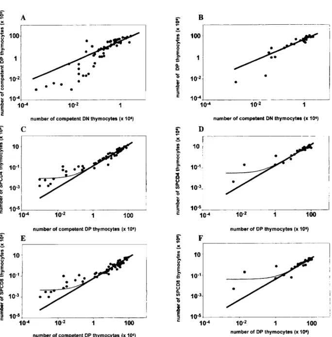
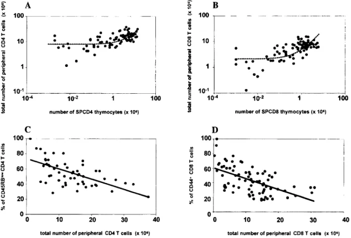
In the first part, the Thymus and its role in the maintenance of T cell numbers were evaluated. We developed a novel experimental system that allowed us to obtain a quantitative assessment of the fraction of competent pre-T cell precursors required to restore thymus function and also to evaluate the contribution of the thymus to the peripheral T cell pools. With the help of a mathematical model we were able to interpret the data obtained in order to demonstrate that there are no compensatory homeostatic mechanisms during thymic development and that the size of the peripheral total T cell pool is fairly independent of thymic output. Thus, peripheral mechanisms compensate for a lack of thymic output. When the naïve and activated/memory T cell compartments were analysed separately, we found that the naïve T cell compartment was more prone to reflect the size of the thymic SP compartment. Thus, we concluded that these compensatory mechanisms are more efficient in the generation of activated/memory T cells.
In the second part, the subject of research was the importance of peripheral T cell interactions for the establishment of peripheral T cell homeostasis. We have studied the interactions between the CD4+CD25+CD45RB|0W T cells (the regulatory/suppressor CD4+ T cells) and CD4+CD25"CD45RBhigh T
observed that the ratio CD4+CD25+CD45RB|0W to CD4+CD25"CD45RBhi9h T cells present in the cells
transferred was determinant for the numbers of cells recovered, and thus this interaction potentially determinant for peripheral T cell homeostasis. We have demonstrated this, re-introducing the CD4+CD25+ T cells in a mouse system (the CD257" BM chimeras) were the peripheral homeostasis is
disturbed and this sub-population is absent. As we observed, the re-introduction of the CD4+CD25+ T
cells in these BM chimeras had as a consequence the normalisation of the peripheral T cell pools. We have found proof that the presence of this sub-population is essential for the existence of homeostasis of the peripheral T cell numbers and thus, that peripheral T cell homeostasis is achieved also through sub-population structure.
In the third part of these studies, the importance of resources for the maintenance of the peripheral T cell sub-population structure was examined. Immediate candidates as resources are interieukins. The IL2/_ mice have reduced numbers of CD4+CD25+ T cells and develop autoimmune
manifestations We postulated that the lack of IL2 was responsible for the decreased survival of the CD4+CD25+ regulatory T cells in the peripheral T cell pools, and thus that the autoimmune
manifestations were again the consequence of a disruption in the peripheral sub-population structure, as these mice are devoid of this specific sub-population. We tested this hypothesis, by sorting the few CD4+CD25+ T cells present in the IL27 mice and testing these cells as suppressors in vivo. These
cells proved to exert suppressor functions, suggesting that the IL2"' mice are able to generate the CD4+CD25+ regulatory T cells. We confirmed this, by establishing BM chimeras using as donor BM
cells a mixture of BM cells from IL2"' and CD257" cells. These chimeras do not develop autoimmune
manifestations and the peripheral T cell pools have the normal representation of the CD4+ T cell
sub-populations, including the CD4+CD25+ T cells. Thus, the IL27 BM precursors were able to generate a
viable population of regulatory T cells, as long as IL2 was present in the periphery. This illustrates the role of cytokines as resources with a major importance for the establishment of the observed peripheral sub-population structure.
Returning to the main subject of this thesis, our results allow us to state that the observed peripheral T cell homeostasis reflects not only the thymic production but also peripheral phenomena, and that these include interactions between different sub-popoulations. Underrepresented peripheral sub-populations, like the CD4+CD25+ regulatory T cells, play a major role in the maintenance of
RESUMO
Os números de células T periféricas são mantidos sob controlo homeostático. Apesar de as células T serem produzidas diariamente em números consideráveis no timo, e de novas células T serem geradas à periferia devido a estimulação antigénica, os números de células T periféricas são mantidos constantes. Assim sendo, uma nova célula poderá apenas integrar o compartimento periférico de células T substituindo uma outra. Por conseguinte, a seleção dos repertórios periféricos de células T não está apenas dependente da interação de cada célula com o antigénio, estando também dependente de interações com outras células (Freitas and Rocha, 2000). Para compreender a homeostasia das células T é necessário identificar os agentes intervenientes no processo e quantificar a contribuição de cada um deles.
As células T sao originadas no timo. A produção túnica de células T será exportada para a periferia, correspondendo a um potencial acrescento diário de novas células T. Por esta razão, é importante ter em conta a contribuição do timo para a homeostasia das células T periféricas.
O compartimento de células T periféricas inclui numerosos outros compartimentos, uma vez que diferentes categorias de células T são geradas no timo e, à periferia, é possível a diferenciação das células T em diversas outras sub-populações com características próprias. Assim, é importante diferenciar os compartimentos de células T CD4+ e CD8+ e, dentro destes, compartimentos naive ou
activados/memória. Estas sub-populações contribuem diferentemente para a imunocompetência do indivíduo. Por esta razão, é também importante ter em conta os mecanismos involvidos no controlo das proporções relativas destas sub-populações.
O objectivo deste trabalho é contribuir para a compreensão dos mecanismos responsáveis pela homeostasia das populações de células T periféricas em geral, e das populações de células T CD4+ em particular. Os trabalhos foram divididos em três partes.
Numa primeira parte, o papel do timo na manutenção dos números de células T foi investigado. Desenvolvemos um novo sistema experimental que nos permitiu não apenas a avaliação da fração de células pre-T competentes necessária para assegurar a função tímica mas também a avaliação da contribuição da produção tímica de células T para a manutenção dos diferentes compartimentos de células T periféricas. Fomos capazes de demonstrar que não existem mecanismos de compensação homeostática durante o desenvolvimento tímico e que os números totais de células T periféricas não reflectem o número de células T exportadas pelo timo, o que implica a existência à periferia de mecanismos capazes de compensar a redução nos números de células exportadas pelo timo. Obtivemos também dados que nos permitiram concluir que os mecanismos compensatórios referidos são mais eficientes na geração de células T activadas/memória.
A segunda parte deste trabalho está relacionada com a relevância das interações entre populações de células T periféricas para a homeostasia das células T periféricas. Investigámos as interações entre as sub-populações CD4+CD25+CD45RBl0W (células reguladoras ou supressoras) e as
células CD4+CD25'CD45RBh'9h (células naives), transferindo as duas populações isoladamente ou em
determinante para o número de células T CD4+ recuperadas nestes animais. Assim, esta interação
pode estar implicada no estabelecimento da homeostasia das células T periféricas. Fomos capazes de demonstrar a importada da presença desta sub-população reguladora para a manutenção do equilíbrio normal dos números de células T CD4+ periféricos, reintroduzindo a população CD4+CD25+
en animais quimeras de medula óssea (Rag2v" reconstituídos com células de medula óssea de
dadores CD257") onde esta população está ausente. Estes animais apresentam distúrbios
homeostáticos graves, que resultam no desenvolvimento de doenças autoimunes acompanhadas de importantes acumulações de linfócitos. A reintrodução da sub-população de células reguladoras é suficiente para impedir estas anomalias, restitutindo a normalidade às populações de linfócitos T CD4+ periféricas.
Na parte final da tese, a relevância dos recursos para a manutenção da estructura populacional das células T CD4+ periféricas foi investigada. A proporção de células CD4+CD25+ é
reduzida em animais IL27". Estes animais apresentam um fenótipo semelhante ao dos animais CD25"'"
. Formulámos a hipótese de a falta de IL2 à periferia nos animais IL2 ' ser a causa da redução na proporção de células CD4+CD25+ observada e de ser esta a causa das anomalias verificadas nos
animais IL2. Testámos esta hipótese, separando as células CD4+CD25+ existentes nos animais IL2'
e testando estas células enquanto células supressoras in viva Verificámos que estas células são capazes de exercer funções supressoras. Estabelecemos também quimeras de medula óssea utilizando animais hospedeiros Rag/" irradiados e reconstituindo estes com misturas de células de medula óssea provenientes de animais CD25"'" e IL2"/_. Estas quimeras não apresentam
manifestações de natureza autoimune e a observação da composição das populações de células T CD4+ periféricas revela a presença da proporção normal de células reguladoras CD4+CD25+, que
apenas podem ter origem nas células de medula óssea proveninetes de dadores IL2"". Demonstrámos assim que os percursores IL2_/ são capazes de gerar uma população normal de
células CD4+CD25+, na condição de a IL2 estar presente à periferia. Estes resultados demonstram o
papel dos recursos, neste caso da IL2, no estabelecimento da estructura populacional observada nos compartimentos de células T periféricos.
No seu conjunto, os resultados obtidos permitem-nos afirmar que a homeostasia das populações de células T observada não é apenas o resultado da exportação de células T por parte do timo, sendo também o resultado de fenómenos que ocorrem à periferia, nomeadamente, interações entre as diferentes sub-populações. Populações com fraca representação, tal como as células reguladoras CD4+CD25+, são determinantes para o estabelecimento do equilíbrio homeostático dos
RÉSUMÉ
Chez les vertébrés adultes, le nombre de cellules T périphériques est soumis à un contrôle strict. Bien qu'un grand nombre de cellules T soient produites chaque jour, le nombre de cellules T à la périphérie reste constant. Chaque nouveau lymphocyte T produit ne pourra donc s'établir à la périphérie qu'après la mort d'un lymphocyte déjà établi. Dans cette optique, la sélection du répertoire des cellules T périphériques n'est pas uniquement dépendant des interactions entre une cellule et son antigène, mais elle est également dépendante d'interactions entre différentes sous-populations cellulaires (Freitas and Rocha, 2000). Dans le but de comprendre comment l'homéostasie des cellules T périphériques est atteinte, et afin de comprendre pourquoi l'homéostasie parvient à un certain
niveau d'équilibre, il est important de déterminer chaque facteur intervenant dans ce processus et la contribution de chacun d'entre eux.
Les cellules T sont générées dans le thymus. La production thymique de cellules T est responsable, chaque jour, de l'export de nouvelles cellules T. Cependant la taille du thymus n'est pas constante, puisque le thymus involue avec l'âge, ce qui peut avoir des conséquences sur l'homéostasie des cellules T périphériques. Il faut donc tenir compte du compartiment T central, le thymus, lorsque l'on étudie l'homéostasie des compartiments périphériques.
Le compartiment T périphérique est composé d'un certain nombre de sous-compartiments, puisque chaque cellule T n'est pas identique, et de nombreuses cellules vont se différencier à la périphérie en des sous-populations spécifiques, possédant des fonctions et des propriétés différentes. Par exemple, les sous-populations CD4+ et CD8+ devront être considérées séparément puisqu'elles
sont impliquées dans différents types de réponses immunitaires et ont des mécanismes d'action différents. De la même façon, les compartiments naïfs, effecteurs et mémoires contribuent différemment à l'immuno-compétence de chaque individu. Les mécanismes impliqués dans le contrôle du nombre de chacune de ces sous-populations sont également importants pour la compréhension de l'homéostasie cellulaire T totale.
L'objectif de cette thèse a été de comprendre les mécanismes responsables de l'homéostasie périphérique T en général, et de l'homéostasie des cellules T CD4+ périphériques en particulier. Ce
travail a été divisé en trois parties.
Dans la première partie de cette thèse, nous avons évalué le rôle du thymus dans la maintenance du nombre de cellules T. Nous avons développé un nouveau système expérimental nous permettant d'obtenir une estimation quantitative de la fraction des cellules précurseures pré-T compétentes, nécessaire pour assurer la fonction thymique mais aussi d'évaluer la contribution thymique à l'établissement du compartiment T périphérique. Nous avons montré qu'il n'existe pas de mécanismes homéostatiques compensatoires au cours du développement thymique. Ce résultat nous a ensuite conduit à évaluer l'effet d'un export thymique réduit sur l'établissement du compartiment T périphérique. Nous avons montré que la taille du compartiment T périphérique est indépendante du thymus, suggérant que des mécanismes compensatoires se mettent en place à la périphérie. Lorsque nous avons étudié les compartiments naïfs et activées/mémoires séparément, nous avons observé
que les mécanismes compensatoires sont plus efficaces pour les sous-populations activées/mémoires.
Dans la seconde partie de cette thèse, nous avons étudié le rôle des interactions entre les cellules T périphériques dans l'établissement de l'homéostasie T périphérique. Nous avons analysé l'interaction entre les cellules T CD4+CD25+CD45RB|0W (également appelées cellules T CD4+
régulatrices) et les cellules CD4+CD25"CD45RBhigh, dans des expériences de transfert chez la souris.
Nous avons observé que le ratio entre les cellules CD4+CD25+CD45RB|0W et les cellules CD4+CD25"
CD45RBhigh transférées était déterminant pour le nombre de cellules recouvrées suggérant donc que
l'interaction entre ces deux populations pourrait être déterminante pour l'homéostasie périphérique T. Nous avons testé cette hypothèse en transférant des cellules T CD4+CD25+ dans un modèle murin
(les chimères de moelle osseuse CD25"'") où l'homéostasie périphérique est perturbée et où cette sous-population CD25+ est absente. Nous avons observé que la présence de ces cellules T
CD4+CD25+ dans ces chimères de moelle osseuse a pour conséquence la normalisation du
compartiment T périphérique. Nous avons montré donc que l'homéostasie des cellules T périphériques est atteinte aussi grâce à la structure des sous-populations qui la constitue.
Dans la troisième partie de cette thèse, nous avons étudié l'importance des ressources pour la maintenance de la structure des sous-populations T périphériques. Il a été montré que le nombre de cellules T CD4+CD25+ est réduit chez les souris invalidées pour le gène de l'IL-2. Il a aussi été montré
que ces souris développent des maladies auto-immunes avec des caractéristiques communes à celles développées par les souris CD257". Nous avons fait l'hypothèse que le manque d'IL-2 serait
responsable de la diminution de la survie des cellules T régulatrices CD4+CD25+ dans le
compartiment T périphérique, et que donc les manifestations auto-immunes seraient la conséquence de la perturbation de la structure des sous-populations périphériques, puisque ces animaux ne contiennent pas cette sous-population spécifique. Nous avons testé cette hypothèse en triant les quelques cellules T CD4+CD25+ présentes chez les animaux IL2"'" et en testant leur fonction de
cellules suppressives in vivo. Ces cellules ont montré leur capacité à exercer une fonction suppressive, suggérant que les souris IL-27" sont capable de produire des cellules régulatrices T
CD4+CD25+. Nous avons confirmé ces résultats en établissant des chimères de moelle osseuse, avec
des cellules provenant de la moelle osseuse d'animaux IL-2"'" et d'animaux CD25"'". Ces animaux chimériques ne développent pas de maladies auto-immunes et le compartiment T périphérique est constitué d'une proportion normale des différentes sous-populations CD4+, notamment les
CD4+CD25+. Les précurseurs issus de la moelle osseuse des animaux IL-2'" ont donc été capables de
générer une population viable de cellules T régulatrices, capable d'utiliser pour leur survie l'IL-2 produite de façon paracrine. Ces résultats illustrent bien le rôle des cytokines comme ressources majeures, notamment pour l'établissement de la structure des populations périphériques.
L'ensemble des résultats obtenus au cours de cette thèse nous a conduit à formuler que l'homéostasie des cellules T périphériques est le résultat, non seulement de l'impact thymique, mais aussi de mécanismes périphériques. Les populations sous représentées, comme la population de cellules T régulatrices CD4+CD25+, pourraient exercer un rôle important dans la maintenance de
ABBREVIATIONS
AICD- Activation Induced Cell Death
Ag- Antigen
APC- Antigen Presenting Cell
BM- Bone Marrow
BrDU- Bromodioxyuridine
CFSE- 5,6-Carboxyfluorescein diacetate succinimidil ester
CLP- Common Lymphoid Precursor
DC- Dendritic Cells
DN- Double Negative
DP- Double Positive
IBD- Inflammatory Bowel Disease
FTOC- Fetal Thymus Organ Culture
HSA- Heat Stable Antigen
HU- Hidroxyurea
MHC- Major Histocompatibility Complex
NK- Natural Killer
RTE- Recent Thymus Emigrants
SP- Single Positive
TCR- T Cell Receptor
Tg - Transgenic
TN- Triple Negative
TREC- Thymus Recombination Excision Circles
WT- Wild Type
SECTION A
1- RELEVANCE OF LYMPHOCYTE HOMEOSTASIS
The ability of the Immune system to cope with infections and to do it safely is directly related to the individual's lymphocyte pool at any given time instant and throughout the individual's lifetime. As the total number of lymphocytes is kept constant, it follows that any new lymphocyte, in order to integrate the peripheral pool, must replace an existing one. As each lymphocyte bears a unique receptor, the specificities present in the peripheral pool of an individual will be the result of this selection. Thus, the immunocompetence of an individual is directly dependent on the mechanisms and rules responsible for the maintenance of lymphocyte numbers.
1.1- Homeostasis in the Immune System
" As in complex ecological systems, the immune system shows a « return tendency, due to density dependent processes, to reach a stationary distribution of population densities » (Hanski, 1999). This is usually referred to as lymphocyte homeostasis (Freitas and Rocha, 2000).
Lymphocytes are produced daily in significant numbers in the Thymus (T cells) and Bone Marrow (B cells), and export from the primary lymphoid organs results in a daily input to the peripheral lymphocyte pools (Scollay and Godfrey, 1995). More cells are generated in the periphery as a result of antigen encounter or non-Ag dependent processes (Doherty et al., 1997; Tough et al., 1996). As numbers are kept constant, we must assume that an equal number of cells leave the peripheral lymphocyte pool, due to migration into tissues or death (Freitas and Rocha, 2000). To predict which cells will be able to integrate the peripheral lymphocyte pools and to understand why these pools are kept constant at size n=x and not n=y we must identify the mechanisms responsible for the homeostatic regulation of peripheral lymphocyte numbers.
1.2- B and T cell pools represent lymphocyte pools with independent
homeostatic regulation
The first sub-division of the peripheral lymphocyte pool is a separation between the peripheral T and B cell pools. These two lymphocyte sub-populations are responsible for the specific immune responses and are thus essential for the efficacy of the immune system, providing an adaptive immune response. They differ in the ontogeny and also in the nature of their effector mechanisms. Thus, B cells are generated in the BM (Bursa in birds) while T cells owe their name to their thymic origin. The B cell effector mechanisms are dependent on the production of antibody, while T cell effector mechanisms are more diverse and include direct cell-cell interactions but also production and release of soluble factors (cytokines) (for an overview see Janeway et al., 1999). In addition, these T and B populations also have independent homeostatic regulations. This conclusion can be drawn from the simple observation of B cell numbers in animals devoid of T cells (CD3c'' (Malissen et al., 1995) or TCRa"A (Mombaerts et al., 1992a) mice). The fact that B cell numbers in these animals do
not significantly differ from those found in normal mice strongly suggests that the homeostatic regulation of the B cell pool is independent of the presence or absence of T cells. This conclusion is further supported by the fact that in the inverse situation, i.e. in animals devoid of B cells (uKO), the size of the T cell pool is not significantly affected either (Kitamura et al., 1991). This observation will allow us to split the problem of peripheral lymphocyte homeostasis in peripheral B cell homeostasis and peripheral T cell homeostasis. The latter will be the object of this thesis.
Most of the information we dispose of today concerning lymphocyte development or even peripheral selection and survival events comes from studies performed in the mouse model. The following sections concern the murine immune system, the direct object of this thesis, though pertinent human data may be referred to. In the final section of this thesis the implications for the human case will be briefly discussed.
PARTI
T CELL GENERATION AND EXPORT
2- GENERATION OF T CELLS
The role of the thymus as the T cell production site was firstly observed after thymectomy studies. The observation was that after thymectomy, specific areas of the peripheral lymphois organs were absent. Subsequent thymic graft studies shwon that cells originating in the grafts were indeed preferentially found in the thymus-dependent areas of the peripheral lympohid organs. The existence of thymus-dependent and thymus-independent areas of the peripheral lymphoid organs led to the designation of the thymus-dependent cells as T cells (see early work revision in Parrott and De Sousa, 1971). Thus, although some T cells with an extrathymic origin exist (e.g. originated in the gut (Saito et al., 1998) the large majority of T cells originate in the thymus, developing from a BM derived precursor. The thymic T cell development occurs in a sequence of steps that correspond to a functional and morphological transformation. The resulting cell has a functional specific receptor and can have different classes of effector mechanisms. These cells are exported to the periphery.
2.1- The Thymus: Histology
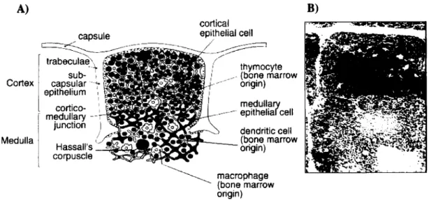
The Thymus consists of numerous lobes. We can distinguish in each an outer cortical region (thymic cortex) and an inner medulla (fig.1). A higher magnification shows the thymic stroma, consisting of epithelial cells and connective tissue, and the presence of scattered Macrophages and Dendritic Cells. The large majority of the other cells present are thymocytes, the developing T cells (fig.1). Thymocytes result from the expansion in the thymus of colonizing cells coming from the BM (see 2.2). During the course of their development thymocytes will distribute along the different areas of the thymus (fig.1). The more immature cells are found in the outer cortex, and the more mature ones in the medulla. A very brief description of the events involved follows in the next sections.
A) B)
Figure 1- The Thymus: Its structure and its cellular content. The figure shows the histology of the organ (B), and the distribution of the different cell types in the identifiable areas (A).
(From Janeway, C. et ai, 1999)
2.2- Colonization of the Thymus: Bone Marrow Precursors
The T cell precursors must migrate from the BM into a functional thymus in order to develop. This conclusion can be drawn from the nude mice analysis, a situation where a failure in the development of the thymus results in the almost complete absence of T cells. BM precursors from these mice are able to develop fully, when transferred into recipients with a normal thymus. Conversely, BM precursors from normal congenic mice are not able to generate T cells after transfer into nude mice (for an overview see Janeway et al., 1999). This sets up the basis for an interactive view of T cell development: the microenvironment has a crucial role on the delivery of the signals that will drive intrathymic T cell development (Anderson and Jenkinson, 2001 ; Savino et al. 2002).
An i.v. injection of 5 x 10⁵ BM cells into irradiated mice is clearly sufficient to
reconstitute the thymus of these mice, and the intrathymic injection of much larger numbers of donor thymocytes provides only transient reconstitution, confirming the requirement for BM derived precursors to achieve permanent thymic reconstitution (Scollay et al., 1986). While it is clear that there must be a thymic colonization by BM derived precursors, whether this occurs continuously (Scollay et al., 1986) or in waves (Foss et al., 2001) is still under debate, though the first view is clearly favoured.
2.3- Lineage commitment in lymphopoiesis
The nature of the BM precursor cell and the signals that drive its differentiation along the T cell pathway have been object of numerous studies. A common lymphoid precursor (CLP) has been identified, first as a cell with the ability to differentiate into the T and B cell lineages (Wu et al., 1991), then also into DCs (Ardavin et al., 1993) and NK cells (Kondo et al., 1997) but that it does not give origin to myeloid cells (reviewed in Akashi et al., 2000). This cell does not seem to have self-renewing ability, as reconstitution with CLPs provides only transient reconstitution (Akashi et al., 2000).
The branching in the development of the CLP into the different lymphoid lineages is dependent on the action of transcription factors (e.g. Pax-5 or GATA-3) (Nutt et al., 1999; Ting et al., 1996), transmembrane ligands or receptors (e.g. Notch and its ligands) (Pui et al.,
1999; Radtke et al., 1999) and on cytokine signalling (e.g. IL7 or IL15) (Di Santo et al., 2000; Peschon et al., 1994). From now, only T cell development will be considered in this introduction. (For a more detailed description of the factors and mechanisms involved in lineage commitment see revisions in Akashi et al., 2000; Busslinger et al., 2000; Deftos and Bevan, 2000; Di Santo et al., 2000; Kuo and Leiden, 1999).
3- DEVELOPMENT OF T CELLS
After colonizing the thymus T cell precursors, upon interaction with the thymic microenvironment, will undergo a number of developmental changes, that are mostly related with the expression of the TCR (ap -the large majority- or yò ) and the assembly of its signalling apparatus (CD3). The TCR is a clonally variable ap heterodimer while the CD3 complex is a group of invariant polypeptides (y, 6, t, t and r| ). The TCR genes (both a and |i chain genes) will go through a series of programmed rearrangements of germ line V D and J genes, a process referred to as V(D)J recombination (for an overview see Janeway et al., 1999). The enzyme responsible for these rearrangements is the RAG (1 and 2) and mice lacking this enzyme lackT (and B) cells (Mombaerts et al., 1992b; Shinkai etal., 1992).
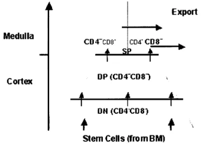
We can characterize the developmental stages by the expression of cell-surface markers. The expression of the coreceptors CD4 and CD8 correlates with the state of development of T cells: from immature (CD4CD8" Double Negative) to mature (CD4+CD8" or
CD4'CD8+ Single Positive), passing through an intermediate immature (CD4+CD8+ Double
Positive) stage (fig.2). When reaching the thymus, precursors do not express the markers characteristic of T cells and their receptor genes are not rearranged. At this stage, the
precursor cell can still differentiate into B cells, NK cells and ap or yõ T lymphocytes (Akashi et al., 2000). Medulla Cortex CD4"CD8" CD4CD8" Export
f
5P
t
DP (CD4XD8-ÏA 1
t-D H (Ct-D4Ct-D8)+ i
Stem Cells (from B M)
Figure 2: Differentiation along the T cell pathway in the Thymus
3.1- The Double Negative Thymic compartment
After the first interactions with the thymic microenvironment, the first rounds of proliferation will take place and characteristic markers (Thy1 and HSA in mice or CD2 in humans) of the T cell lineage will be expressed. At this point thymocytes are immature triple negative (CD4" CD8CD3"), or Double Negative (DN), regarding the coreceptors. DN thymocytes account for about 5% of the total number of thymocytes and comprise the more immature stages of T cell development, along with some other cells (some yò and some minor populations of ap T cells). 60% of DN thymocytes will develop into ya or ap T cells (for an overview see Janeway et al., 1999) (yôT cells are a minority that we are not going to discuss further as they are not the object of this thesis).
The DN (or TN) immature thymocyte stage can be subdivided further with the help of cell surface markers, corresponding to sequential stages of T cell development. These markers are the adhesion molecule CD44 and the a chain of the IL2 receptor -CD25. The maturation sequence is: CD44+CD25" — CD44+CD25+ — CD44"CD25+ — CD44CD25".
These stages are also known as DN1, DN2, DN3 and DN4, respectively (or TN1, TN2, TN3 and TN4) (Godfrey and Zlotnik, 1993) (fig.3).
As mentioned previously, most of the changes occurring at this stage are related to the expression of the TCR. TCRp gene rearrangement precedes TCRu rearrangement and starts at the DN3 stage, or at the transition into the DN3 stage (Godfrey and Zlotnik, 1993). It occurs in two consecutive steps, involving an initial D-^J joining event, followed by a V-» DJ rearrangement. Depending on the ability to form a productive TCR|5 rearrangement, a cell will proceed or not in it's development ((3 selection) (Godfrey and Zlotnik, 1993). The monitoring of this process is done through the expression of a preTCR, resulting from the association of the rearranged TCRp chain with a preTa (pTa) chain and the CD3 complex molecules (von Boehmer and Fehling, 1997). The pTa is a surrogate chain that is encoded by a non-rearranging gene. The signalling provided by the preTCR seems to rescue DN3 cells from apoptosis (von Boehmer and Fehling, 1997). If the preTCR is not successively assembled, the T cell development is blocked at this DN3 stage. Consistent with this, the Rag2_/" (Shinkai et al., 1992), unable to perform TCR gene rearrangement, and the CD3e ~
mice (Malissen et al., 1995), unable to mount a functional CD3 complex, both display a developmental arrest at this stage.
The fraction of thymocytes producing in-frame p rearrangements has been calculated to be 5/9 (Malissen et al., 1992), so a large proportion of thymocytes fail in preTCR formation and do not proceed further in development. If the conditions apply, developing thymocytes will loose the CD25 expression and will acquire low levels of coreceptor expression. These events, along with extensive proliferation (see below for the quantitative aspects of thymic differentiation) characterize the DN4 stage that precedes the DP immature stage.
A summary of the events occurring in the DN Thymic compartment is presented in figure 3, including some markers not discussed in detail here.
DN1 DN2 DN3 DN4
Commitment: T,B,NK,DC(?) T (ap a n dYò ) T (ap a n dYs ) Tap(yS/) T«p (CD4 or CD» a[J genes: Germline > (Rearranging p. u Rearranging
Events
checkpoints T cellcommitment pT" required preTCRrequired ( M III I Hill
3.2- The Double Positive Thymic compartment
After the final stage of development in the DN compartment, the TCRp chains are rearranged and expressed, the CD3 complex is apparent at the cell surface and the coreceptors are expressed, rendering thymocytes Double Positive.
Each of the p selected cells can independently start to rearrange their a chain genes so that each productive TCRp rearranged chain can test many different a chains for positive selection (see below). The expression of the preTCR is also responsible for the phenomenon of allelic exclusion, meaning that only one chromosome TCRp chain will be expressed (von Boehmer and Fehling, 1997), and implying that T cells will express one single TCRp chain. The TCR a chain genes do not have D gene segments, so recombination is done with Vu and Ja genes only and there is no allelic exclusion on the TCR a chain locus, so the two chromosome sequences will have the opportunity to rearrange, increasing the probability of producing a functional a chain. Therefore, many T cells will produce valid a chain rearrangements from both chromosomes, and will be able to express two different a chains (Malissen et al., 1992). This is also referred to as allelic inclusion of the TCRa chain locus. The TCR expressing cells will then pass through the processes of thymic selection.
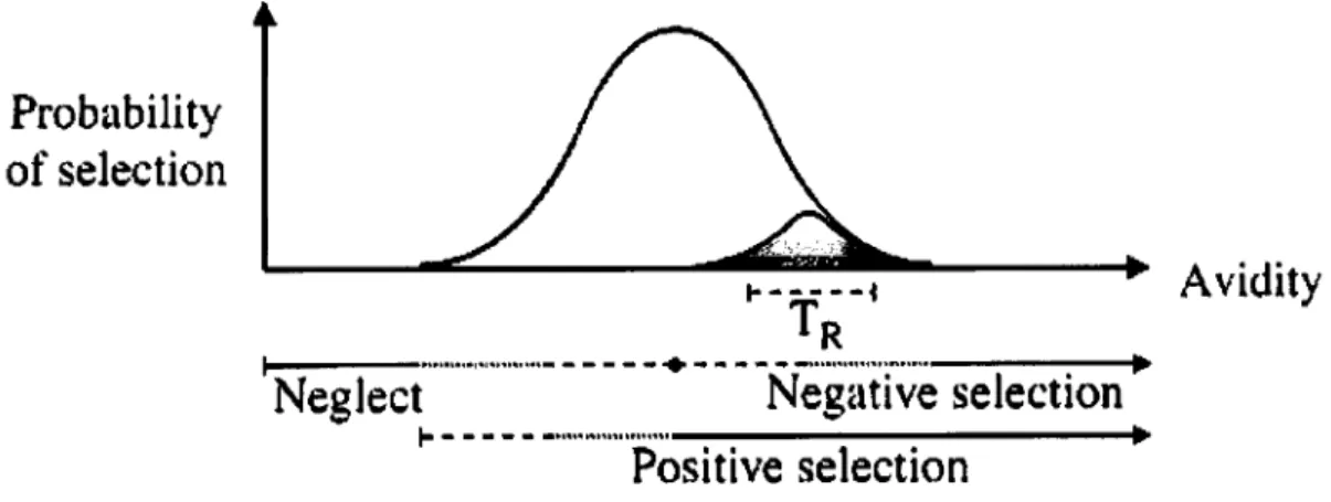
3.2.1-Positive and Negative Selection
The T cell function is dependent upon the recognition by a given TCR of specific peptides bound to specific MHC molecules (see review on the history of the discovery of MHC restriction in Zinkemagel and Doherty, 1997). For a T cell to respond to a given Ag, the Ag must first be processed in the intracellular compartments of an Antigen Presenting Cell (APC) where it is coupled to MHC molecules. There are two classes of MHC molecules: Class I MHC molecules, which present peptides derived from intracytosolic antigens, and Class II MHC molecules that present peptides derived from antigens captured in vesicles (for an overview see Janeway et al., 1999). Class I molecules will be recognized by CD8+ T cells.
Class II MHC molecules will be recognized by CD4+ T cells. MHC recognition determines the
characteristic types of responses of T cells. Thus, the TCR must be MHC restricted, recognizing the presenting MHC molecules of the individual (Zinkemagel and Doherty, 1997) and should allow for enough diversity to be able to respond to unpredictable Ags. However, as the MHC can equally bind peptides derived from the individual itself (self peptides) it is equally important that the selected TCRs are selected in such a way that they do not respond to presented self peptides, as this would result in the destruction of the individual. The selected TCRs must be self- tolerant.
The processes that ensure both conditions occur in the thymus and are named positive (matching TCR with self MHC) and negative (deleting TCRs specific for self peptide-self MHC complexes) selection (for an overview see Janeway et al., 1999). The exact mechanisms and processes responsible for positive and negative selection, as well as the
mechanisms that are responsible for CD4+ vs CD8+ T cell lineage commitment are complex,
have been object of a large body of work and are still under intense investigation (see revisions in Benoist and Mathis, 1997; Sebzda et al., 1999; Amsen and Kruisbeek, 1998; Marrack and Kapler,1997; Hogquist, 2001). As these subjects are not directly related to the results and concepts that are central to this thesis, I will not describe them here.
3.3- The Single Positive Thymic Compartment
The final stages of thymic differentiation occur in the thymic medulla, where thymocytes are found after downregulation of one of the coreceptors. Phenotypically, SP thymocytes have been shown to undergo changes in the expression of cell-surface markers like CD24 (HSA), CD62L, Qa-2, CD69 and CD45RB (Lucas et al., 1994), and in chemokine receptors like CCR7 or CCR9 (Campbell et al., 1999; Norment et al., 2000; Wurbel et al., 2000). Thus, some differentiation events take place in the SP thymic stage. The thymic medulla has also been identified as a location of tolerance induction (Anderson and Jenkinson, 2001; Klein and Kyewski, 2000), though it is not clear if this means that positive/negative selection can occur at this stage or if tolerance in the thymic medulla is achieved through non-deletional processes (Anderson and Jenkinson, 2001). Interestingly, SP thymocytes have been shown to proliferate before export to the peripheral pool, an event that should increase the numbers of exportable cells (Ernst et al., 1995; Penit and Vasseur,
1997).
Hence, the final stages of the Single Positive pool represent the pool of selected lymphocytes and receptors that will be exported to the periphery and that will be responsible for the T cell immunocompetence of the individual. The importance of the size of this compartment and of the export rate will be discussed below.
3.4- Kinetics of T cell Development
The duration of all the developmental processes necessary to complete T cell development is of 4 to 5 weeks. Most studies, relying on DNA labelling techniques (namely [H3] Thymidine
(Egerton et al., 1990) or BrDU incorporation (Huesmann et al., 1991; Penit et al., 1995; Penit and Vasseur, 1997), have allowed the study of the duration of thymic development and have also allowed the study of the magnitude of expansion and the identification of the developmental stages where expansion is occurring.
The BrDU technique is now the most widely spread method to determine the proportion of cycling cells in a population. BrDU is a halogenated nucleotide that incorporates into the DNA as a thymidine analogue. As antibodies against the BrDU are available, the cycling cells can be identified. A single pulse of labelling is used to determine the fraction of cycling cells in a population, as well as the time taken for these pulse-labelled cells to progress to subsequent developmental stages. Continuous labelling studies are useful to determine the turnover time of a population, as labelled cells replace their unlabelled counterparts (Scollay and Godfrey, 1995). The principle of the [H3] Thymidine technique is
the same, but the detection method is different (Egerton et al., 1990). The majority of the most recent studies on these subjects use the BrDU incorporation method.
3.4.1- The DN compartment
During the early stages of thymocyte development, a small number of precursor cells will not only undergo developmental changes and choices but also massive expansion. The minimal number of BM cells capable of providing precursors for thymic reconstitution in irradiated animals has been estimated at about 3 x 10⁵ cells (Scollay et al., 1986). The number of real
T cell precursors responsible for thymus colonization has not been easy to evaluate, as reliable markers for these precursors are not available and they may develop into T cell committed precursors already in the thymus (Akashi et al., 2000). The DN developmental process seems to take about 2 weeks. It was found that after one BrDU pulse, 20 to 30 % of
the whole DN compartment was BrDU+ (Penit et al., 1995). At the most immature DN1 stage
(CD44+CD25"), little division is taking place, with only 4% BrDU+ cells (Penit et al., 1995). An
increase was found to occur at the DN2 (CD44+CD25+) stage, with 20 % of BrDU+ cells after
a single BrDU pulse. The majority of the labelled DN thymocytes were equally distributed
between the DN3 (CD44"CD25+) and DN4 (CD44CD25) stages, as a result of the higher
representativity of these later subsets. However, in the DN3 stage, a more reduced
percentage of the cells incorporated BrDU, with only 10% of BrDU+ cells. The highest
proportion (35%) of dividing cells was found in the later DN4 stage, and this value was no different from the one found for the earliest of the DP cells (CD4l0WCD8l0W) (Penit et al., 1995).
Theese results suggest that cell proliferation starts during or just after CD25 expression, stops after CD44 down-regulation (this fits with the fact that only a fraction of the DN3 cells will make productive prearrangements and with the observed disappearance of an important
progeny of these DN3 stage cells) and restarts during CD25 loss (Penit et al., 1995). It had been calculated that in the early stages of Thymic development thymocyte precursors will expand in such a way that one single precursor will give rise to up to 4 000 daughter cells (Shortman et al., 1990). However, estimates made after analysis of the BrDU data referred above give an estimate in the order of a 300 fold expansion, corresponding to a total of 9 to 10 divisions (Penit et al., 1995). The differences found can be partially due to the different methods used (thymidine incorporation vs. BrDU) or to the difficulty in the evaluation of the number of thymic precursors that colonize the thymus (Shortman et al., 1990). A summary is shown in figure 4.
3.4.2- The DP compartment
After ^selection, and as a direct consequence of |3selection (Fehling and von Boehmer, 1997) the last proliferative stage of DN4 and of early DP thymocytes will take place. As a result of these early stage proliferative phases, the DP compartment will make up for about 85% of the total thymocyte number. The effects of positive and negative selection on the thymic transit duration and on thymocyte number have been evaluated in Tg and WT mice, using the BrDU technique. BrDU incorporation studies in normal C57BI6 mice have clearly shown that the daily generation of DP thymocytes largely exceeds the generation of mature SP thymocytes (Huesmann et al., 1991). Similar studies performed using Tg mice have also revealed that the lifespan or the transit time of cells in the DP compartment is between 3 and 4 days, and the value of 3.5 days has been used to describe the duration of this procedure. It was possible too, to conclude from these studies, that positive selection occurs without cell division and that the same holds true for the DP-^SP transition (Ernst et al., 1995; Huesmann et al., 1991). The linear kinetics observed in the cell-labelling experiments suggests that the bulk of the cells moves in the DP compartment as if in a conveyor belt, on a first in - first out basis (Scollay and Godfrey, 1995). When evaluating the efficiency of the selection processes, it is also obvious that the large majority of the cells will not be able to reach final maturation. Most DP thymocytes will die by neglect, as a result of a failure to produce a TCR that is able to react with self MHC-peptide complexes to originate TCR mediated signalling above lower limit threshold levels (reviewed in Sebzda et al., 1999). These thymocytes will undergo death by apoptosis (Surh and Sprent, 1994). The proportion of DP thymocytes that die by neglect has been calculated to be 90% (Egerton et al., 1990; Huesmann et al., 1991).
As referred above, the final SP repertoire is dependent on positive and negative selection. The proportion of the positively selected thymocytes transiting to the SP compartment has been estimated to be below 5% (Egerton et al., 1990).
3.4.3- The SP compartment
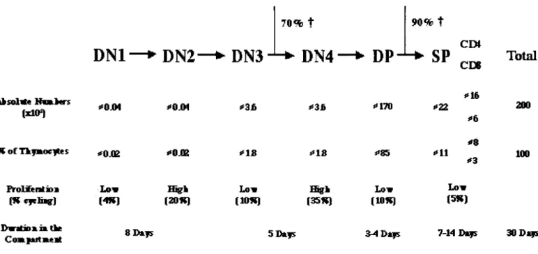
Thymocytes remain for as long as 2 weeks in the medullary stage of T cell development, before exit to peripheral pools (Egerton et al., 1990). The daily rate of production of SP thymocytes seems to represent 1% of the thymus cellularity (Egerton et al., 1990), a value that corresponds approximately to the daily rates of thymic export into the peripheral compartments (Scollay et al., 1980 and see chapter 6). It has been shown that thymocytes in the SP Thymic compartment can proliferate, a last thymic expansion phase that has been suggested to be responsible for an increase in the positively selected repertoire numbers before peripheral colonization (Ernst et al., 1995; Penit and Vasseur, 1997). This post selection expansion phase was suggested to be independent of TCR-MHC interactions and dependent on IL7R expression (Hare et al., 2000; Hare et al., 1998) and could originate an increase in the thymic output of up to 30% (Penit and Vasseur, 1997). In absolute terms, the rate of production of mature thymocytes has been calculated as 3% of the number of DP thymocytes, equivalent to 1% of the total thymocyte number (Egerton et al., 1990). This number is in agreement with estimates on thymic export (Scollay et al., 1980) (chapter 6). An attempt to give a general overview of T cell developmental kinetics is shown in figure 4.
DN1
DN2—* DN3
70% ÎDN4
DP
90% t CE»SP Total
CE* 1*10") "0J04 *0JM *3fi *3b *i70 «22 " l l > * 6 200 R o i Tkfmoeftits "0J02 *»M *18 "IS «*85 I ' l l *8 *3 100 FmlifentiDB(Kcfclbr) (4K) Lav (20*) [10«) Lav (35«) (10*) Low
Low (5*J
I > W H M > » i» the
C o m j t t r t m t H 8 DATS 5D&J5 3-4 D**s 7 14 D*)5 30 D*r
Figure 4: Kinetics and quantitative aspects of T cell development. Absolute numbers are calculated for a young adult mouse (200xl06 thymocytes). The % of cycling cells is given as found after one single pulse labeling of BrDU. Data compiled from
4-HOMEOSTASIS WITHIN THE THYMUS
Though the sequence of events in thymocyte development is characterized in some detail, less detailed information is available that relates to the existence of homeostasis within the thymus. If the idea that the thymocyte number is kept under control is questioned by the fact that the thymus involutes with age (chapter 5), it is equally true that the events in thymic selection take place at a considerably faster time-scale. The many selection and expansion phases in thymic development could thus be the "target" for homeostatic processes, if the number of thymocytes was under control in any of the developmental stages. One group has assessed this directly, and reached the conclusion that the mature CD8+ but not the CD4+ SP
compartment was under homeostatic control (van Meerwijk et al., 1998). The magnitude of the "homeostatic" compensation found was, however, not very significant.
Another piece of information that could be related to homeostasis-like phenomena inside the thymus is the availability of selection "niches" for positive selection (Huesmann et al., 1991; Merkenschlager, 1996; Merkenschlager et al., 1994). When most of the DP thymocytes express a selectable transgenic TCR, the formation of mature SP cells is 10 to 20 times more efficient than observed in normal mice. However, this means that only 20% of the DP thymocytes mature (Huesmann et al., 1991). This is due to the limited availability of stromal cells (Merkenschlager, 1996; Merkenschlager et al., 1994) capable of mediating positive selection, as most DP thymocytes with a selectable transgenic TCR will undergo maturation when they represent only 5% or less of the total DP pool (Huesmann et al., 1991). This observation suggests that there is a rate-limiting step for the number of positively selectable thymocytes. It is not clear if this is a mechanism that could be responsible for the maintenance of thymocyte numbers (or of SP thymocyte numbers), as the transgenic mouse situation may be too dissimilar to the physiological condition, and this kind of competition can be extremely rare in the physiological situation. Thus, the existence of homeostasis inside the thymus is (was) still an open question. We have investigated into this, developing a novel system and analysis for this purpose. (Section B, article #1).
5- THE THYMUS AND AGING
The thymus involutes with age. This important observation has been verified in several animal models and in the human situation. It has received attention not only because of it's implications for the reconstitution of the immune system in situations where the lymphocyte pool is depleted due to irradiation or disease but also because thymic involution correlates
with an immunological decline, reflected in an increase in both the susceptibility to infections and in the incidence of autoimmune disorders. We therefore knew for a long time that the thymus exerts some functional activity even in the adult (Metcalf, 1965a; Miller, 1965; Miller, 1962; Taylor, 1965). The ability to reconstitute an individual's lymphocyte pool after peripheral depletion also correlates inversely with age. Older people and animals do not completely reconstitute the peripheral lymphocyte pool, while much younger patients and animals do (Mackall and Gress, 1997). In humans, the reduction of Thymic mass starts at the age of 1 year (when the organ attains its maximal size) and results in an important reduction of Thymic mass by the time of puberty (George and Ritter, 1996). In mice, declines in the capacity to promote thymocyte proliferation are noted as early as 2 weeks after birth (Hirokawa et al., 1994) and a reduction in the thymic size is visible from week 6 after birth (Hirokawa and Makinodan, 1975). However, children of up to 15 years and mice of 3-4 months are still able to regenerate the peripheral T cell pool to a normal size, which has contributed to the general idea that Thymic involution starts at puberty, an idea that has been challenged (George and Ritter, 1996; Steinmann et al., 1985).
In humans, the decrease in thymus size is masked by changes in the architecture of the organ. In a child, the thymic lobes are separated by thin septa of connective tissue. In the thymus of an elderly person, the septa have greatly expanded and mostly comprise fat cells. Adipose tissue also develops under the capsule, separating it from the true thymic tissue. Thus, this increase in fat, connective tissue and perivascular space counterbalances the diminution of the lymphoepithelial areas of the thymus and the overall size of the organ remains constant throughout life. In mice this does not happen and the size of the thymus decreases with age. In the thymus of an old (24 month) mouse, the thymic T cell production has been estimated to be 0,7% of the number of T cells produced by a newborn mouse (George and Ritter, 1996).
Thymic involution can be derived from factors intrinsic to the immune system or can be a response to extrinsic factors. In the first situation, thymic involution could be either due to a deficient supply of BM precursors or secondary to alterations on thymic stroma. A third possibility was that these two factors were acting at the same time. This was tested in BM chimera systems, where BM from old donors was grafted into irradiated young hosts or where BM from young donors was grafted into irradiated old hosts (Hirokawa et al., 1994). In other experimental setups, neonatal thymi were grafted under the kidney capsule of old mice (Mackall and Gress, 1997; Metcalf, 1965b). All the results obtained point to a more relevant role of the thymic stroma, even if the capacity of old BM to repopulate the thymus seems to be slightly reduced (Mackall and Gress, 1997; Metcalf, 1965b). Hence, when aged irradiated mice were reconstituted with BM from young donors, the thymic abnormalities were not reversed and the thymic size and cellularity remained reduced (Hirokawa et al., 1994;
Mackall et al., 1998). When aged mice received a neonatal Thymic transplant and were then irradiated and reconstituted with neonatal BM cells, normal Thymic regenerative capacity was observed (Mackall and Gress, 1997). Thus, the age of the thymus and not extrathymic factors present in the aged milieu is the major factor contributing to the reduced generation of T cells from aged thymi.
The general notion that thymic involution is linked to puberty had suggested that hormonal factors could be the primary cause for age related thymic involution. Interactions between the gonadal steroids and the immune system have been documented and include the occurrence of thymic hyperplasia after gonadectomy or after destruction of the anterior portion of the hypothalamus (Hirokawa et al., 1994). Additional data suggest that age-related changes within the thymus itself may increase the susceptibility to inhibition via the extrathymic hormonal milieu (Mackall and Gress, 1997). Thus, though the extrathymic milieu does exert some influence on thymic involution, the primary cause seems to lie in the thymus itself. When irradiated aged mice were reconstituted with BM from young donors, the thymic reconstitution was reduced but the thymocyte subset representation was normal, confirming that the aged thymus is able to function and to generate substantial numbers of T cells. This is also being confirmed in aged humans, where recent measures of TRECs or of TCR rearrangement in old individuals or HIV infected adults has also provided evidence for the continuous production of T cells late in life (Douek et al., 1998; Jamieson et al., 1999). This last point is of major relevance for the reconstitution after depletion of the peripheral T cell pool and points out that thymic export is a component of peripheral homeostasis that is present throughout life. The next section deals with thymic export.
6- THYMIC EXPORT AND MIGRATION
After the developmental processes referred above (see chapter 3), mature T cells are exported to the periphery where they will constitute the peripheral T cell pool. Thymic emigrants will be part of the daily input of T cells incorporating into the peripheral T cell pool. As the T cell number is kept constant, it follows that a newcoming T cell will only integrate into these peripheral pools if another T cell is being replaced. The quantification of the thymic output is thus crucial for the understanding of the T cell homeostasis' dynamics. The number of cells exported each day will not only be responsible for the renewing of the available repertoire, adding new specificities to the peripheral pools, but will also be responsible for the replacement of at least part of the cells previously installed in the peripheral pools. The question that arises is: to what extent? To advance in the resolution of this problem, the
thymic output and its impact on the observed peripheral homeostasis were evaluated. Five basic strategies have been used:
1- The evaluation of the impact of thymic ablation (thymectomy) on the maintenance of peripheral numbers (Metcalf, 1965a; Miller, 1965; Miller, 1962; Rocha et al., 1983; Mackall, 1993; Parrottand de Sousa, 1971; Taylor, 1965).
2- The ability of peripheral T cells to expand after transfer into athymic hosts (Rocha et al., 1989; Tanchot and Rocha, 1995).
3- The evaluation of the impact of an increase in thymic mass (or thymic export) on the peripheral T cell numbers (Berzins et al., 1998; Berzins et al., 1999; Leuchars et al.,
1978; Metcalf, 1965b).
4- The direct measurement of the number of thymic emigrants after intrathymic injection of fluorescent dyes (Graziano et al., 1998; Kelly et al., 1993; Scollay et al., 1980) or after the identification of the thymic migrant phenotype (Douek et al., 1998; Kong et al., 1998; Kong et al., 1999; McFarland et al., 2000).
5- The evaluation of the thymic output after the induction of peripheral T cell depletion by administration of anti-thyl antibodies. (Gabor et al., 1997).
In parallel, and as seen above (Chapter 5), the thymic emigration was evaluated in the aging thymus situation.
6.1- Quantitative aspects of thymic output
Adult thymectomy was shown to be responsible for a 40% reduction in the size of the peripheral pools (Rocha et al., 1983) and the presence of a thymus was shown to be essential for peripheral T cell reconstitution after T cell depletion (Mackall et al., 1997; Metcalf, 1965a; Miller, 1965; Miller, 1962; Parrott and de Sousa, 1967). Accordingly, direct measurements of thymic export using intrathymic FITC injection in wild type (Scollay et al., 1980) or in TCR Tg mice (Kelly et al., 1993) have shown that a relatively constant fraction of the thymocyte number (1%) is exported daily into the peripheral pools in young adult mice. This translates into a number between 1 to 2 x 10⁶ cells that are exported daily. With age, the
fraction of thymocytes exported daily is reduced (0,1% at 6 months of age), and thus, an increasingly smaller number of thymocytes are exported (Scollay et al., 1980). These estimates of thymic export have been directly or indirectly confirmed in a number of later studies (Berzins et al., 1998; Berzins et al., 1999; Gabor et al., 1997; Tanchot and Rocha, 1997) and studies using CFSE intrathymic injection gave slightly higher but comparable
values for daily export in young adult mice (2 - 3 x 10⁶) (Graziano et al., 1998). Thus, thymic
export alone is responsible for an input of 50 x 107 cells per month into the peripheral T cell
pool. In order to achieve homeostasis, the incorporation of these T cell emigrants into the peripheral T cell pool must either be restrained, by some kind of feedback mechanism acting on thymic export or by pre-emptive selection at the time of incorporation, or compensated by death of T cells from the already established peripheral pools.
In studies of hyperthymic mice (mice receiving grafts of thymic lobes under the kidney capsule), it was found that the rates of thymic export by individual grafted lobes were independent of the number of thymuses grafted and were constant, independently of the degree of replenishment of the peripheral T cell pool (Berzins et al., 1998; Leuchars et al., 1978). This suggests that there is no feedback control of the peripheral T cell pool over thymic export (Berzins et al., 1998; Leuchars et al., 1978; Tanchot and Rocha, 1997). Studies on the reverse situation, where thymic export was evaluated after T cell depletion was induced by the administration of anti Thy 1 antibodies, also suggested that in a situation of demand due to peripheral depletion, the thymus is not able to compensate by increasing the thymic output (Gabor et al., 1997). We have developed a system that allows the study of thymic export in a situation where the peripheral compartment is not full and the thymus should be able to support an increase in thymic export (see results section, article #1).
Another issue is the incorporation of thymic emigrants into the peripheral T cell pools. To study how the peripheral T cell pool reacts to thymic export, experiments were performed using the thymic graft protocol. The major conclusion is that the size of the peripheral pool is largely independent of the thymic output or mass. Mice receiving an additional thymus, grafted under the kidney capsule, will double the number of T cells exported daily, yet peripheral T cell numbers are kept at similar levels (Berzins et al., 1998). This situation is overcome by grafting a much larger thymic tissue (9 thymic lobes). These results were interpreted as proof for the existence of peripheral homeostasis mechanisms, that were responsible for the non-increase of peripheral T cell number in the first experiment but that these mechanisms could be overcome in extreme situations, as in the second experiment (Berzins et al., 1998). In a complementary study, the same group has suggested the existence of a separate peripheral pool for Recent Thymic Emigrants (RTE), and that these are "exempt from peripheral T cell homeostasis", for a period of three weeks (Berzins et al., 1999). It could also be true that homeostasis was just reset for an equilibrium value around a higher steady-state number.
It was further suggested that this "exclusion" of RTEs from peripheral T cell homeostasis allows repertoire turnover throughout adult life, an important role for thymic output (Berzins et al., 1999). This leads to a second issue on the relevance of thymic output for the peripheral T cell pool: the qualitative role of thymic output.
6.2- Qualitative aspects of thymic output
It is known that peripheral T cells are capable of considerable expansion, of a magnitude similar to that of colony forming units (Miller and Stutman, 1984; Rocha et al., 1989). The evaluation of the proportion of peripheral T cells in cycle, incorporating BrDU in a 24 hour period, shows that peripheral expansion is an important mechanism of mature T cell production in the adult mouse (Rocha et al., 1990; Sprent, 1993), independently of whether cycling cells represent a large (Rocha et al., 1990) or a small (Tough and Sprent, 1994) fraction of the total peripheral T cells. However, peripheral division mechanisms are not able to generate new TCRs, thus, peripheral division will give rise to a less diverse repertoire. Accordingly, it has been shown that the peripheral compartments obtained after peripheral expansion are biased towards an activated/memory phenotype (Mackall et al., 1993). Thus, thymic output seems to be essential for the renewing of the specificities present at the peripheral pools, being the only provider of naïve T cells. Importantly, as we will see below, the naive and the activated peripheral T cell pool sizes are independently regulated (Tanchot and Rocha, 1995).
It has been discussed whether, inside this peripheral naive pool, RTEs are preferentially selected for entry into the peripheral naïve T cell pool (Berzins et al., 1998), representing a sub-division in the peripheral naive pool for a period of seeding of 3 weeks (Berzins et al., 1999) or whether the entry of RTEs into the peripheral naive pool is a random event (Tanchot and Rocha, 1997), being the replacement of the naive pool T cells independent of cell age. The cell-age independent replacement of the peripheral naïve T cells would enable rapid contraction of large clones and a longer survival of rare ones, reinforcing the role of continuous thymic outputs in the maintenance of repertoire diversity in the naive pools (Tanchot and Rocha, 1997). In the absence of thymic output, the size of the naive pool would not necessarily decrease, as peripheral expansion concerns the activated/memory pool but has no influence on the size of the naive pools (Tanchot and
Rocha, 1995) but the individual life-spans of the naive cells will increase as a result of a lack of competing cells (Freitas et al., 1996). However, as larger clones are allowed to persist, the diversity of the naive pool will decrease.
Thus, although thymic export does not seem to play a very important role for the maintenance of peripheral T cell numbers after initial seeding, it does seem to play an essential role for the peripheral naïve T cell pool, being the only source of new specificities.
6.3- Migration
The information concerning the signals triggering mature thymocyte migration from the thymus is sparse. Chemokines are obvious candidates for molecules involved in thymocyte exit from the thymus. Indeed, a recent report (Ueno et al., 2002) has shown a role for the chemokine CCL19 in the emigration of mature thymocytes in Fetal Thymus Organ Cultures (FTOCs). Importantly, mature thymocytes express CCR7, the receptor for CCL19 and CCL21 and neutralization of CCL19 but not of CCL21 was shown to result in impaired thymic emigration (Ueno et al., 2002). In CCR7A mice, thymic emigration and peripheral seeding are
reduced in newborn mice, when compared to the wild type situation (Ueno et al., 2002). In adult CCR7"7" mice, however, the circulating T cell pool is not reduced (Forster et al., 1999;
Ueno et al., 2002), suggesting that alternative pathways operate and allow the emigration of mature thymocytes (Ueno et al., 2002).
Thus, more information is required to identify the signals involved and elucidate the mechanisms resulting in thymic emigration. Whatever these are, a fraction of the mature SP T cells generated will be exported and will be confronted with the peripheral T cell compartment. How this peripheral T cell compartment is organized is the subject of the next chapter.