Contents lists available atScienceDirect
Biomedicine & Pharmacotherapy
journal homepage:www.elsevier.com/locate/biophaReduction of in
flammation and colon injury by a Pennyroyal phenolic
extract in experimental in
flammatory bowel disease in mice
João Rocha
a,d, Rosa Direito
a,d, Ana Lima
b,d, Joana Mota
b,d, Margarida Gonçalves
c,d,
Maria Paula Duarte
e,d, João Solas
a,d,f, Bruno Felício Peniche
a,d, Adelaide Fernandes
a,d,
Rui Pinto
a,d,g, Ricardo Boavida Ferreira
b,d, Bruno Sepodes
a,d, Maria-Eduardo Figueira
a,d,⁎aResearch Institute for Medicines and Pharmaceutical Sciences (iMed.UL) and Faculdade de Farmácia da Universidade de Lisboa, Av. Prof. Gama Pinto, 1649-003 Lisboa,
Portugal
bDisease & Stress Biology Group, LEAF, Instituto Superior de Agronomia, Universidade de Lisboa, 1349-017 Lisbon, Portugal
cDCTB/MEtRICs/Faculdade de Ciências e Tecnologia da Universidade NOVA de Lisboa, Campus de Caparica, 2829-516 Caparica, Portugal dVALORIZA, Instituto Politécnico de Portalegre, Campus Politécnico, 10, Portalegre, Portugal
eH&TRC– Health and Technology Research Center, ESTeSL – Lisbon School of Health Technology, Instituto Politécnico de Lisboa, Lisbon, Portugal fH&TRC– Health and Technology Research Center, ESTeSL – Lisbon School of Health Technology, Instituto Politécnico de Lisboa, Lisbon, Portugal gJCS, Dr Joaquim Chaves Lab Analises Clínicas, Miraflores-Algés, Portugal
A R T I C L E I N F O Keywords: Pennyroyal Mentha pulegium Colitis IBD Inflammation Proliferation A B S T R A C T
Purpose: Little is known about the pharmacological effects of the phenolic compounds of Pennyroyal (Mentha pulegium). This Mediterranean aromatic plant, used as a gastronomic spice and as food preservative by the food industry has been studied mainly due to its essential oil antibacterial properties, composed primarily by monoterpenes. With this work, we aimed to evaluate the effects of a phenolic extract of pennyroyal in the impairment of inflammatory processes in Inflammatory Bowel Diseases (IBD) and in the potential inhibition of progression to colorectal cancer (CRC).
Methods: To that purpose, we evaluated the effect of pennyroyal extract administration in a model of TNBS-induced colitis in mice and further determined its effect on human colon carcinoma cell proliferation and in-vasion.
Results: The phenolic extract of pennyroyal exhibited antioxidant properties in in vitro assays and administration of the extract in a rat model of carrageenan-induced paw oedema led to significant anti-inflammatory effects. Further results evidenced a beneficial effect of the phenolic extract in the attenuation of experimental colitis and a potential antiproliferative effect on cultured colon cancer cells, effects not previously described, to our knowledge. A reduction in several markers of colon inflammation was observed following administration of the extract to colitis-induced mice, including functional and histological indicators. A successful inhibition of cancer cell invasion and proliferation was also observed in in vitro studies with HT-29 cells. Furthermore, the extract also led to a reduced expression of iNOS/COX-2 in the colon of colitis-induced mice, both being crucial med-iators of intestinal inflammation.
Conclusions: Taking into consideration the central role of inflammation in the pathophysiology of CRC and the recognised connection between inflammatory events and cancer, these results enlighten the relevance of the phenolic constituents of pennyroyal as important pharmacological sources in the investigation of new treatment options for patients with inflammatory bowel diseases.
1. Introduction
Inflammatory Bowel Diseases (IBD) is a term given to generally characterize two types of intestinal diseases: Crohn’s Disease and
ulcerative colitis. Despite decades of extensive research on these dis-eases, a specific aetiological cause is yet to be established, although the aetiology of both diseases appear to be related to a dysregulated mu-cosal immune response to environmental factors in genetically
https://doi.org/10.1016/j.biopha.2019.109351
Received 1 May 2019; Received in revised form 13 August 2019; Accepted 14 August 2019
⁎Corresponding author at: Research Institute for Medicines and Pharmaceutical Sciences (iMed.UL) and Faculdade de Farmácia da Universidade de Lisboa, Av. Prof. Gama Pinto, 1649-003 Lisboa, Portugal.
E-mail address:efigueira@ff.ulisboa.pt(M.-E. Figueira).
0753-3322/ © 2019 The Authors. Published by Elsevier Masson SAS. This is an open access article under the CC BY-NC-ND license (http://creativecommons.org/licenses/BY-NC-ND/4.0/).
susceptible hosts [1,2].
Colorectal cancer (CRC) is one of the most common cancers worldwide, with over 1.8 million new colorectal cancer cases and 881000 deaths estimated to have occurred in 2018, accounting for about 1 in 10 cancer cases and deaths. Overall, colorectal cancer ranks third in terms of incidence but second in terms of mortality [3]. Ad-ditionally, CRC is the third most incident cancer in the male population (10.9%) and the second most common in the female population (9.5%) [3]. Interestingly, only ˜5–6% of CRC cases are related to germline mutations [4], whereas˜70% of CRC tumours are sporadic [5].
Taking this information into consideration, an opportunity might exist to increase CRC prevention by decreasing its risk factors. In fact, since the tumour initiation and progression processes are multifactorial, they are also influenced by several external factors, especially regarding the environment and diet, with the latter being able to either ameliorate or even increase CRC risk [6]. In patients with IBD, chronic in-flammation is known to be a major risk factor for the development of gastrointestinal malignancies [7] with inflammatory signalling path-ways associated with increased intestinal production of reactive oxygen species being considered to be the main driving factor bridging IBD into CRC [8,9]. The chronic inflammation found in IBD often leads to ab-normal cell growth (dysplasia). Although these cells cannot be called malignant, the probability of gaining anaplastic characteristics and developing into cancer cells is higher [5,9].
Taking into account the beneficial role of phenolic acids in in-flammatory processes and in cell proliferation, an opportunity exists for an adjuvant therapy that not only reduces the intestinal inflammation of IBD patients but may also have a potential preventive effect in the evolution of highly proliferative, inflammatory and dysplastic cells into carcinoma cells [10–12].
Pennyroyal [Mentha pulegium L. (Lamiaceae)] is a Mediterranean aromatic plant, commonly used in gastronomy as a spice that recently sparkled the interest of the food industry [13]. There is a growing in-terest in replacing synthetic chemicals by natural products with bioactive properties from plant origin. In that context, the application of essential oils is becoming increasingly important as natural additives for shelf-life prolongation of food products, substituting the use of synthetic preservatives [14]. In fact, studies have shown that M. pule-gium essential oil exhibited antibacterial activity against several bac-terial strains [13,15]. Essential oils are a mix of volatile compounds, and are regarded as a prime source of bioactive compounds, mainly with antioxidative and antimicrobial properties [14], with the main compounds studied with relation to those effects being of terpenoid structures (e.g. pulegone, pinene, limonene) and have been the main focus of research regarding this plant [13,15,16]. The toxicological profile of pennyroyal preparations has also been subjected to extensive research, and several constituents have been identified as hepatotoxic and abortifacient. However, those effects are characteristic of terpenoid compounds, specific constituents of the essential oil and therefore not present in phenolic extracts [17].
In contrast, little is known about the pharmacological effects of the phenolic compounds of M. pulegium. Chemically, the phenolic content of M. pulegium is comparable to plants known to exhibit pharmacolo-gical properties in experimental models of inflammation which in-creases the interest regarding its potential to exert anti-inflammatory actions [18–23].
In this study we evaluated the beneficial effect of a phenolic extract of M. pulegium (pennyroyal) as a potential pharmacological tool in the management of inflammatory processes associated to IBD and aiming to assess the potential inhibition of cell progression to colorectal cancer and its relation with the anti-inflammatory effect. Therefore, we as-sessed the effect of M. pulegium extract in an experimental TNBS-in-duced colitis model with further evaluation of a potential inhibitory effect on proliferation and invasion properties of human colon carci-noma cell.
2. Materials and methods 2.1. Reagents and chemicals
Ketamine (Imalgene® 1000) and xilazine (Rompun® 2%) were ac-quired from Bio2 Produtos Veterinários (Lisboa, Portugal). Unless otherwise stated, all remaining substances were acquired from Sigma-Aldrich, Portugal.
2.2. Plant material and extract preparation
Fresh samples of Mentha pulegium L. were obtained from a local cultivar (Lisboa, Portugal). Plants (leaves and stems) were washed under running tap water and chopped into thin slices. Chopped plant material (20 g) was than extracted with 100 mL of ethanol (70%, v/v), for 24 h, in the dark, at room temperature and under stirring. The re-sulting extracts werefiltered (Whatman, nº1) and ethanol was elimi-nated in rotary evaporator at 40 °C (Heidolph LABOROTA 4001). Then, extracts were centrifuged (6000g, 15 min, 4 °C) (Sigma 4K-15C) and the supernatants were divided into 1 mL aliquots and stored at−50 °C until future analyses.
2.3. Total phenolic and totalflavonoid content
Total phenolic compounds were determined according to Kosar et al. [24] and totalflavonoid content was determined according to Barros et al. [25]. Results of total phenolic content were expressed as mg gallic acid equivalents (GAE) per mL extract and per g of dry plant and results of totalflavonoid content were expressed in μmol equiva-lents of catechin (CE) per mL of extract and per g of dry plant. 2.4. High-performance liquid chromatography (HPLC)
The phenolic components of the aqueous extract were isolated using solid phase extraction C18 columns. Briefly, 2 mL of the aqueous extract was added to the column and the adsorbed phenolic components were eluted with aqueous formic acid (0.1% v/v) and acetonitrile. The chromatographic separation was performed in a HPLC system (SpectraSystem, Thermo), equipped with a diode array detector (DAD), and a Thermo C-18 column. The eluents used were 0.1% formic acid (solvent A) and a mixture of 90% acetonitrile + 9.9% water + 0.1% formic acid (solvent B), and the flow rate was 0.8 mL/min. Identification of the main functional groups present in the extract was performed by comparison of their UV spectra with those of re-presentative standards analysed in the same conditions.
2.5. Antioxidant capacity
2.5.1. Cupric reducing antioxidant capacity (CUPRAC) assay
CUPRAC assay was performed according to the normal sample measurement procedure as previously described [26]. Results were expressed asμmol ascorbic acid equivalents (AAE) per mL of extract. 2.5.2. Ferric reducing antioxidant power (FRAP) assay
The FRAP assay was carried according to the procedure as pre-viously described [27]. A calibration curve of ferrous sulphate (0–1.25 mM) was used and results were expressed as μmol Fe2+
per mL of extract.
2.5.3. DPPH radical-scavenging assay
The DPPH assay was carried according to the procedure previously described [28]. Results were expressed as mg ascorbic acid equivalents (AAE) per mL of extract.
2.5.4. Superoxide anion radical-scavenging assay
according to the procedure previously described [29]. Results were expressed asμmol equivalents of gallic acid per mL of extract. 2.6. Carrageenan-induced paw oedema in rat
2.6.1. Animals
The paw oedema study was carried out using 48 male Wistar rats (150–200 g) (Harlan - Spain). All rats had free access to water and food until 12 h of the study.
2.6.2. Oedema induction and evaluation
Paw oedema was induced by intradermal (sub-plantar) injection into the rat left hind paw of 100μL of a λ-carrageenan solution (1% in saline) as previously described [30]. Paw volume measurements were as following: V0or basal volume is the volume of the hind paw mea-sured immediately after carrageenan injection and V6is the volume at 6 h post carrageenan administration. The increase in paw volume was measured as the oedema volume and was expressed as a relative per-centage of the increase in the volume at 6 h compared to the initial volume, according to the following formula: % paw volume increase = [(V6-V0) / V0] x 100.
2.6.3. Experimental groups
Animals were randomly allocated into the following six groups: (i) control group - animals were subjected to the oedema protocol de-scribed above, with the exception of 100μL of sterile saline being ad-ministered via subplantar injection instead of carrageenan. Animals were also administered with water (1 mL/kg) by oral gavage (n = 6); (ii) carrageenan group - animals with paw oedema induction as de-scribed above and administered with 1 mL/kg of water by oral gavage (n = 8); (iii) M. pulegium group - animals with paw oedema induction as described above and administered with M. pulegium extract (15 mg of phenolic acids/kg by oral gavage) 30 min before injection of carra-geenan (n = 8); (iv) indomethacin group - animals with paw oedema induction as described above and administered with indomethacin (10 mg/kg by oral gavage) 30 min before injection of carrageenan (n = 8); (v) tempol group - animals with paw oedema induction as described above and administered with tempol (30 mg/kg by oral gavage) 30 min before injection of carrageenan (n = 8); (vi) trolox group -animals with paw oedema induction as described above and adminis-tered with trolox (10 mg/kg by oral gavage) 30 min before injection of carrageenan (n = 8). The dose of the extract was selected according to previous studies by our group regarding the evaluation of the beneficial effects of different phenolic extracts in several models of inflammation, and the dose of 15 mg/kg of phenolic acids has generated consistent results and is also within the range of possibility for clinical translation and use, considering a human adult of 70 kg [31–34].
2.7. TNBS-induced ulcerative colitis model in mice 2.7.1. Animals
Male mice (CD-1 strain), weighing 28–33 g (5–6 weeks of age) (Harlan, Spain), were maintained according to the standard housing guidelines with free access to water and food, in a room with en-vironmental conditions automatically controlled (22 ± 1 °C with a 12/ 12 h light/dark cycle) at the Animal Facility of the Faculty of Pharmacy - University of Lisbon.
2.7.2. Induction of colitis
Induction of colitis was performed by administration of TNBS as previously described [31]. Briefly, a 50% ethanolic solution of TNBS (2.5% m/v) was administered by intracolonic administration (4 cm above the anus). At day 4 post-induction, blood samples were collected by cardiac puncture under surgical anaesthesia, followed by euthanasia by cervical dislocation and subsequent necropsy. The colon was re-moved and was observed for classification of diarrhoea severity.
Furthermore, the colon was washed with PBS for a macroscopic ob-servation of lesions andfixed in PFA for histological studies.
2.7.3. Experimental groups
Mice were allocated in a randomized way into the following ex-perimental groups:
1 Sham group (n = 6): the colitis induction protocol was followed as described above with the exception of the intracolonic administra-tion being performed with 100μL of saline solution instead of the alcoholic TNBS solution. Animals were administered with 10 mL/kg of water by oral gavage throughout the four days of the experiment. 2 Ethanol group (n = 6): the colitis induction protocol was followed as described above with the exception of the intracolonic adminis-tration being performed with 100μL of 50% (v/v) ethanol solution instead of the alcoholic TNBS solution. Animals were administered with 10 mL/kg of water by oral gavage throughout the four days of the experiment.
3 TNBS group (n = 10): the colitis induction protocol was followed as described above, with the administration of 100μL of a TNBS so-lution (2.5% TNBS in 50% ethanol). Animals were administered with 10 mL/kg of water by oral gavage throughout the four days of the experiment.
4 TNBS + M. pulegium group (n = 10): the colitis induction protocol was followed as described in the previous experimental group. Animals were administered with M. pulegium extract (15 mg/kg of phenolic acids by oral gavage) throughout the four days of the ex-periment.
2.7.4. Macroscopic evaluation of colitis severity
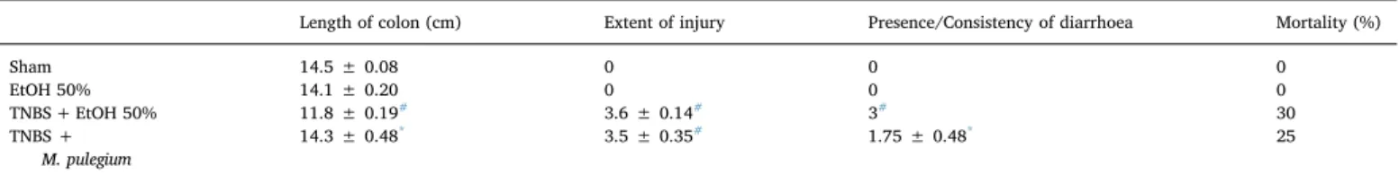
Diarrhoea severity was classified by an observer blinded to the ex-perimental groups according toTable 1. A microscope observation of the tissue was performed followed by measurement of the entire colon and injury extent.
2.7.5. Histology and immunohistochemistry procedures
Haematoxylin & Eosin (H&E) staining was performed as previously described [31], as well as the immunohistochemistry studies, for measurement of COX-2 and iNOS expression. Briefly, the colons were fixed in 4% PFA in PBS during 72 h at room temperature, decalcified, dehydrated and embedded in paraffin. Immunostaining was done in sections cut to a thickness of 6 mm and application of the primary an-tibodies, rabbit anti-COX-2 and rat anti-iNOS was performed, followed by the secondary antibodies incubation: anti-rabbit and anti-mouse antibodies for horseradish peroxidase (antibody catalogue numbers are: COX2 – rabbit, Cell signalling #4842, d1:500; iNOS – mouse, BD Biosciences #610329, d1:1000). Colon histological damage was scores as follows: score 0 - normal colon with no lesions, the mucosa is of uniform thickness, and the crypts are straight, normal crypt architecture, there is no cellular infiltration, oedema, or exudate; score 1 -colon with mild lesions, there are mucosal erosion and small superficial ulcers scattered along the length of the colon, with slight crypt loss and mononuclear cell infiltration; score 2 - colon with moderate lesions, intestines have extensive erosion and ulceration, with moderate crypt loss and neutrophil infiltration; score 3 - colon with very severe ul-ceration, much of the mucosa is thin with loss of crypts and markedly
Table 1
Score of diarrhoea severity.
Score Faeces consistency 0 Normal (hard pellets) 1 Slightly mucous
2 Soft
increased infiltration of neutrophils and acute inflammatory exudate. The intensity of the protein staining is relatable to the level of expres-sion of iNOS and COX-2. The level of iNOS or COX2 staining was quantitatively evaluated by determining the percentage of tissue area that was stained in brown, using the ImageJ (Fiji Is Just) software. 2.8. In vitro anti-proliferative cell assays evaluation
2.8.1. HT29 cell culture
The HT29 colon adenocarcinomas from Homo sapiens sapiens, cell line ECACC, nº 91072201, was used in in vitro experiments, as pre-viously described [31].
2.8.2. HT29 cell proliferation assay
HT29 cell proliferation assay was performed according to Carmichael et al. [35] with modifications previously described [31]. 2.8.3. Wound healing assay
The selected pennyroyal phenolic extract corresponding to an EC50 level of activity was assessed for its inhibitory activities in HT29 colon adenocarcinoma cells using standard cell migration analysis (wound healing assay) as previously described [31]. Data is presented as the mean ± SD.
2.9. Evaluation of inhibition of matrix metalloproteinases (MMPs) gelatinolytic activity
2.9.1. Minimal Inhibitory Concentrations (MICs) and half maximal effective inhibition (IC50) inhibition
MMP-inhibition was tested using the DQ gelatin assay as described in Lima et al. [36]. Minimal Inhibitory Concentrations (MICs) were assessed using the micro dilution method as previously described [37]. 2.9.2. MMP activity in HT29 colon cancer cells
MMP activity in HT29 cancer cells was performed by gelatin zy-mography according to standard methods [38], with the modifications previously described [31].
2.10. Animal in vivo experiments
Experiments were conducted according to the Home Office Guidance in the Operation of Animals (Scientific Procedures) Act 1986, published by Her Majesty's Stationary Office, London, UK, and the Institutional Animal Research Committee Guide for the Care and Use of Laboratory Animals published by the US National Institutes of Health (NIH Publication no. 85–23, revised 1996), as well as to the currently adopted EC regulations (Directive 2010/63/EU). The studies were performed in compliance with the ARRIVE Guidelines for Reporting Animal Research summarized at http://www.
2.11. Statistical analysis
In the in vivo animal experiments the results were expressed as mean ± standard error of the mean (SEM) of n observations (n re-presenting the number of animals). Comparison of results was per-formed by a one-factorial ANOVA test, followed by a Bonferroni's post hoc test (Prism 6.0 software– GraphPad). Statically significances were considered for P values less than 0.05.
In the in vitro and ex vivo studies with the HT-29 cells all experi-ments were executed as triplicates (a 3 independent experiexperi-ments) and results were expressed as the mean ± standard deviation (SD). Comparison of results was performed by the use of the software SigmaPlot (12.5 version), with one-factorial ANOVA test followed by a Tukey test for comparison between groups. Statistical differences were considered significant when P < 0.05.
3. Results
3.1. Phenolic profile and antioxidant capacity of the M. pulegium phenolic extract
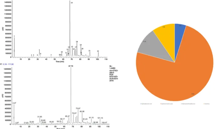
Total phenolic andflavonoid content of M. pulegium phenolic extract is shown in Table 2. The chromatographic profile obtained for the phenolic extract of M. pulegium is depicted inFig. 1and reveals that there is a predominance of hydroxycinnamic acids (74.4%), followed by hydroxymethoxyflavones (11.2%), catechins (9.6%) and hydro-xybenzoic acids (4.8%).
The main component of the extract was identified as rosmarinic acid, and accounts for 36.7% of the relative chromatographic area. The main constituents of the M. pulegium phenolic extract are represented in the graph depicted inFig. 1.
In order to determine the antioxidant potential of M. pulegium ex-tract reducing capacity (FRAP and CUPRAC) and free radical scaven-ging activity (DPPH and O2−assay) assays were employed (Table 3). The antioxidant capacity of M. pulegium extract was detected in all the assays. Results obtained are in accordance with previous studies which also demonstrated that ethanolic extracts of M. pulegium exhibit redu-cing capacity and are able to scavenge reactive oxygen species [13,39,40].
3.2. Paw oedema evaluation
Induction of oedema by carrageenan injection expectedly led to a significant increase in the volume of the hind paw at 6 h post-carra-geenan compared to the control group. Administration of M. pulegium phenolic extract was able to reduce the oedema formation in a sig-nificant way (Fig. 2). When comparing this effect to the ones exhibited by administration of tempol (30 mg/kg), trolox (30 mg/kg) and in-domethacin (10 mg/kg), it became evident that M. pulegium phenolic extract administration reduced oedema formation at only a slightly lower magnitude as those known antioxidant and anti-inflammatory substances (Fig. 2) at a dose of 12.5 mg of phenolic acids/kg. 3.3. Macroscopical and functional signs of colitis injury
Animals from both the Sham Group and the Ethanol Group ex-hibited no macroscopic evidence of colon lesion and mortality was of 0%. Rectal administration of TNBS/EtOH led to a statistically sig-nificant: colon length decrease, length of injury formed (ulcer) increase and diarrhoea severity increase, with a 30% mortality rate. In the groups treated with the M. pulegium extract all macroscopic signs of colon injury were significantly reduced comparing to the colitis-in-duced animals (Table 4,Fig. 3). Macroscopical observations and pho-tographs of the removed colon also confirmed these beneficial effects, with a flaccid appearance and an abundance of liquid faeces. Ob-servation of the colons through a surgical microscope was able to evi-dently demonstrate that colon injury was attenuated in animals ad-ministered with the extract when compared to the untreated colitis animals (Fig. 3).
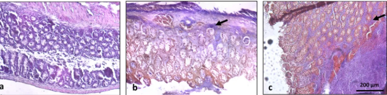
3.4. Histological features and inflammatory markers of colitis injury The evaluation of histologic signs of injury (Fig. 4) showed that Table 2
Total phenolic andflavonoid contents of M. pulegium extract.
Total phenolics Totalflavonoids
mg GAE/mL mg GAE/ g dry plant mg CE/mL mg CE/g dry plant 1.09 ± 0.05 23.55 ± 1.12 2.79 ± 0.10 60.45 ± 2.15
healthy animals exhibited a colon with normal appearance, with no evidence of lesions, a mucosa with a uniform thickness, crypts with normal architecture and no observable signs of inflammatory events (score 0). On the contrary, untreated colitis mice exhibited severe ul-ceration with abnormal architecture crypts (with evidences of complete loss) and a thinner mucosa with a clear evidence of immune cells in-filtration, equivalent to a score of 3. Colons from animals administered with M. pulegium extract show slight to moderate lesions with minimal crypt alteration with less immune cell infiltration resulting in a damage
score of 1. As evidenced inFig. 8, induction of colitis also led to a clear increase of iNOS and COX-2 expression along the existing crypts (shown in brown colour). In animals treated with M. pulegium extract the pression of both markers was significantly reduced with the iNOS ex-pression being totally reduced in these animals, which further sup-ported the histological observations (Fig. 5).
3.5. Cell invasion properties of HT-29 cells
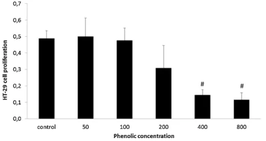
Figs. 6 and 7, show that exposure to 200, 400 and 800μg of phe-nolics/mL of M. pulegium phenolic extracts reduced the proliferative ability of HT-29 cells to invade their wounds, with a dose-dependent effect. When using the EC50 concentration (regarding MMP inhibition) the cell migration property assessed in the wound healing assay was reduced by over 70% when compared to control.
Fig. 1. Main constituents and phenolic profile of the Mentha pulegium extract with identification of the functional groups from the main components: 1. HB -hydroxybenzoic acid; 2-8. and 14-20. HC– hydroxicinnamic acids; 9-13. HMF – hydroxymetoxyflavones; 21,22. C - catequines.
Table 3
Antioxidant capacity of M. pulegium extract.
CUPRAC FRAP DPPH O2−.Assay
μmol AAE/mL μmol Fe2+/mL mg AAE/mL μmol GAE/mL
1.71 ± 0.07 10.29 ± 0.63 0.88 ± 0.02 9.23 ± 3.29
All values are mean ± standard deviation of triplicates.
Fig. 2. Effect of M. pulegium extract on the rat paw edema development elicited by carra-geenan 6 h after edema induction. Effect of a single administration of M. pulegium extract (12,5 mg of phenolic acids/kg, p.o.) in com-parison with the effect of a single administra-tion of indomethacin (10 mg/kg, p.o., n = 8), lycopene (50 mg/kg, n = 8; p.o.), tempol (30 mg/kg, n = 8; p.o.) and trolox (30 mg/kg, n = 8; p.o.) [1]. The data is presented as means with their standard errors. *P < 0.01 vs Con-trol Group;#P < 0.01 vs Carrageenan group; θP < 0.001 vs Carrageenan group.
3.6. Cell MMP activity in cultured HT-29 cells and colon tissue from the in vivo colitis model
Gelatinolytic activity (Fig. 8a) showed that MMPs in HT-29 cells were inhibited by the phenolic extracts from M. pulegium. Since the DQ-gelatin assay provided evidence of total DQ-gelatinolytic activity in the extracellular matrix, we further analysed the activity of MMP-2 and -9 through zymography (Fig. 8b). When observing the effects of the ex-tract in MMP-2 expression, a visible inhibition was observed, sup-porting the results evidenced in Figure 11a. Minimal inhibitory con-centrations (MIC) for HT-29 cell growth and gelatinase activity was quantified and are expressed inTable 5.
4. Discussion
Although the food industry has been mainly focused on the essential oil of pennyroyal due to its known antibacterial properties and use as food preservative, few studies have focused on phenolic extracts of pennyroyal and, until now, no study has specifically investigated its
beneficial effects on IBD.
Zaidi et al isolated a series of hydroxylated metoxyflavones from dried leaves of M. pulegium and M. suaveolens, using alcohol solvents and purification by column chromatography [41]. Proestos et al. [42] and Fatiha et al. [40] studied the crude ethanolic extracts obtained from the dried leaves of M. pulegium and concluded that their main phenolic components are hydroxybenzoic and hydroxycinnamic acids,flavones andflavanones.
Taking into account the relative chromatographic areas of the main components detected in the HPLC-DAD profile of the M. pulegium ex-tract used in this work, it is evident that there is a predominance of hydroxycinnamic acids (74.4%), followed by hydroxymethoxyflavones (11.2%), catechins (9.6%) and hydroxybenzoic acids (4.8%) (Fig. 1). The main component of the extract is rosmarinic acid, a hydro-xycinnamic acid, and accounts for 36.7% of the relative chromato-graphic area (Fig. 1).
This in line with other publications regarding the characterization of the phenolic content of M. pulegium leaves extract. In 2018, Politeo et al performed a phytochemical analysis as well as an evaluation of the Table 4
Morphologic and functional observations of the colon, immediately after collection.
Length of colon (cm) Extent of injury Presence/Consistency of diarrhoea Mortality (%)
Sham 14.5 ± 0.08 0 0 0 EtOH 50% 14.1 ± 0.20 0 0 0 TNBS + EtOH 50% 11.8 ± 0.19# 3.6 ± 0.14# 3# 30 TNBS + M. pulegium 14.3 ± 0.48* 3.5 ± 0.35# 1.75 ± 0.48* 25 # P < 0.05 vs Sham. * P < 0.05 vs TNBS + EtOH 50%.
Fig. 3. Effect of M. pulegium extract administration on the lenght of colon (cm), extent of intestine injury (cm) and visual macroscopic observation of the colon. Sham group (n = 6), EtOH group (n = 6), TNBS group (n = 10), TNBS + M. pulegium (n = 10).#P < 0.001 vs Sham group, *P < 0.001 vs TNBS group.
antioxidant and anticholinesterase potential of hot water and metha-nolic extracts from M. pulegium L. [43]. Rosmarinic acid was the most abundant compound in the tested extracts, followed by ellagic acid, eriodictyol, naringenin and chlorogenic acid. The authors found that the phenolic-rich extracts demonstrated good radical scavenging po-tential, reducing power and ability to inhibit lipid oxidation. The tested extracts also showed low ability to inhibit protein oxidation and low or
no acetylcholinesterase and butyrylcholinesterase inhibition potential. In order to determine the antioxidant potential of M. pulegium ex-tract, reducing capacity (FRAP and CUPRAC) and free radical scaven-ging activity (DPPH and O2- assay) assays were employed (Table 3). The antioxidant capacity of M. pulegium extract was detected in all the screening assays (FRAP, CUPRAC, DPPH and superoxide assays) and the results obtained are in accordance with previous studies which also Fig. 4. Effect of M. pulegium extract administration on the histological features of colon inflammation. Effect of M. pulegium administration on the histological features of colon inflammation. (A) Sham group (n = 6) shows a normal colon with no lesions and normal crypt architecture. (B) TNBS group (n = 10) exhibits severe ulceration with altered crypts and increased infiltration of immune cells (black arrow, score 2,5). (C) TNBS + M. pulegium group (n = 10), exhibits colon with mucosal erosion and small superficial ulcers scattered along the length of the colon, with slight crypt alteration and immune cell infiltration (black arrow, score 1). Original magnification 100 × . Scale bar equals 200 microns.
Fig. 5. Effect of M. pulegium extract administration on the colon tissue expression of COX-2 and iNOS. (A) – COX-2 expression; B) – iNOS expression. (1) Sham group (n = 6), (2) TNBS group (n = 10), (3) TNBS + M. pulegium group (n = 10). The samples from animals subjected to intestinal colitis showed marked expression of COX-2 and iNOS (brown staining, arrows), while samples from M. pulegium extract treated animals exhibited marked reduction for both markers. Original mag-nification x400. Scale bar equals 200 microns. Percentage of tissue area stained in brown, was obtained using the ImageJ (Fiji Is Just) software. *P < 0.05 vs Sham, **P < 0.01 vs Sham,##P < 0.01 vs TNBS.
demonstrated that phenolic extracts of M. pulegium exhibit reducing capacity and are able to scavenge reactive oxygen species [13,39,40].
Although, as previously mentioned, very few studies have focused on the evaluation of biological activities of phenolic extracts of pen-nyroyal (as well as other aromatic plants), the ones actually performed and published appear to corroborate the results obtained in our study. In a 2011 study by Moussaid and colleagues,five ethanolic extracts of plants used in Moroccan traditional medicine were tested for their in vitro antioxidant and in vivo anti-inflammatory activities (using a carrageenan-induced ear oedema model in mice). The most active plant in both properties was M. pulegium, which was also the plant that ex-hibited a higher concentration of phenolics andflavonoids.
A study from Brahmi and colleagues [44] evaluated the antioxidant, anti-inflammatory and cytoprotective effects of ethanolic extracts from 3 Mentha species from Algeria (M. spicata, M. pulegium and M. ro-tundifolia) on murine RAW 264.7 LPS-treated macrophages. All the
extracts strongly reduced IL-6 secretion and two of them (M. pulegium and M. rotundifolia) also decreased MCP-1 and TNF-α secretion. The same authors previously characterized the phenolic content of the M. pulegium extract, having identified as the main constituents rosmarinic acid and diosmin [45].
Our results showed that the phenolic extract of M. pulegium (pen-nyroyal) possesses a significant anti-inflammatory effect, identifiable in an acute setting, as demonstrated by the reduction of paw oedema in the carrageenan-induced oedema model. Furthermore, we were able to demonstrate that administration of this extract to animals subjected to colitis induction resulted in a significant reduction of several para-meters that are mimicked in human IBD: intestinal inflammatory injury (macroscopical and histological findings); diarrhoea severity and ex-pression of inflammatory markers.
Previous studies by our group have shown that phenolic extracts exhibited a beneficial effect in this model of IBD [31,34], where Fig. 6. Effect of Pennyroyal extract administration on HT29 cell proliferation after exposure to 0, 50, 100, 200,400 and 800 μg total phenolics/mL of pennyroyal extracts, quantified by the MTT method. Results are expressed as represent an average of at least three replicate experiments (n = 3) ± SD.#P < 0,001 vs Control.
Fig. 7. HT29 cell migration after exposure to phenolic extracts of pennyroyal as determined by wound healing assays. The histogram reports the relative migration rates, where values are the means of at least three replicate experiments ± SD, and are expressed as % wound closure in relation to day 0. Cells were grown until reaching 80% confluence and the monolayer was scratched with a pipette tip (day 0). Cells were then exposed to the phenolic concentrations equivalent of the determined EC50 of MMP activity (500 g/mL). Cell migration was recorded after 48 h. ***P < 0.001.
administration of persimmon and spearmint phenolic extracts at similar doses also led to similar results. Although some of the more species-specific compounds are not common to these plants, it proves the concept that the same dose of total phenolic compounds has a beneficial effect in the same experimental model of disease.
In the carrageenan-induced paw oedema model we observed a sig-nificant reduction of oedema caused by local inflammation, in a mag-nitude of effect that was comparable to substances used as positive controls, a non-steroidal anti-inflammatory drug (indomethacin) and two reactive oxygen species scavengers (trolox and tempol).
Although no direct correlation can me made regarding the me-chanisms responsible for these effects, our results did show that not
only the pennyroyal extract possessed a high antioxidant capacity, but the chemical profiling revealed a high concentration of rosmarinic acid, a common aromatic herb constituent. Rosmarinic acid has also been studied by our group and has revealed to be one of the main responsible for the anti-inflammatory effect of a rosemary extract in local models of acute inflammation in rodents and when administered isolated has shown to reveal important anti-inflammatory effects in acute models of critical care settings [30]. Recently, two studies have reported the effect of administration of rosmarinic acid in another model of IBD, the DSS-induced colitis in mice. In one of these studies, rosmarinic acid ad-ministration (30 mg/kg p.o. for 7 days after induction of colitis) sig-nificantly reduced the severity of colitis and results suggested that the suppression of colonic inflammation was related to a dual inhibition of NF-κB and STAT3 pathways activation [46]. In our study we demon-strated that two of the parameters that were reduced by administration of rosmarinic acid were the protein expression of COX-2 and iNOS. Studies have been shown that diet polyphenols were able to inhibit several transcription factors known to induce COX-2 and iNOS ex-pression (NF-κB, JAK/STAT and MAPK), in general processes of in-flammation and immunomodulation [10], intestinal inflammation [47] and CRC [48]. In a second study, administration of rosmarinic acid (25, Fig. 8. Effect of pennyroyal extract on the gelatinase activity of MMP-2 and MMP-9.
a) Effect of pennyroyal extract after exposure of isolated MMPs to 0, 50, 100, 200,400 and 800 μg total phenolics/mL of ppennyroyal extracts, as quantified by the DQ fluorogenic method. Results are expressed as relative fluorescence as a % of controls and represent an average of at least three replicate experiments (n = 3) ± SD. b) Effect of Pennyroyal extract administration on the gelatinase activity of MMP-2 and MMP-9 in HT-29 cells.
b1 - Proteolytic activity of gelatinases present in the HT29 extracellular media after a 48 h exposure to 0 and 500μg total phenolics.mL-1 of pennyroyal extracts, as quantified by the DQ fluorogenic method. Results are expressed as relative fluorescence as a % of controls and represent an average of at least three replicate experiments (n = 3) ± SD.⁎P < 0.05.
b2 - Representative image of the zymographic profiles of MMP-9 and MMP-2 activities in HT29 extracellular media after a 48 h-exposure to 0 and 500 μg/mL). HT29 extracellular media was loaded in 12.5% (w/v acrylamide) polyacrylamide gels co-polymerized with 1% (w/v) gelatin. C = controls and P = pennyroyal extract. * *P < 0.001.
Table 5
Minimal inhibitory concentrations against HT29 cell growth and gelatinase activity. Values are inμg total phenolics.mL-1.
MIC (μg total phenolics/mL) HT29 cell growth 5.2
50 and 100 mg/kg p.o. 3 days before colitis induction and then daily for 8 days) significantly decreased disease severity and histological score of colons in DSS-induced colitis mice [49]. Particularly for the higher dose, rosmarinic acid administration was able to reduce nitric oxide (NO) production and, among other inflammatory mediators, also re-duced the expression of iNOS and COX-2. Inhibition of NO production can be directly correlated to reduction of iNOS expression and is an important target in inflammatory processes since NO is an important initiator of oxidant and proinflammatory pathways. Its role in human IBD has also been investigated and studies have shown that the level of NO production by iNOS exhibits a high correlation with the intensity of disease in IBD [50] and was in fact proposed as a new and useful bio-marker in the clinical setting, namely in diagnostic protocols and in the monitoring of patients with colitis or Crohn's disease [51]. Pro-in-flammatory cytokines at the site of inflammation can induce COX-2 expression and increase the synthesis of prostaglandins that conse-quently are able to stimulate cancer cell proliferation, promote angio-genesis, inhibit apoptosis, and increase metastatic potential [48]. The influence of increased COX-2 expression and upstream pathways that regulates it have been shown to be of significant relevance in IBD and CRC in humans [52,53].
Our results show that administration of a pennyroyal extract at a dose of 15 mg/kg (of phenolic acids) was able not only to reduce the severity of colitis but also to reduce the expression of COX-2 and iNOS (among other parameters) with just 4 days of treatment.
Another inflammatory mediator that might also contribute to the beneficial effect of pennyroyal administration in this animal model of colitis, is the expression of matrix metalloproteinases (MMP). Matrix metalloproteinases (MMPs) are considered to be relevant proteases in-volved in the pathogenesis of IBD through their ability to remodel the extracellular matrix in response to inflammatory stimuli and by their immunomodulating effects [54]. Also, genetic studies performed in humans show that genetic variations in MMPs may be associated with increased risk of ulcerative colitis and may play a role in interindividual differences in UC susceptibility and clinical outcome [55]. Additionally, MMPs are enzymes that play a crucial role in the transformation and progression of tumours, especially during the invasion and metastasis stages [56] and some preclinical and even clinical studies have already shown that MMP-2 and MMP-9 expression and activation might be of great relevance for migration and invasion of CRC cells, leading to fa-cilitation of angiogenesis and metastasis [57,58]. Here we evaluated the inhibitory effect of the pennyroyal phenolic extract in the gelatinolytic activity of both MMP-2 and MMP-9 in HT29 CRC cells. Our results showed that pennyroyal extract was able to reduce the activity of both MMP-2 and MMP-9. Furthermore, we were able to observe a significant inhibitory effect of both the proliferation and invasibility properties of HT-29 CRC cells.
Given the relevance of MMP activity in inflammatory and onco-genesis processes, studies have been performed to investigate the multiple pathways upstream MMP expression that are able to regulate it. Not surprisingly, some of the most influent pathways involved in the increased expression of MMPs are also crucial pathways and tran-scription factors closely related to inflammatory pathways responsible for intestinal inflammation and progression to colorectal cancer, such as the above mentioned NF-κB, STAT and MAPK pathways [59,60], AP-1 and PKC [60,61].
As mentioned in the introduction section, a well-established con-nection exists between IBD and CRC. Dyson and colleagues have ex-tensively characterized the risk factors involved in this connection, having identified the following risk factors has being the most relevant: the duration and extent of colitis, the severity of inflammation and the existence of family history of sporadic colorectal cancer [62]. Kraus and colleagues have pointed out the cumulative effect of chronic in-flammation and its correlation with inin-flammation severity and disease extent and duration as the main link between IBD and the elevated CRC risk [63]. Although the exact mechanisms and pathways are not yet
elucidated, mainly due to a pleiotropic effect and cross-link between several inflammatory pathways, chronic inflammation is considered to be the main driver towards initiation and promotion of cancerous processes in the colon. Increasing evidence points to two key genes in the inflammatory process, cyclooxygenase-2 (COX-2) and nuclear factor kappaB (NF-kB), that may provide a mechanistic link between in-flammation and cancer while other factors such as, TNF-a and IL-6-induced signaling have been recently shown to promote tumor growth in experimental models of colitis-associated cancer [63]. Therefore, colitis-associated cancer appears to be influenced by inflammatory processes, an imbalance in intestinal microbiota, and a crosstalk be-tween various signaling pathways. A clear mechanistic origin is not yet identified but a link through a molecular crosstalk of multiple in-flammatory loops including TGFβ, NFKB, TNFα and ROS are a few possibilities [64].
In conclusion, our results show, to our knowledge for thefirst time, a beneficial effect of M. pulegium (pennyroyal) phenolic extract on the amelioration of IBD severity and on the impairment of processes re-levant for progression into CRC. Given the role of inflammatory pro-cesses in the progression of colorectal cancer and the important link between inflammation and cancer, these results might open new re-search opportunities for the adjuvant therapeutic management of IBD patients.
Funding
This work was supported by Universidade de Lisboa (PhD grant REITORIA/BD/FF01/2015) and by FCT/MCTES as the funding entity of the MEtRiCs unit under the project UID/EMS/04077/2019.
Declaration of Competing Interest
On behalf of all authors, the corresponding author states that there is no conflict of interest.
References
[1] A. Salaritabar, B. Darvishi, F. Hadjiakhoondi, A. Manayi, A. Sureda, S.F. Nabavi, L.R. Fitzpatrick, S.M. Nabavi, A. Bishayee, Therapeutic potential offlavonoids in inflammatory bowel disease: a comprehensive review, World J. Gastroenterol. 23 (2017) 5097,https://doi.org/10.3748/wjg.v23.i28.5097.
[2] D. Corridoni, K.O. Arseneau, F. Cominelli, Inflammatory bowel disease, Immunol. Lett. 161 (2014) 231–235,https://doi.org/10.1016/j.imlet.2014.04.004. [3] F. Bray, J. Ferlay, I. Soerjomataram, R.L. Siegel, L.A. Torre, A. Jemal, Global cancer
statistics 2018: GLOBOCAN estimates of incidence and mortality worldwide for 36 cancers in 185 countries, CA Cancer J. Clin. 68 (2018) 394–424,https://doi.org/10. 3322/caac.21492.
[4] E.M. Stoffel, F. Kastrinos, Familial colorectal cancer, beyond lynch syndrome, Clin. Gastroenterol. Hepatol. 12 (2014) 1059–1068,https://doi.org/10.1016/j.cgh. 2013.08.015.
[5] I. Mármol, C. Sánchez-de-Diego, A. Pradilla Dieste, E. Cerrada, M.J. Rodriguez Yoldi, Colorectal carcinoma: a general overview and future perspectives in color-ectal cancer, Int. J. Mol. Sci. 18 (2017) 197,https://doi.org/10.3390/ ijms18010197.
[6] M.G. Donovan, O.I. Selmin, T.C. Doetschman, D.F. Romagnolo, Mediterranean diet: prevention of colorectal Cancer, Front. Nutr. 4 (2017) 59,https://doi.org/10.3389/ fnut.2017.00059.
[7] J.E. Axelrad, S. Lichtiger, V. Yajnik, Inflammatory bowel disease and cancer: the role of inflammation, immunosuppression, and cancer treatment, World J. Gastroenterol. 22 (2016) 4794,https://doi.org/10.3748/wjg.v22.i20.4794. [8] M. Romano, F. DE Francesco, L. Zarantonello, C. Ruffolo, G.A. Ferraro, G. Zanus,
A. Giordano, N. Bassi, U. Cillo, From inflammation to cancer in inflammatory bowel disease: molecular perspectives, Anticancer Res. 36 (2016) 1447–1460 (Accessed April 4, 2018),http://www.ncbi.nlm.nih.gov/pubmed/27069120.
[9] G. Rogler, Chronic ulcerative colitis and colorectal cancer, Cancer Lett. 345 (2014) 235–241,https://doi.org/10.1016/j.canlet.2013.07.032.
[10] N. Yahfoufi, N. Alsadi, M. Jambi, C. Matar, The immunomodulatory and anti-in-flammatory role of polyphenols, Nutrients 10 (2018) 1618,https://doi.org/10. 3390/nu10111618.
[11] F. Shahidi, J. Yeo, Bioactivities of phenolics by focusing on suppression of chronic diseases: a review, Int. J. Mol. Sci. 19 (2018) 1573,https://doi.org/10.3390/ ijms19061573.
[12] A. Kapinova, P. Kubatka, O. Golubnitschaja, M. Kello, P. Zubor, P. Solar, M. Pec, Dietary phytochemicals in breast cancer research: anticancer effects and potential
utility for effective chemoprevention, Environ. Health Prev. Med. 23 (2018) 36,
https://doi.org/10.1186/s12199-018-0724-1.
[13] B. Teixeira, A. Marques, C. Ramos, I. Batista, C. Serrano, O. Matos, N.R. Neng, J.M.F. Nogueira, J.A. Saraiva, M.L. Nunes, European pennyroyal (Mentha pule-gium) from Portugal: chemical composition of essential oil and antioxidant and antimicrobial properties of extracts and essential oil, Ind. Crops Prod. 36 (2012) 81–87,https://doi.org/10.1016/J.INDCROP.2011.08.011.
[14] P. Tongnuanchan, S. Benjakul, Essential oils: extraction, bioactivities, and their uses for food preservation, J. Food Sci. 79 (2014) R1231–R1249,https://doi.org/10. 1111/1750-3841.12492.
[15] N. Silva, S. Alves, A. Gonçalves, J.S. Amaral, P. Poeta, Antimicrobial activity of essential oils from Mediterranean aromatic plants against several foodborne and spoilage bacteria, Food Sci. Technol. Int. 19 (2013) 503–510,https://doi.org/10. 1177/1082013212442198.
[16] N. Ceyhan-Güvensen, D. Keskin, Chemical content and antimicrobial properties of three different extracts of Mentha pulegium leaves from Mugla Region, Turkey, J. Environ. Biol. 37 (2016) 1341–1346 (Accessed April 4, 2018),http://www.ncbi. nlm.nih.gov/pubmed/29261270.
[17] P. Gordon, S.C. Khojasteh, A decades-long investigation of acute metabolism-based hepatotoxicity by herbal constituents: a case study of pennyroyal oil, Drug Metab. Rev. 47 (2015) 12–20,https://doi.org/10.3109/03602532.2014.990032. [18] R.F. Hurrell, M. Reddy, J.D. Cook, Inhibition of non-haem iron absorption in man
by polyphenolic-containing beverages, Br. J. Nutr. 81 (1999) 289–295 (Accessed April 4, 2018),http://www.ncbi.nlm.nih.gov/pubmed/10999016.
[19] F. Brahmi, F. Dahmoune, N. Kadri, M. Chibane, S. Dairi, H. Remini, S. Oukmanou-Bensidhoum, L. Mouni, K. Madani, Antioxidant capacity and phenolic content of two Algerian Mentha species M. rotundifolia (L.) Huds, M. pulegium L., extracted with different solvents, J. Complement. Integr. Med. 14 (2017),https://doi.org/10. 1515/jcim-2016-0064.
[20] L. Alpsoy, H. Sahin, S. Karaman, Anti-oxidative and anti-genotoxic effects of me-thanolic extract of Mentha pulegium on human lymphocyte culture, Toxicol. Ind. Health 27 (2011) 647–654,https://doi.org/10.1177/0748233710393402. [21] A. Taamalli, D. Arráez-Román, L. Abaza, I. Iswaldi, A. Fernández-Gutiérrez,
M. Zarrouk, A. Segura-Carretero, LC-MS-based metabolite profiling of methanolic extracts from the medicinal and aromatic species Mentha pulegium and Origanum majorana, Phytochem. Anal. 26 (2015) 320–330,https://doi.org/10.1002/pca. 2566.
[22] M. Shekarchi, H. Hajimehdipoor, S. Saeidnia, A.R. Gohari, M.P. Hamedani, Comparative study of rosmarinic acid content in some plants of Labiatae family, Pharmacogn. Mag. 8 (2012) 37–41,https://doi.org/10.4103/0973-1296.93316. [23] S.E. Çelik, A.N. Tufan, B. Bekdeser, M. Özyürek, K. Güçlü, R. Apak, Identification
and determination of phenolics in Lamiaceae species by UPLC-DAD-ESI-MS/MS, J. Chromatogr. Sci. 55 (2017) 291–300,https://doi.org/10.1093/chromsci/bmw184. [24] M. Koşar, F. Göger, K.H. Can Başer, In vitro antioxidant properties and phenolic
composition of Salvia virgata Jacq. from Turkey, J. Agric. Food Chem. 56 (2008) 2369–2374,https://doi.org/10.1021/jf073516b.
[25] L. Barros, A.M. Carvalho, J.S. Morais, I.C.F.R. Ferreira, Strawberry-tree, blackthorn and rose fruits: detailed characterisation in nutrients and phytochemicals with antioxidant properties, Food Chem. 120 (2010) 247–254,https://doi.org/10.1016/ J.FOODCHEM.2009.10.016.
[26] R. Apak, K. Güçlü, M. Ozyürek, S.E. Karademir, Novel total antioxidant capacity index for dietary polyphenols and vitamins C and E, using their cupric ion reducing capability in the presence of neocuproine: CUPRAC method, J. Agric. Food Chem. 52 (2004) 7970–7981,https://doi.org/10.1021/jf048741x.
[27] D. Ramful, T. Bahorun, E. Bourdon, E. Tarnus, O.I. Aruoma, Bioactive phenolics and antioxidant propensity offlavedo extracts of Mauritian citrus fruits: potential pro-phylactic ingredients for functional foods application, Toxicology 278 (2010) 75–87,https://doi.org/10.1016/j.tox.2010.01.012.
[28] N. Miceli, A. Trovato, P. Dugo, F. Cacciola, P. Donato, A. Marino, V. Bellinghieri, T.M. La Barbera, A. Güvenç, M.F. Taviano, Comparative analysis offlavonoid profile, antioxidant and antimicrobial activity of the berries of Juniperus communis L. var. communis and Juniperus communis L. var. saxatilis Pall. from Turkey, J. Agric. Food Chem. 57 (2009) 6570–6577,https://doi.org/10.1021/jf9012295. [29] P. Valentão, E. Fernandes, F. Carvalho, P.B. Andrade, R.M. Seabra, M.L. Bastos,
Antioxidant activity of Centaurium erythraea infusion evidenced by its superoxide radical scavenging and xanthine oxidase inhibitory activity, J. Agric. Food Chem. 49 (2001) 3476–3479 (Accessed December 20, 2018),http://www.ncbi.nlm.nih. gov/pubmed/11453794.
[30] J. Rocha, M. Eduardo-Figueira, A. Barateiro, A. Fernandes, D. Brites, R. Bronze, C.M.M. Duarte, A.T. Serra, R. Pinto, M. Freitas, E. Fernandes, B. Silva-Lima, H. Mota-Filipe, B. Sepodes, Anti-inflammatory effect of rosmarinic acid and an extract of Rosmarinus officinalis in rat models of local and systemic inflammation, Basic Clin. Pharmacol. Toxicol. 116 (2015) 398–413,https://doi.org/10.1111/ bcpt.12335.
[31] R. Direito, A. Lima, J. Rocha, R.B. Ferreira, J. Mota, P. Rebelo, A. Fernandes, R. Pinto, P. Alves, R. Bronze, B. Sepodes, M.-E. Figueira, Dyospiros kaki phenolics inhibit colitis and colon cancer cell proliferation, but not gelatinase activities, J. Nutr. Biochem. 46 (2017) 100–108,https://doi.org/10.1016/j.jnutbio.2017.03. 002.
[32] M.E. Figueira, M.B. Câmara, R. Direito, J. Rocha, A.T. Serra, C.M.M. Duarte, A. Fernandes, M. Freitas, E. Fernandes, M.C. Marques, M.R. Bronze, B. Sepodes, Chemical characterization of a red raspberry fruit extract and evaluation of its pharmacological effects in experimental models of acute inflammation and col-lagen-induced arthritis, Food Funct. 5 (2014) 3241–3251,https://doi.org/10.1039/ c4fo00376d.
[33] M.-E. Figueira, M. Oliveira, R. Direito, J. Rocha, P. Alves, A.-T. Serra, C. Duarte,
R. Bronze, A. Fernandes, D. Brites, M. Freitas, E. Fernandes, B. Sepodes, Protective effects of a blueberry extract in acute inflammation and collagen-induced arthritis in the rat, Biomed. Pharmacother. 83 (2016) 1191–1202,https://doi.org/10.1016/ j.biopha.2016.08.040.
[34] R. Direito, J. Rocha, A. Lima, M.M. Gonçalves, M.P. Duarte, V. Mateus, C. Sousa, A. Fernandes, R. Pinto, R. Boavida Ferreira, B. Sepodes, M.-E. Figueira, Reduction of inflammation and colon injury by a spearmint phenolic extract in experimental bowel disease in mice, Medicines 6 (2019) 65,https://doi.org/10.3390/ medicines6020065.
[35] J. Carmichael, W.G. DeGraff, A.F. Gazdar, J.D. Minna, J.B. Mitchell, Evaluation of a tetrazolium-based semiautomated colorimetric assay: assessment of radio-sensitivity, Cancer Res. 47 (1987) 943–946 (Accessed April 4, 2018),http://www. ncbi.nlm.nih.gov/pubmed/3802101.
[36] A.I.G. Lima, J. Mota, S.A.V.S. Monteiro, R.M.S.B. Ferreira, Legume seeds and col-orectal cancer revisited: protease inhibitors reduce MMP-9 activity and colon cancer cell migration, Food Chem. 197 (2016) 30–38,https://doi.org/10.1016/j. foodchem.2015.10.063.
[37] S. Bouhdid, J. Abrini, M. Amensour, A. Zhiri, M.J. Espuny, A. Manresa, Functional and ultrastructural changes in Pseudomonas aeruginosa and Staphylococcus aureus cells induced by Cinnamomum verum essential oil, J. Appl. Microbiol. 109 (2010) 1139–1149,https://doi.org/10.1111/j.1365-2672.2010.04740.x.
[38] M. Toth, A. Sohail, R. Fridman, Assessment of gelatinases (MMP-2 and MMP-9) by gelatin zymography, Methods Mol. Biol. (2012), pp. 121–135,https://doi.org/10. 1007/978-1-61779-854-2_8.
[39] A.T. Mata, C. Proença, A.R. Ferreira, M.L.M. Serralheiro, J.M.F. Nogueira, M.E.M. Araújo, Antioxidant and antiacetylcholinesterase activities offive plants used as Portuguese food spices, Food Chem. 103 (2007) 778–786,https://doi.org/ 10.1016/J.FOODCHEM.2006.09.017.
[40] B. Fatiha, H. Didier, G. Naima, M. Khodir, K. Martin, K. Léocadie, S. Caroline, C. Mohamed, D. Pierre, Phenolic composition, in vitro antioxidant effects and tyr-osinase inhibitory activity of three Algerian Mentha species: M. spicata (L.), M. pulegium (L.) and M. rotundifolia (L.) Huds (Lamiaceae), Ind. Crops Prod. 74 (2015) 722–730,https://doi.org/10.1016/J.INDCROP.2015.04.038.
[41] F. Zaidi, B. Voirin, M. Jay, M.R. Viricel, Freeflavonoid aglycones from leaves of mentha pulegium and mentha suaveolens (labiatae), Phytochemistry 48 (1998) 991–994,https://doi.org/10.1016/S0031-9422(97)01042-X.
[42] C. Proestos, N. Chorianopoulos, G.-J.E. Nychas, M. Komaitis, RP-HPLC analysis of the phenolic compounds of plant extracts. Investigation of their antioxidant capa-city and antimicrobial activity, J. Agric. Food Chem. 53 (2005) 1190–1195,https:// doi.org/10.1021/jf040083t.
[43] O. Politeo, M. Bektašević, I. Carev, M. Jurin, M. Roje, Phytochemical composition, antioxidant potential and cholinesterase inhibition potential of extracts from Mentha pulegium L, Chem. Biodivers. 15 (2018) e1800374, ,https://doi.org/10. 1002/cbdv.201800374.
[44] F. Brahmi, T. Nury, M. Debbabi, S. Hadj-Ahmed, A. Zarrouk, M. Prost, K. Madani, L. Boulekbache-Makhlouf, G. Lizard, Evaluation of antioxidant, anti-inflammatory and cytoprotective properties of ethanolic mint extracts from Algeria on 7-Ketocholesterol-treated murine RAW 264.7 macrophages, Antioxidants 7 (2018) 184,https://doi.org/10.3390/antiox7120184.
[45] B. Fatiha, H. Didier, G. Naima, M. Khodir, K. Martin, K. Léocadie, S. Caroline, C. Mohamed, D. Pierre, Phenolic composition, in vitro antioxidant effects and tyr-osinase inhibitory activity of three Algerian Mentha species: M. spicata (L.), M. pulegium (L.) and M. rotundifolia (L.) Huds (Lamiaceae), Ind. Crops Prod. 74 (2015) 722–730,https://doi.org/10.1016/J.INDCROP.2015.04.038.
[46] B.-R. Jin, K.-S. Chung, S.-Y. Cheon, M. Lee, S. Hwang, S. Noh Hwang, K.-J. Rhee, H.-J. An, Rosmarinic acid suppresses colonic inflammation in dextran sulphate sodium (DSS)-induced mice via dual inhibition of NF-κB and STAT3 activation, Sci. Rep. 7 (2017) 46252,https://doi.org/10.1038/srep46252.
[47] T. Guina, M. Deiana, S. Calfapietra, B. Cabboi, M. Maina, C.I. Tuberoso, G. Leonarduzzi, P. Gamba, S. Gargiulo, G. Testa, G. Poli, F. Biasi, The role of p38 MAPK in the induction of intestinal inflammation by dietary oxysterols: modulation by wine phenolics, Food Funct. 6 (2015) 1218–1228,https://doi.org/10.1039/ C4FO01116C.
[48] S.J. Desai, B. Prickril, A. Rasooly, Mechanisms of phytonutrient modulation of cy-clooxygenase-2 (COX-2) and inflammation related to Cancer, Nutr. Cancer 70 (2018) 350–375,https://doi.org/10.1080/01635581.2018.1446091.
[49] L. Zhao, Y. Zhang, G. Liu, S. Hao, C. Wang, Y. Wang, Black rice anthocyanin-rich extract and rosmarinic acid, alone and in combination, protect against DSS-induced colitis in mice, Food Funct. 9 (2018) 2796–2808,https://doi.org/10.1039/ c7fo01490b.
[50] R.K. Cross, K.T. Wilson, Nitric oxide in inflammatory bowel disease, Inflamm. Bowel Dis. 9 (2003) 179–189 (Accessed December 20, 2018),http://www.ncbi. nlm.nih.gov/pubmed/12792224.
[51] J.O. Lundberg, P.M. Hellström, M.K. Fagerhol, E. Weitzberg, A.G. Roseth, Technology Insight: calprotectin, lactoferrin and nitric oxide as novel markers of inflammatory bowel disease, Nat. Clin. Pract. Gastroenterol. Hepatol. 2 (2005) 96–102,https://doi.org/10.1038/ncpgasthep0094.
[52] D. Wang, R.N. DuBois, The role of COX-2 in intestinal inflammation and colorectal cancer, Oncogene 29 (2010) 781–788,https://doi.org/10.1038/onc.2009.421. [53] S.A. Azer, Overview of molecular pathways in inflammatory bowel disease
asso-ciated with colorectal cancer development, Eur. J. Gastroenterol. Hepatol. 25 (2013) 271–281,https://doi.org/10.1097/MEG.0b013e32835b5803.
[54] M. de Bruyn, J. Vandooren, E. Ugarte-Berzal, I. Arijs, S. Vermeire, G. Opdenakker, The molecular biology of matrix metalloproteinases and tissue inhibitors of me-talloproteinases in inflammatory bowel diseases, Crit. Rev. Biochem. Mol. Biol. 51 (2016) 295–358,https://doi.org/10.1080/10409238.2016.1199535.
[55] A.R. Morgan, D.-Y. Han, W.-J. Lam, C.M. Triggs, A.G. Fraser, M. Barclay, R.B. Gearry, S. Meisner, P. Stokkers, G.E. Boeckxstaens, L.R. Ferguson, Genetic variations in matrix metalloproteinases may be associated with increased risk of ulcerative colitis, Hum. Immunol. 72 (2011) 1117–1127,https://doi.org/10.1016/ j.humimm.2011.08.011.
[56] M.Z. Banday, A.S. Sameer, A.H. Mir, T.A. Mokhdomi, N.A. Chowdri, E. Haq, Matrix metalloproteinase (MMP) -2, -7 and -9 promoter polymorphisms in colorectal cancer in ethnic Kashmiri population— a case–control study and a mini review, Gene 589 (2016) 81–89,https://doi.org/10.1016/j.gene.2016.05.028. [57] S. Zeynali-Moghaddam, M. Mohammadian, F. Kheradmand, A. Fathi-Azarbayjani,
Y. Rasmi, O. Esna-Ashari, H. Malekinejad, A molecular basis for the synergy be-tween 17‑allylamino‑17‑demethoxy geldanamycin with Capecitabine and Irinotecan in human colorectal cancer cells through VEFG and MMP-9 gene ex-pression, Gene 684 (2019) 30–38,https://doi.org/10.1016/j.gene.2018.10.016. [58] Y. Wang, N. Wu, B. Pang, D. Tong, D. Sun, H. Sun, C. Zhang, W. Sun, X. Meng, J. Bai, F. Chen, J. Geng, S. Fu, Y. Jin, TRIB1 promotes colorectal cancer cell mi-gration and invasion through activation MMP-2 via FAK/Src and ERK pathways, Oncotarget 8 (2017) 47931–47942,https://doi.org/10.18632/oncotarget.18201. [59] M. Fanjul-Fernández, A.R. Folgueras, S. Cabrera, C. López-Otín, Matrix
metalloproteinases: evolution, gene regulation and functional analysis in mouse models, Biochim. Biophys. Acta - Mol. Cell Res. 1803 (2010) 3–19,https://doi.org/ 10.1016/J.BBAMCR.2009.07.004.
[60] C. Yan, D.D. Boyd, Regulation of matrix metalloproteinase gene expression, J. Cell. Physiol. 211 (2007) 19–26,https://doi.org/10.1002/jcp.20948.
[61] S. Chakraborti, M. Mandal, S. Das, A. Mandal, T. Chakraborti, Regulation of matrix metalloproteinases: an overview, Mol. Cell. Biochem. 253 (2003) 269–285 (Accessed March 1, 2019),http://www.ncbi.nlm.nih.gov/pubmed/14619979. [62] J.K. Dyson, M.D. Rutter, Colorectal cancer in inflammatory bowel disease: What is
the real magnitude of the risk? World J. Gastroenterol. 18 (2012) 3839,https://doi. org/10.3748/wjg.v18.i29.3839.
[63] S. Kraus, N. Arber, Inflammation and colorectal cancer, Curr. Opin. Pharmacol. 9 (2009) 405–410,https://doi.org/10.1016/j.coph.2009.06.006.
[64] A. Jurjus, A. Eid, S. Al Kattar, M.N. Zeenny, A. Gerges-Geagea, H. Haydar, A. Hilal, D. Oueidat, M. Matar, J. Tawilah, I.H. Hussein, P. Schembri-Wismayer, F. Cappello, G. Tomasello, A. Leone, R.A. Jurjus, Inflammatory bowel disease, colorectal cancer and type 2 diabetes mellitus: the links, BBA Clin. 5 (2016) 16–24,https://doi.org/ 10.1016/j.bbacli.2015.11.002.