UNCORRECTED
PROOF
Materials Today Chemistry xxx (2017) xxx-xxx
Contents lists available at ScienceDirect
Materials Today Chemistry
journal homepage: www.elsevier.com
Biomimetic click assembled multilayer coatings exhibiting responsive properties
Maria P. Sousa
a, b, 1, Israel Gonzalez de Torre
c, d, Mariana B. Oliveira
a, b, 1, José C. Rodríguez-Cabello
c, e,
João F. Mano
a, b,∗, 1a3B's Research Group- Biomaterials, Biodegradables and Biomimetics, University of Minho, Headquarters of the European Institute of Excellence on Tissue Engineering and Regenerative Medicine, AvePark, 4806-909 Taipas, Guimarães, Portugal
bICVS/3B's – PT Government Associate Laboratory, Braga, Guimarães, Portugal
cG.I.R. Bioforge, University of Valladolid, Edificio I+D, Paseo de Bel en, 1, 47011, Valladolid, Spain dTechnical Proteins Nanobiotechnology S.L., Valladolid, Spain
eNetworking Research Center on Bioengineering, Biomaterials and Nanomedicine CIBER-BBN, Valladolid, Spain
A R T I C L E I N F O
Article history:
Received 12 January 2017
Received in revised form 6 April 2017 Accepted 6 April 2017 Available online xxx Keywords: Elastin-like recombinamers Click chemistry Layer-by-layer Stimuli-responsive Myogenic differentiation A B S T R A C T
Stimuli-responsive polymers are capable of changing their physico-chemical properties in a dynamic way, to respond to variations on the surrounding environment. These materials have gained increasingly importance for different areas, such as drug delivery, biosensors, microelectronic systems and also for the design and modification of biomaterials to apply on tissue engineering field. In the last years, different strategies have been envisaged for the development of stimuli-re-sponsive biomaterials. Layer-by-layer (LbL) is a promising and versatile technique to modify biomaterials' surfaces, and has allowed tailoring interactions with cells. In this study, LbL is used to construct biomimetic stimuli-responsive coat-ings using elastin-like recombinamers (ELRs). The recombinant nature of ELRs provides the ability to introduce specific bioactive sequences and to tune their physicochemical properties, making them attractive for biomedical and biological applications. By using complementary clickable ELRs, we were able to construct multilayer coatings stabilized by cova-lent bonds, resulting from the Huisgen 1,3-dipolar cycloaddition of azides and alkynes. Herein, we exploited the switch-able properties of the ELRs-based coatings which are dependent on lower critical solution temperature (LCST) transition. Above LCST, the polymers collapsed and nanostructured precipitates were observed on the surface's morphology, in-creasing the water contact angle. Also, the influence of pH on prompting reversible responses on coatings was evaluated. Finally, in vitro cell studies using a C2C12 myoblastic cell line were performed to perceive the importance of having bioactive domains within these coatings. The effect of RGD incorporation is clearly noted not only in terms of adhesion and proliferation but also in terms of myoblast differentiation.
© 2016 Published by Elsevier Ltd.
1. Introduction
Scientists have been increasingly applying efforts to imitate mate-rials, systems or elements present in Nature, in the pursue of solutions for some healthcare concerns. Particular focus has been given to bioin-spired systems; for instance, a basic process of living systems is the ability to respond or adapt to different stimuli [1,2]. In these adapta-tion processes, different length scales can be considered; from mole-cular interactions triggering a cascade of cellular events like cell sig-naling, endocytosis and exocytosis, to macroscopic interactions with external stimuli like temperature, prompting cascades of nervous sig-nals transmitted to the brain and causing a physiological response [3]. Therefore, developing polymeric materials capable of responding to environmental changes represents a challenge with high impact. Different stimuli, such as temperature, pH, chemical composition, me-chanical forces or even light or magnetic abilities, can trigger mor-phological, chemical and physical changes on polymeric materials,
∗Corresponding author. ICVS/3B's – PT Government Associate Laboratory, Braga,
Guimarães, Portugal.
Email address: [email protected] (J.F. Mano)
1Current address: Department of Chemistry, CICECO, University of Aveiro, 3810-193 Aveiro, Portugal.
[3–5]. A diversity of architectures, from 2-dimensional to 3-dimen-sional, has been suggested to develop stimuli-responsive systems. Examples include thin films [6,7], membranes [8,9], nanoparticles [10,11], gels or even capsules [10]. A wide-range of techniques has been employed to produce such kind of architectures, and LbL ap-peared as one of the strongest candidates to fabricate structures with distinct geometries [12]. It is an inexpensive and versatile tool for biomaterials surface modification and has allowed tailoring cell-ma-terial interactions [13–15]. Intermolecular interactions are in the base of LbL methodology, with electrostatic forces assuming a major role [16]; typical LbL is mainly based on the deposition of oppositely charged polyelectrolytes. However, also non-electrostatic forces can be involved [17]; van der Waals, hydrogen, coordination and cova-lent bonds are some of them. In recent years, significant increase of stability has been reported when considering covalent bonds as the major player on the LbL construction [18,19]. For instance, Seo J. et al. [20] developed multifunctional polymer multilayer thin films with good physicochemical stability using the LbL deposition based on covalent bonds between activated esters and amine groups. Other strategies based on covalent interactions have been suggested. Click chemistry based LbL was reported to produce ultrathin films on silica particles, developing pH-responsive click capsules [21]. Herein, we take advantage of a catalyst-free click technology, in which specific
http://dx.doi.org/10.1016/j.mtchem.2017.04.001 2468-5194/© 2016 Published by Elsevier Ltd.
UNCORRECTED
PROOF
interactions are a result from Huisgen 1,3-dipolar cycloaddition ofazides and alkynes [22,23].
Nowadays, polymeric materials have been the most studied class to produce LbL nanostructured assemblies, either being of synthetic or natural origin [12]. Polysaccharides [24,25], proteins [26–28] and even DNA [29] have been explored. Using LbL methodology, Costa R. et al. [26] produced responsive thin multilayer coatings based on electrostatic interactions assembly of chitosan and elastin-like recom-binamers (ELRs). Recombinant elastin has been reported as biocom-patible, with mechanical properties comparable to those of native hu-man elastin and with a thermosensitive behavior dependent of their lower critical solution temperature (LCST) [26,30,31].
In this study, we propose the production of stable thin coatings to be used in biomedicine, combining ELRs with LbL technology and click chemistry. Taking advantage of recombinant technologies, different ELRs were synthetized. This technology allows to intro-duce specific bioactive moieties and tailor physicochemical and bioac-tive properties of the polymers, making them attracbioac-tive for biomed-ical and tissue engineering proposes. Therefore, we investigate the re-sponsive abilities of these coatings and the influence of having the arginine-glycine-aspartic acid (RGD) motif on the chemical structure of the coatings. The presence of RGD tripeptide has gained partic-ular interesting due to its well-known support to cellpartic-ular adhesion [26,32,33].
We hypothesize that the presence of RGD motifs on the surface of the films can be of extreme relevance to apply those kind of films to-wards tissue engineering strategies or in the design of new microenvi-ronments for cell culture.
2. Experimental section 2.1. Materials
The bioproduction, purification and chemical modification of the different clickable ELRs (cyclooctyne- and azide-modified ELRs) can be found elsewhere [34]. Low glucose Dulbecco's modified Eagle's medium (DMEM) without phenol red and sodium bicarbonate and DMEM with phenol red were purchased as powder from Sigma-Aldrich, as well as the sodium bicarbonate, sodium acetate tri-hydrate, PBS tablets, 4′,6-Diamidine-2′-phenylindole dihydrochloride (DAPI), Phalloidin–Tetramethylrhodamine B isothiocyanate, Triton ×100 and bovine serum albumin (BSA). Sodium hydroxide (NaOH) was purchased from Fisher Scientific and glacial acetic acid from VWR. The glass coverslips (13 mm diameter) were provided by Agar Scientific (UK). Fetal bovine serum (FBS), penicillin-streptomycin, Alexa Fluor 488, and DPBS (PBS without calcium and magnesium) were supplied by Life Technologies. CellTiter 96®Aqueous One So-lution was purchased from Promega, horse serum (New Zealand ori-gin) from Invitrogen and skeletal muscle troponin T antibody from Acris Antibodies.
2.2. Bioproduction of the ELRs
Genetic engineering techniques were used to synthetize the ELRs of interest, following a procedure already described elsewhere [34]. The obtained ELRs were purified, dialyzed and then lyophilized. In the end, two ELRs were used: the ELR HRGD6, which contains the adhesion sequence RGD and the ELR VKVx24, which contains a similar structure to the first one but without the bioactive sequence. To allow the click chemistry reaction, reactive groups were required; for that, the bioproduced ELRs were chemically modified at their ly-sine amino acids by transformation of the ε-amine group present in
the lateral chain of the lysine residue. This process was achieved with a relatively mild and easy-to-perform reactions. A diazo transfer reac-tion to amines was performed on the two different ELRs, in order to introduce azides directly at the lysine positions of proteins, following a methodology described before [34]. The resulting azide conversion was in the order of 70–90% of substitution and the two working ELRs were, thus, chemically modified: VKV-N3and RGD-N3. Also alkyne
cyclooctyne groups were introduced at the lysine positions of proteins with a substitution degree in the order of 60%, following a procedure already described [34]. From cyclooctyne modification we obtained the clickable ELRs: VKV-cyclo.
2.3. Size distribution and zeta potential measurement of the polymeric solutions
The modified ELRs solutions were prepared in serum-free DMEM without phenol red at 0.5 mg ml−1. After that, the solutions were
main-tained overnight at different temperatures: 4 °C, room temperature (RT, 20 °C) and 37 °C. The single size distribution of the prepared solutions was then measured through dynamic light scattering (DLS), using a Nano-ZS equipment from Malvern (United Kingdom). The measurements were made in the equipment at 4 °C, RT and 37 °C, ac-cordingly to the different overnight incubations.
ELR solutions were prepared at same concentrations, and their pH was adjusted to 6.5, 7.0 or 7.5 using NaOH 1 M and acetic acid 1% (v/v). The zeta (ζ)-potential of the different solutions were also deter-mined using a Nano-ZS equipment from Malvern.
2.4. Build-up kinetics construction
A quartz crystal microbalance with dissipation monitoring sys-tem (QCM-D, Q-Sense, Sweden) was used to follow up the absorp-tion of the ELRs above crystal gold-coated crystals. ELRs soluabsorp-tions were prepared at concentrations of 0.5 mg ml−1in serum-free DMEM without phenol red, pH of 7. This water-based solvent was used as washing solution. The multilayer construction of combinations of (i) VKV-cyclo with RGD-N3and (ii) VKV-cyclo with VKV-N3was
in-vestigated for the deposition of 4 bilayers. Each polymer solution was pumped during 20 min with washing steps between each layer (15 min). The working temperature was defined as RT and the flow rate as 50 μl min−1. The thickness of the multilayer films was esti-mated based on the Voigt model [35], using the Q-tools software (Q-Sense, Sweden).
2.5. Assembly of ELRs clickable multilayers
Glass coverslips were cleaned with 5 min cycles of acetone, ethanol and isopropanol (all from Sigma-Aldrich) in a ultrasonication bath and activated using an UV-Ozone Cleaner (ProCleaner 220, Bio-force Nanoscience) during 10 min. The polymer solutions were pre-pared at concentrations of 0.5 mg ml−1 in serum-free DMEM with-out phenol red and sodium bicarbonate, pH of 7. Different formula-tions were produced; the combination of VKV-cyclo with RGD-N3
and VKV-cyclo with VKV-N3. For both, we started adsorbing the
cy-clooctyne-modified ELR during 20 min, followed by a washing step with serum-free DMEM. Then the click reaction was completed in-troducing the azide-modified polymers, through the immersion of the surfaces in the RGD-N3or VKV-N3, again during 20 min and then
a quick washing step. The process was repeated four times, at RT, in order to form a 4 bilayer elastin-based film. Note that, in between each incubation time, the solutions were maintained at 4 °C. In the
UNCORRECTED
PROOF
end, two click assembled multilayer coatings were obtained: i)((VKV- cyclo/RGD- N3)4and ii) (VKV-cyclo/VKV- N3)4.
2.5.1. Fluorescence microscopy
Fluorescence microscopy was used to investigate the effectiveness of the coating process. The addition of fluorescent probes to ELRs was described elsewhere [34]. Acetylene Fluor 488 was added to azide modified ELRs, providing them with fluorescence. Labelled ELRs were used to construct the ELR-based multilayers at RT, instead of using the non-fluorescent forms. After 4 bilayers, the (VKV- cyclo/ RGD- N3)4and (VKV-cyclo/VKV- N3)4films were dried at RT,
pro-tected from light and visualized under transmitted and reflected light microscope with apotome 2 (Axio Imager Z1m, Zeiss, Germany).
2.5.2. AFM
Atomic force microscopy (AFM) was used to investigate the topography of the multilayer-coated surfaces. These studies were per-formed using AFM equipment (Dimension Icon, Bruker, USA) op-erated in a tapping mode at a frequency of 1 Hz. The samples were immersed in PBS during 30 min and their topography was evaluated at RT, with an analyzed area of 5 × 5 μm2. Coatings performed with 1 bilayer and 4 bilayers of (VKV- cyclo/RGD- N3) and
(VKV-cy-clo/VKV- N3) coatings were imaged. Values of arithmetic averaged
roughness (Ra) surface were determined analyzing 5 samples of each
condition.
2.6. Stimuli-responsive properties investigation
2.6.1. SEM
Scanning electron microscopy (SEM) was used to investigate the morphology of the resulting multilayer-coated surfaces. Surface mi-crographs were obtained using a high-resolution field emission SEM with focused ion beam (Auriga Compact, Zeiss, Germany). (VKV-cy-clo/RGD-N3)4and (VKV-cyclo/VKV-N3)4coatings, produced at RT,
were observed after overnight incubations at different temperatures: 4 °C, RT and 37 °C. Prior to observation the samples were dried and coated with platinum using a sputter coater (EM ACE 600, Leica, Austria).
2.6.2. WCA
The water contact angle (WCA) of the elastin-coated surfaces was investigated to study the influence of the temperature and the pH on their wettability. The WCA values were measured using a OCA20 system (DataPhysics, Germany). WCA of cleaned glass cov-erslips, (VKV-cyclo/RGD-N3)4and (VKV-cyclo/VKV-N3)4coatings,
obtained at RT, were investigated firstly at RT and then for repeat-ing cycles of temperatures of 37 °C and 4 °C. This was possible us-ing a liquid temperature control unit (TFC 100, DataPhysics, Ger-many), which could be coupled with OCA20 system and linked to a temperature-controlled bath system. This system allowed a protective atmosphere to surround the sample working area, maintaining tem-perature and humidity homogeneously distributed over working area. The cycle temperatures were continuously repeated 10 times in or-der to evaluate the reversibility of the temperature responsiveness of elastin-coated surfaces.
The pH responsiveness of the surfaces was also investigated. WCA measurements were made after sample immersion at working pH 7. Then, the elastin-based surfaces were submitted to sequential mersions on different pH solutions, wherein after each 60-min im-mersion the samples were withdraw and the WCA measured. Basi-cally, acid and alkaline sodium acetate solutions were prepared at a
150 mM concentration; the samples were sequentially and continu-ously immersed in solutions with pH values of 4, 10, 3, 11, 2 and 12.
2.6.3. Cascade of pH effect after film construction
QCM-D analysis was performed to investigate the influence in situ of acidic and alkaline cascades after the film construction. The proce-dure described for the build-up kinetics construction was repeated. Af-ter the film build-up, the elastin multilayers were flushed with acidic or alkaline sodium acetate solutions, where the pH was adjusted us-ing appropriated volumes of NaOH (0.5 M) and acetic acid (2%(v/v)). These solutions were injected into the system for 30 min, followed by a short injection with a sodium acetate solution at pH 7 to evaluate the reversibility of the process. The influence of the pH on the elastin mul-tilayers was evaluated by varying the pH in a cyclic way in an acidic range (pH 4, pH 3 and pH 2) and, also, in an alkaline range (pH 10, pH 11 and pH 12). The respective frequency and dissipation changes were recorded in real time.
2.7. Cellular in vitro studies
Cell studies were performed onto elastin-coated surfaces (surface area around 133 mm2) using C2C12 myoblast cell line (ATCC,
CRL-1772). This cell line is a mouse myoblast cell line which has al-ready been well-characterized on literature [36], presenting some in-teresting features; these cells can rapidly differentiate into myotubes, expressing different characteristic muscle proteins and being a well-known model to study in vitro cell differentiation. Tissue culture polystyrene surfaces (TCPS) were used as positive control and clean and activated glass coverslips as reference control. The cells were cul-tured at passages 5, 6 and 7 on 150 cm3flasks and maintained in
cul-ture with DMEM with phenol red supplemented with 10% fetal bovine serum (FBS) and 1% penicillin/streptomycin, until achieving 65/70% confluence. Then, the cells were enzymatically detached from the flasks using TrypleExpress (Life Technologies ™) and seeded on the surfaces at a density of 2.0 × 10⁴cells per sample, by dropping 200 μl of the cell suspension right above the samples. The samples were in-cubated at 37 °C and 5% CO2. After 3 h, 1 ml of culture medium was
added to each sample. The cells coated on the samples were analyzed at different time points, using the methodologies described below.
2.7.1. Cellular metabolic activity
C2C12 were seeded on 13 mm diameter circular glass coverslips uncoated and coated with modified ELRs and incubated for 1, 2 and 5 days. The respective metabolic activity was evaluated using MTS (3-(4,5-dimethylthiazol-2-yl)-5-(3-carboxymethoxyphenyl)-2-(4-sul-fophenyl)-2H-tetrazolium) reagent (Promega), according with the manufacturer's instructions. The cells adhered to the elastin-based coatings were washed with sterile DPBS and incubated with 500 μl of a 4:1 mixture of serum-free DMEM and MTS reagent. The samples were incubated at 37 °C and 5% CO2during 3 h and, after this time,
the absorbance was read at 490 nm using a microplate reader (Synergy HT, BioTek). The blank values of absorbance for the materials incu-bated without cells were subtracted to the absorbance values when in presence of the cells.
2.7.2. Cellular proliferation
Cell proliferation assay was performed using a double stranded DNA (dsDNA) quantification kit (Picogreen®, Invitrogen).
C2C12 cells seeded above the elastin-coated surfaces and incubated for 1, 2 and 5 days of culture were washed twice with DPBS and transferred into Eppendorf tubes containing 1 ml of ultra-pure water.
UNCORRECTED
PROOF
The samples were left to incubate for 1 h at 37°C and 5% CO2humid-ified atmosphere and then frozen at -80°C until analysis. For the DNA quantification, the samples were thawed and sonicated for 20 min. The DNA standards were prepared at concentrations 0 μl ml−1, 0.2 μl ml−1, 0.5 μl ml−1, 1 μl ml−1and 1.5 μl ml−1. The reacting reagent, TE buffer and the samples were added in triplicate to a 96-well opaque plate (Falcon). The fluorescence was measured using a microplate reader, with an excitation wavelength of 480 nm and an emission wavelength of 528 nm. For each sample, the DNA concentration was calculated using a standard curve that relates DNA concentration with fluores-cence intensity.
2.7.3. Cellular morphology
At 1, 2 and 5 days of cell culture, C2C12 cells seeded on the elastin-coated and TCPS were washed with DPBS and then fixed with formalin 10% ((v/v) in DPBS) during 30 min at 4 °C. The fixed sam-ples were then stained with rhodamine phalloidin (1:200 in DPBS) for cellular F-actin, and DAPI (1:1000 in DPBS) for cell nuclei. A trans-mitted and reflected light microscope with apotome 2 (Axio Imager Z1m, Zeiss, Germany) was used to image the stained cells.
2.7.4. Myogenic differentiation
To evaluate the potential of the developed coatings to allow myo-genic differentiation, C2C12 cells were seeded at 1.5 × 10⁴cells per sample on the ELR-coated and uncoated glass coverslips, following the same procedure described before. The cells were maintained at 37 °C and 5% CO2in DMEM culture medium. When 90% of
con-fluence was achieved, the culture medium was exchanged by differ-entiation medium, composed of DMEM supplemented with 2% of horse serum and 1% of antibiotics/antimicotics, to induce cell differ-entiation. After one week, the cells were stained by an immunocyto-chemistry protocol to identify troponin T-positive cells. After fixing the cells with formalin 10% (v/v), the samples were washed and per-meabilized with 1% Triton ×100 ((v/v) in DPBS) and blocked with 0.1% of BSA. After washing with DPBS, the samples were incubated overnight at 4 °C with the mouse troponin T antibody (1:100 in DPBS) and after this time the samples were washed and incubated with the secondary antibody anti-mouse Alexa Fluor 488 (1:800 in DPBS) dur-ing 1 h at RT. Then the cells were incubated with DAPI (1:1000 in DPBS) to counterstain the nuclei. The samples were then extensively washed with DPBS to remove the excess of fluorescence probes. A transmitted and reflected light microscope with apotome 2 was used to image the stained cells.
2.7.5. Morphometric parameters
Different morphometric parameters can be determined from the immunofluorescence images; fusion index and average number of tro-ponin T-positive myotubes per area were determined using ImageJ (National Institute of Health, USA) tools. A cell containing 3 or more nuclei was considered a myotube. The fusion index was calculated, as the ratio of the nuclei number within the troponin T-positive myotubes versus the total number of nuclei in the same area. Also, the average of myotubes area, perimeter, length and elongation factor were deter-mined using ImageJ tools. The elongation factor describes to what ex-tent the equimomental ellipse is lengthened or stretched out [37].
2.8. Statistical analysis
Unless referred, all quantitative results were obtained in tripli-cated and considered as mean ± standard deviation (SD). Statistical
analysis was done with the help of GraphPad 6.0 software, using the one –way analysis of variance (ANOVA) with Bonferroni post-test multiple comparison; differences were considered statistically signifi-cant with a p value less than 0.05, 0.01 and 0.001.
3. Results and discussion
3.1. Size distribution and zeta potential measurements
ELRs have been explored for biomedical applications [38–40] due, not only to the ability of tailoring amino acid contents, mechanical stiffness and degradation ratio, but also its thermoresponsive proper-ties. The proposed ELRs were obtained using genetic engineering in
E. choli; whose bioproduction, purification and modification are well
established [41]; the proton nuclear magnetic resonance (NMR), the Fourier transform infrared spectroscopy (FTIR) and the differential scanning calorimetry (DSC) spectra were collected for each modified ELR (VKV-cyclo, RGD-N3and VKV-N3) [34]. For the present
inves-tigation, DCS results had particular interest since these materials could present different behaviors below and above LCST This important pa-rameter was already investigated for these modified ELRs, using wa-ter as solvent [34]. The cyclooctyne modified VKV (VKV-cyclo) pre-sented LCST around 15 °C; the azide modified RGD (RGD-N3) and VKV (VKV-N3) showed LCST around 21 °C and 24 °C, respectively [34]. Besides we used a water-based solvent, DMEM is composed of salts, which can slightly change the LCST values. Nonetheless we used these values as reference. Size and zeta potential measurements were carried out to perceive if some changes in ELR aggregates size or net charge happen with temperature or pH variation.
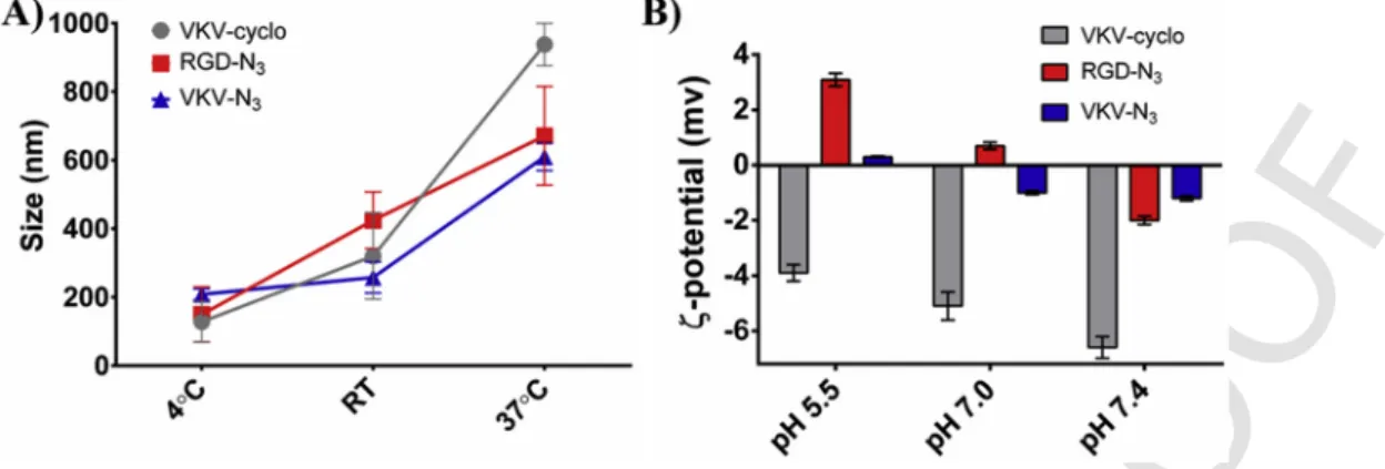
Size measurements of the structures in solution were carried out for each ELR – see Fig. 1A. These measurements were made at 4 °C, which is far below the reported LCST, RT, which is close to LCST, and 37 °C, which is far above LCST. At 4 °C, moderate polydispersity was found for VKV-cyclo (Pdi = 0.5 ± 0.05), RGD-N3
(Pdi = 0.5 ± 0.06) and VKV-N3 (Pdi = 0.6 ± 0.15). The size
ution of VKV-cyclo was about 128 ± 59.6 nm; similar size distrib-utions were obtained for RGD-N3and VKV-N3(150 ± 80.1 nm and
208 ± 15.9 nm, respectively). At RT, ELRs polydispersion slightly in-creased (Pdi = 0.6 ± 0.18 for VKV-cyclo). The single size tion of the different ELRs also increased; the single size distribu-tion for VKV-cyclo was about 320 ± 125.6 nm while for azide-mod-ified ELRs was about 425 ± 82.8 nm for RGD-N3and 258 ± 45.4 nm
for VKV-N3. At 37 °C, we observed a significant increase on the
size distribution of the different ELRs, with heterogeneous diameters found (Pdi = 0.5 ± 0.04 for VKV-cyclo). VKV-cyclo presents a sin-gle size distribution of 938 ± 61.8 nm and a little bit lower values were obtained for azide-modified ELRs (671 ± 144.4 nm for RGD-N3
and 610 ± 40.6 nm for VKV-N3). The obtained results are consistent
with the solubility in water-based solvents of the ELRs below LCST and their precipitation above the LCST. As described above, modified ELRs have a LCST close to RT. We believe that at RT occurs the tran-sition phenomenon and the polymers start to collapse. Even so, and as we were working in the transition temperature range, the phase separa-tion was not clearly visible yet and there was no significant differences on the size distribution results when compared with the ones obtained at 4 °C. Below LCST, at 4 °C, the ELRs solutions are hydrated and dispersed in the solvent, mainly in a linear form, while above LCST (37 °C) the polymer solutions started to precipitate in a folded globu-lar organization with higher diameters [34,42].
In order to perceive the best pH to construct the clickable elastin-based coatings, the ζ-potentials of the different ELRs in
UNCORRECTED
PROOF
Fig. 1. Effect of temperature on A) zeta sizer and B) zeta potential of VKV-cyclo, RGD-N3and VKV-N3solutions (0.05%(w/v)). determined for different pH values, at RT - see Fig. 1B. For RGD-N3
and VKV-N3solutions, the decrease of pH implied the protonation of
the solution. Besides the nature of ELRs being essentially hydropho-bic, the proposed ELRs were designed to contain lysine residues, which have positively charged amine groups [26,43]. At pH below 7.0, the RGD-N3solution presented a ζ-potential of 3.1 ± 0.23 mV,
being protonated and, naturally, positively charged. At higher pH val-ues, the amine groups started to deprotonate and the ζ-potential de-creased to −2.0 ± 0.15 mv. When pH was equal to 7.0, RGD-N3
so-lution charge was closer to 0 (ζ-potential = 0.7 ± 0.13 mv). The ζ-po-tential of VKV-N3solutions presented similar behavior to RGD-N3
solutions, at the different pH. Although the solutions were differently charged at pH 6.5 and 7.5, the differences between the respective ζ-po-tentials were not significant. Overall, at pH 7.0 the ζ-poζ-po-tentials of the different azide solutions were closer to 0 and we hypothesize that the different ELRs were almost discharged. For this reason, we decided to construct the coatings at pH 7.0 to minimize the effect of electrostatic interactions on the construction of the LbL-based coatings.
3.2. Build-up kinetics construction
After optimizing the working pH and temperature, the build-up of the elastin-based multilayers was assessed using QCM-D monitoring. Fig. 2A and B represents the frequency (Δfn) and dissipation (ΔDn)
variations at third overtone (n = 3) above a gold crystal, when flushed by the different ELRs solutions. These variations were monitored ac-cordingly to the time of depositions. Two constructions were evalu-ated: the one containing the VKV-cyclo and the RGD-N3(see Fig.
2A) and the other one containing the VKV-cyclo and the VKV-N3
(see Fig. 2B). The first six minutes correspond to the establishment of the baseline. In both graphs, the next 20 min show the deposition of the VKV-cyclo and the subsequent washing until removing the ex-cess of polymer, which was not adsorbed at the surface. The follow-ing 20 min correspond to the RGD-N3or the VKV-N3adsorptions.
For both cases, Δf3decreased with time; this observation can be
re-lated with the time of deposition/adsorption of the polymers above the surface of the gold crystal. On the other hand, ΔD3increased with
time indicating that elastin-based films did not present a rigid behav-ior and started to dissipate energy. In fact, this non-rigid/viscoelas-tic behavior is common for macromolecular systems [44]. The sub-sequent steps show the same trend: the ELRs deposition was strong for the first layers but it decreased for the next ones. Within the mul-tilayers construction, Δf3resultant of washing steps became smaller,
showing that ELRs were strongly linked and formed a stable coating for the LbL build-up. As already referred above, at pH 7.0 both azide
ELRs showed close to neutral zeta potential, and we hypothesize that there is no surface charge overcompensation by the formation of poly-cation–polyanion pairs. Therefore, we can consider the covalent bond-ing resultbond-ing from the click reaction (azide-alkyne cycloaddition) as the main force involved in the LbL construction. Four bilayers were constructed, with good indications of the effectiveness of the click chemistry reaction. A chemical scheme of this reaction is presented in Fig. 2C, where alkyne group links to azide group by means of a cy-cloaddition reaction, being the basis of the ELR-based film build-up. The first layer of VKV-cyclo was adsorbed to the substrates, allowing the further construction of the remaining layers through covalent link-age between cyclooctyne and azide groups, under mild aqueous con-ditions. Caruso's research group [45] used cycloaddition chemistry to build-up LbL multilayer systems by dipping different inert substrates into poly(acrylic acid) copolymerized with azide or alkyne groups. They further took advantage of this technology to fabricate pH respon-sive capsules that can serve as a versatile platform for further func-tionalization [21]. Other advantages were reported using such technol-ogy: producing high stable films, with no need of post cross-linking processes and with the possibility of incorporation of a wide range of functionalized materials [19,46].
Other information could be attained from the QCM-D data. The es-timated thickness of the elastin-based coatings was calculated based on the Voigt Model, using an appropriated software. The estimated thickness after each deposition was plotted over the number of lay-ers. For both constructions (VKV-cyclo/RDG-N3)nand (VKV-cyclo/
VKV-N3)n (where n represents the number of bilayers), the film
growth showed a non-linear behavior - see Fig. 2D and E, respec-tively. We used a non-linear regression to generate a mathemati-cal model which fits both (VKV-cyclo/RDG- N3)n and
(VKV-cy-clo/VKV- N3)nthickness growth. After 4 bilayers, the (VKV-cyclo/
RDG-N3)4 has an estimated thickness of 598 ± 8.5 nm, while the
[VKV-cyclo/VKV- N3]4 presented an estimated thickness of
586 ± 91.2 nm. Taken a hyperbolic model as base, we hypothesize that after reaching the double of bilayers (16 layers), (VKV-cyclo/ RDG- N3)4will present an estimated thickness around 739 nm while
(VKV-cyclo/VKV- N3)4will exhibit an estimated thickness of
ap-proximately 636 nm. Interestingly, after 16 bilayers we will observe a decrease of the rate of the thickness growth. Therefore, we as-sume that after a certain number of layers the film growth achieved a plateau. Comparing the proposed modified ELR-based films with other ELR-based systems already reported in literature based on elec-trostatic interactions [26], we believe that covalent interactions allow the deposition of higher amounts of polymer and, thus, the construc-tion of thicker films with less number of bilayers. Moreover, compar-ing our clickable based multilayer system with other covalent-driven
UNCORRECTED
PROOF
Fig. 2. Build-up assessment of ELR-based films. QCM-D monitoring of normalized frequency (Δfn) and dissipation (ΔDn) obtained at the third overtone, to assess the build-up of A) (VKV-cyclo/RGD-N3)4and B) (VKV-cyclo/VKV-N3)4films. C) Chemical scheme representing the click chemistry reaction, that results from the Huisgen 1,3-dipolar cycloaddition of azides and alkynes. Cumulative thickness evolution and thickness increase for 4 bilayers, estimated using the Voigt model for D) (VKV-cyclo/RGD-N3)4film and E) (VKV-cyclo/ VKV-N3)4films. The cumulative thicknesses follow non-linear growth model.
systems [21,47] we are able to produce thicker films, which means that we can control more precisely the final thickness of the system.
3.3. Elastin-based films production and characterization
The same procedure as the one described for QCM-D build-up was implemented over cleaned and activated glass coverslips, at RT. The solutions were maintained at 4 °C, until use, as well as during the in-cubation steps, to avoid the collapse process of ELRs in solution.
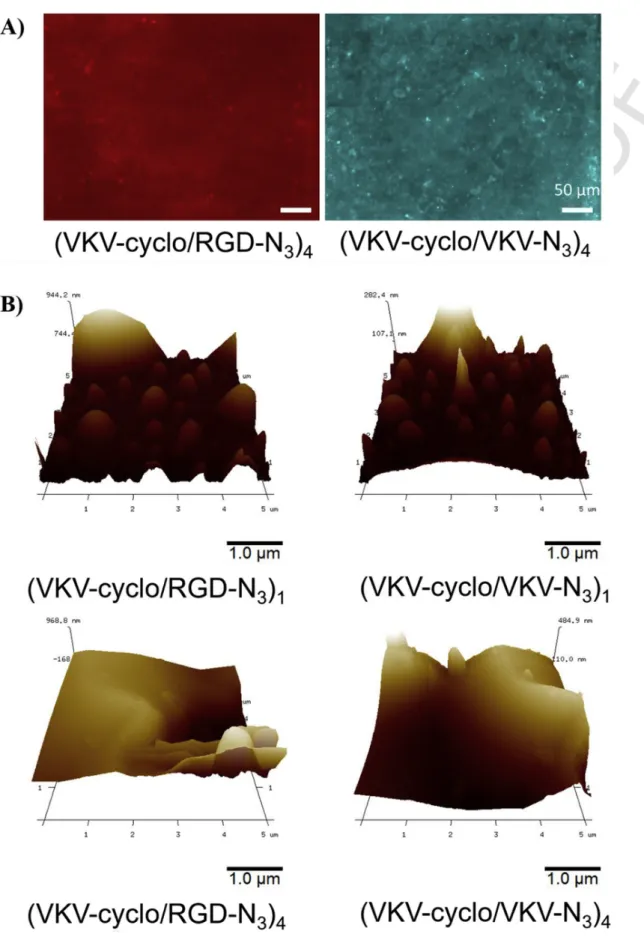
The fluorescence images of the 4 bilayers coatings are presented in Fig. 3A. Following the intensity of the fluorescence of Acetylene Fluor 488 (absorption at 501 nm and emission at 525 nm), it could be observed quite uniform distribution of the intensity on (VKV-cyclo/ RGD-N3)4films, in red, and (VKV-cyclo/VKV-N3)4films, in blue.
The surfaces of the glass coverslips were visibly covered by a thin film. This observation was shared for both (VKV-cyclo/RDG-N3)4
and (VKV-cyclo/VKV-N3)4coatings. This result is in accordance with
the observations retained from QCM-D monitoring, where 4 bilayer systems were constructed with success.
Additionally, the topography of 1 bilayer and 4 bilayers of (VKV-cyclo/RDG-N3) and (VKV-cyclo/VKV-N3) systems processed
and maintained at RT were evaluated under AFM observation-see Fig. 3B. As we worked at a temperature close to LCST nano-sized poly-mer agglopoly-merates can be clearly observed on the surface of the coat-ings, resulting from the collapse of adjacent ELRs chains. For both 1
bilayer systems, a high density of irregularities was perceived and the films presented higher values of roughness (Ra= 22 ± 5.0 nm for
(VKV-cyclo/RDG-N3) and Ra= 22 ± 9.8 nm for (VKV-cyclo/
VKV-N3)) when compared with other related reported systems
[45,48,49]. No significant differences were detected between the roughness of (VKV-cyclo/RDG-N3)4 and (VKV-cyclo/VKV-N3)4
films. Moreover, the roughness significantly increased with the in-creasing number of bilayers with Ra= 72 ± 55.5 nm for (VKV-cyclo/
RDG-N3)4, which could be a result of an increasing of mass adsorbed
on the surface of the glass coverslips. This observation was already reported in literature for other LbL systems [50,51]. Nonetheless, this increase of roughness was smaller for (VKV-cyclo/VKV-N3)4, with
Ra= 25.4 ± 14.0 nm. QCM-D results are in accordance with AFM
observation since rough surfaces induce larger hydrodynamic thick-nesses [50], as the ones estimated based on the Voigt Model.
3.4. Stimuli-responsiveness properties
We investigated the ability of the (VKV-cyclo/RDG-N3)4 and
(VKV-cyclo/VKV-N3)4coatings to respond to changes in the medium
such as pH and temperature, which are parameters that influence the adsorption of proteins at solid/liquid interface [52], among other physicochemical processes. This ability has been gaining importance and different works have been reported towards tissue engineering [53], sensors [54] and drug release systems [55].
UNCORRECTED
PROOF
Fig. 3. A) Fluorescence images of (VKV-N3/RGD-cyclo)4and (VKV-N3/VKV-cyclo)4coatings (azide-modified ELRS were labelled with Acetylene Fluor 488, before LbL con-struction). The coatings were produced and dried at RT. The scale bar is representative for both images. B) AFM images of (VKV-N3/RGD-cyclo)1and (VKV-N3/VKV-cyclo)1and (VKV-N3/RGD-cyclo)4and (VKV-N3/VKV-cyclo)4coatings.
UNCORRECTED
PROOF
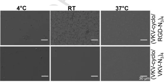
After drying, (VKV-cyclo/RDG-N3)4and (VKV-cyclo/VKV-N3)4surfaces were maintained at RT. SEM images were used to evaluate the morphology of the coatings-see Fig. 4. At RT, both coatings seem to be well distributed over the glass coverslips, even though with some small precipitated polymer. This result was already expected since we constructed and maintained the coatings at a temperature close to LCST. Other conditions were evaluated to study the response to tem-perature. For that, after constructing the films, at RT, onto glass cov-erslips the drying process was made at different temperatures: 4 °C, RT and 37 °C. Some morphological differences are noticed on SEM images; with the significant increase of the temperature above LCST (37 °C) the morphology of (VKV-cyclo/RDG-N3)4and (VKV-cyclo/
VKV-N3)4films seem less homogeneous with small aggregated
poly-mer precipitates adhered all over the glass surfaces and even some salt precipitation. Working at 4 °C, below LCST, the morphology of the coatings seems to be uniformly distributed on the surfaces, with less rough topography. These morphological changes are related with the thermosensitive behavior of these polymers, which are dependent of their LCST [7,56], even after the film construction.
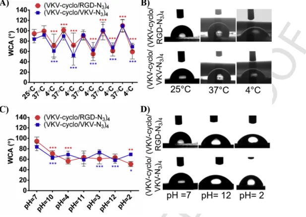
WCA measurements assess the effect of temperature and pH on the wettability of the (VKV-cyclo/RDG-N3)4and (VKV-cyclo/VKV-N3)4
coatings. To investigate the temperature effect, the WCA measure-ments were made under controlled temperature and humidity - see Fig. 5A. At RT, (VKV-cyclo/RDG-N3)4and (VKV-cyclo/VKV-N3)4
coatings presented WCA values of 94 ± 8.1° and 84 ± 7.3°, respec-tively. These values are closed to the threshold of hydrophobicity (WCA>90°). Therefore, we assumed that at RT the coatings have a moderate hydrophobic nature. The WCA of the uncoated glass slides is 59 ± 1.6°. The effectiveness of the coatings was also confirmed by the differences in the WCA, comparing the pre- and the post-coating values. By varying the temperature from 37 °C to 4 °C in repeating cy-cles, we observed switchable values of WCA. Higher values of WCA were observed when the samples were incubated at 37 °C (above the LCST); on the contrary, at temperature below LCST (4 °C) the WCA values were consistently lower. For instance, in the last cycle, the WCA at 37 °C was 110 ± 11.5° for (VKV-cyclo/RDG-N3) films and
110 ± 12.5° for (VKV-cyclo/VKV-N3)4. On the other hand, for the
last cycle at 4 °C the WCA value was about 59 ± 14.1° for (VKV-cy-clo/RDG-N3)4 films and 69 ± 6.7° for (VKV-cyclo/VKV-N3)4. The
images acquired for the calculation of WCA during these tempera-ture cycles are also depicted – see Fig. 5B. These observations can be a result of temperature and individual properties of the
modified ELRs. The three ELRs employed in the films construc-tion showed similar physicochemical characteristics; the competiconstruc-tion between intra and intermolecular hydrogen bonding above and be-low the LCST confers a thermosensitive nature to each individual ELR. When temperature was above LCST, the conformation of the ELRs chains started to collapse excluding water and adopting a type-II β-turns stabilized by intramolecular electrostatics forces between dif-ferent groups within the polymer chains. Two consequences could de-rive from this phenomenon: the interaction between hydrophilic car-boxyl and amine groups and water molecules became more diffi-cult and rounded polymer nano-precipitates were observed all over the surface. The presence of the nano-precipitates impelled the in-crease of the roughness of the coatings. Based on Cassie and Baxter model [57], which describes the entrapment of air-pockets between the grooves and the liquid droplet, we could extrapolate what hap-pens to WCA with the presence of rougher surfaces. With the polymer collapse process, ELRs chains fold and the coatings became rougher; when a droplet is dispensed in a rough surface, the volume of wa-ter infiltrated in the nanostructure decrease and the volume of wawa-ter on the surface increase; this phenomenon resulted in the increase of WCA values. While working at temperatures below LCST, the hy-drophilic groups could easily interact with the water molecules, form-ing water clathrates surroundform-ing the backbone of the ELR. Besides that, as already observed, at 4 °C the surfaces became smoother, with the absence of collapse structures on their surface morphology. The combination of these two effects results on more hydrophilic films. Moreover, playing with temperature below and above LCST could also promote the reconfiguration of the hydrophobic domains: above LCST the hydrophobic chains could be exposed to the outside of the films, decreasing the surface affinity to water. The results ex-posed a strong dependency on temperature indicating the ability to produce smart coatings with switchable wettability using these recom-binant materials [5]. Both (VKV-cyclo/RDG-N3)4and (VKV-cyclo/
VKV-N3)4coatings presented an apparent WCA switchability upon
temperature fluctuations. ELRs are well-known as protein-based poly-mers which present a phase transition in solution above a critical temperature [58]. Responsive polyelectrolyte coatings including ELRs were reported before [7]. However, contrasting with these results, we obtained elastin-based coatings that present a hydrophobic behavior above LCST and a hydrophilic behavior below LCST. Our thermo-re-sponsive system can be interesting for tissue engineering field where, for example, modified surfaces with PNIPAAm have been
Fig. 4. SEM images of (VKV-N3/RGD-cyclo)4and (VKV-N3/VKV-cyclo)4,subjected to incubation at different temperatures. The coatings were produced at RT and then stored at 4 °C, RT and 37 °C, overnight. The scale bar is representative for all images.
UNCORRECTED
PROOF
Fig. 5. A) Temperature dependences of WCA for the different ELR-based films and B) the respective representative pictures for temperature of 25 °C, 37 °C and 4 °C. C) pH
depen-dences of WCA for the different ELR-based films and D) the respective representative photographs for pH = 7, 12 and 2. Each WCA shown on A) and C) were statistical significant from the previous one, for p < 0.05 (*), p < 0.01 (**) and p < 0.001.
broadly reported [53,59] to produce cell sheets based on similar hy-drophilic-to-hydrophobic reversible effect of temperature on wettabil-ity.
To investigate the effect of the pH, ELRs-coated surfaces were immersed in sodium acetate solutions at different extreme acidic and alkaline pH values, fixing temperature the temperature at RT. WCA measurements – see Fig. 5C - were performed after incubations of at least 1 h and a small step for drying of 30 s. The representative im-ages of the WCA for the different conditions is shown in Fig. 5D. At pH 7, (VKV-cyclo/RDG-N3)4presented a WCA value of 94 ± 8.1°
and (VKV-cyclo/VKV-N3)4coatings presented a WCA of 84 ± 7.3°.
Some deviations from the initial WCA were obtained; for (VKV-cy-clo/RDG-N3)4and (VKV-cyclo/VKV-N3)4films, both acidic and
al-kaline pathways meant a more hydrophilic behavior. This could be un-derstood by the isoelectric point; as already suggested by the ζ-poten-tial measurements; close to pH 7, the electrostatic charges were almost neutralized. For (VKV-cyclo/RGD-N3)4 coatings on acidic or
alka-line environments no significant differences were found in the WCA presented at acidic or alkaline routes, but a slightly increase on hy-drophilicity was detected at acidic pH. Indeed, at pH 2, (VKV-cy-clo/RDG-N3)4films exhibited a significantly more hydrophilic
behav-ior. At extreme acidic pH, amine groups were protonated and positive electrostatic forces came to be dominant: the ELRs chains expanded and the films became more hydrophilic. Despite that, for (VKV-cyclo/ VKV-N3)4coatings on acidic or alkaline environments, slightly higher
WCA values were obtained at acidic pH values. This could be related to the balance between charged amine and acids being different from (VKV-cyclo/RDG-N3)4films; probably, for (VKV-cyclo/VKV-N3)4
there was a higher content of charged acid groups at lower pH.
Overall, results can be explained by the balance between hy-drophobic interactions and charged repulsion [60,61], and the respec-tive competition between protonation and deprotonation at alkaline and acidic pH value. When environment conditions like pH change, the ELR-based films, which contain ionizable amine and acid groups, are capable of accepting or donating protons. Therefore, altering the pH can lead to changes on the degree of ionization and, subsequently, on the hydrodynamic volume of the ELRs chains [62,63]. In literature, different wettable behaviors of ELRs-modified surfaces can be found [7,26,64]. This variability is linked with the ability to introduce dif-ferent genetically modified sequences, charges and molecular weight, that can alter the folding behavior at the surfaces [65].
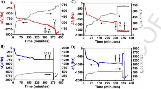
To a better understanding of the pH effect on the stability of the systems immediately after the films construction, we also per-formed QCM-D monitoring studies. After the construction of both (VKV-cyclo/RDG-N3)4and (VKV-cyclo/VKV-N3)4systems, the
re-sulting multilayers were flushed with a cyclic cascade of acidic and al-kaline sodium acetate solutions, separately. QCM-D results show the build-up of four bilayers and their response to changes in pH in terms of Δf and ΔD - see Fig. 6. For all cases, we took as reference the initial working pH 7. QCM-D data showing (VKV-cyclo/RDG-N3)4
and (VKV-cyclo/VKV-N3)4multilayers flushed with cyclic alkaline
cascade of solutions is presented in Fig. 6A and B, respectively. For both cases, a decrease of Δf3upon flushing the film with a solution
at pH 10 was observed; the decrease in Δf3was reversible when the
pH returned to 7. Naturally, ΔD3increased and Δf3decreased when
the coating was flushed with the alkaline solutions. When (VKV-cy-clo/RGD-N3)4multilayers were flushed with pH 11 and pH 12, Δf3
decreased with partially reversibility when pH returned to 7. On the other hand, when (VKV-cyclo/VKV-N3)4 multilayers were flushed
UNCORRECTED
PROOF
Fig. 6. Build-up assessment of the effect of a cascade of pHs on ELR-based films. QCM-D monitoring of normalized frequency (Δfn) and dissipation (ΔDn) obtained at the third overtone of A) (VKV-cyclo/RGD-N3)4film flushed with alkaline pHs, B) (VKV-cyclo/VKV-N3)4film flushed with alkaline pHs, C) (VKV-cyclo/RGD-N3)4film flushed with acidic pHs and D) (VKV-cyclo/VKV-N3)4film flushed with acidic pHs.
with the solution with pH 11 and pH 12, the Δf3abruptly decreased
with no reversibility. The same happened to ΔD3, which showed a
great increase. Therefore, at the pH 11 and 12, the changes in Δf3and
ΔD3seemed to be irreversible and could indicate that (VKV-cyclo/
VKV-N3)4multilayers started to loose structural integrity [66].
Fig. 6C shows the QCM-D data of the (VKV-cyclo/RDG-N3)4
multilayers when flushed with a cyclic acidic cascade of solutions. It can be observed an abrupt decrease of Δf3when the film is flushed
with solutions at pH 4; the decrease in Δf3was irreversible when the
pH returned to 7. At pH 3 and pH 2, there was no changes on Δf3.
Fig. 6D presents the QCM-D results for (VKV-cyclo/VKV-N3)4
mul-tilayers when flushed with an acidic cascade of solutions. For solu-tions with pHs 4 and 3, the behavior of the film was similar to the one obtained for (VKV-cyclo/RDG-N3)4multilayers at pH 4.
Notwith-standing, when (VKV-cyclo/VKV-N3)4films were flushed with the
solution at pH 2, the decrease on the Δf3was very abrupt and higher
than the others. The same happens with ΔD3, which exhibited a
sud-den increase. At this extreme acidic pH, the changes in Δf3and ΔD3
seemed to be irreversible and could indicate some loss of multilay-er's structural integrity. The stability and integrity of these smart coat-ings seemed to be maintained in a wide-range of pH values, being (VKV-cyclo/VKV-N3)4films more susceptible at extreme pH (2 and
12).
Overall, properties like morphology, topography, wettability and degradability of the produced ELR-based films can be modulated through different stimuli, including temperature and pH.
3.5. In vitro cellular response
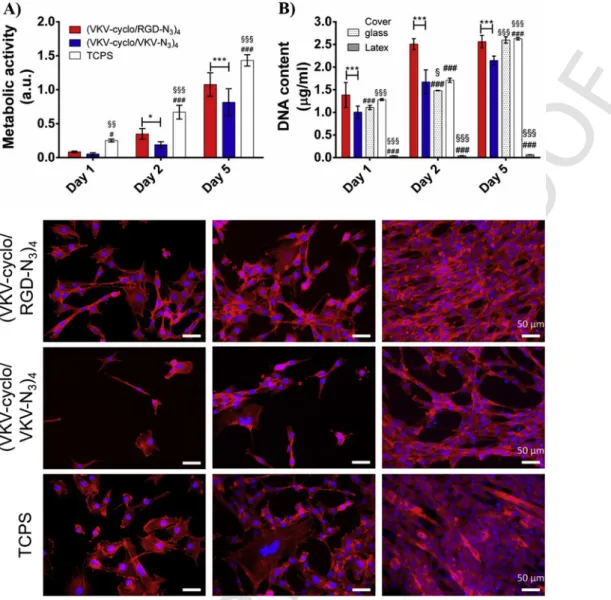
ELR-coated films were cultured with C2C12 cells, in order to eval-uate their biomedical and tissue engineering potential. Adhesion, vi-ability and proliferation are important parameters that depend on the interaction between material and cells [67–69]; MTS assay was used to determine the metabolic activity of C2C12 adhered on the sam-ples - see Fig. 7A. After 2 days of culture some differences started to be noticed, with C2C12 cultured on (VKV-cyclo/RDG-N3)4
films presenting significantly higher values of absorbance and, thus, higher metabolic activity. This trend was maintained and even ampli-fied after 5 days of culture. The total amount of dsDNA on the sam-ples was also investigated-see Fig. 7B. In the first day of culture sig-nificant differences were found between the (VKV-cyclo/RDG-N3)4
and (VKV-cyclo/VKV-N3)4coatings, with significant higher C2C12
density above the surfaces coated with (VKV-cyclo/RDG-N3)4films.
This result was also observed after 2 and 5 days of culture, be-ing in accordance with the results obtained for metabolic activity. As expected, the presence of the RGD motif seemed to influence positively the cellular performance, including adhesion and prolifer-ation, on the ELRs-coated film [26,33,70]. For instance, Picart, C. et al. [70] previously suggested the functionalization of polyelec-trolyte multilayer films with RGD motifs in order to enhance pri-mary human osteoblasts adhesion. We also investigated the morphol-ogy of C2C12, analyzing the F-actin expression of cells adhered to the (VKV-cyclo/RGD-N3)4and (VKV-cyclo/VKV-N3)4coatings (see
Fig. 7C). Some differences were observed on C2C12 morphology and density as a function of culturing time. As observed in Fig. 7C, cell density on (VKV-cyclo/RGD-N3)4films increased with the time of
culture; these results match the DNA quantification and MTS results. In the first day of culture, adhered myoblasts already acquired the star-like shape, which is characteristic of C2C12 cells [36]. This phe-notype could be observed more clearly on cells adhered to (VKV-cy-clo/RGD-N3)4surfaces. At 2 days of culture, myoblasts continued to
proliferate and, naturally, started to fuse one with each other, creat-ing a kind of cellular network [71]. This phenomenon was observed for both (VKV-cyclo/RGD-N3)4and (VKV-cyclo/VKV-N3)4surfaces,
with cells being better distributed for (VKV-cyclo/RGD-N3)4
coat-ings and more clustered in (VKV-cyclo/VKV-N3)4 surfaces. At 5
days of culture, the cells occupied the entire area, forming an orga-nized cellular monolayer above the (VKV-cyclo/RGD-N3)4surface.
The cells adhered to (VKV-cyclo/VKV-N3)4films had a similar
be-havior but, as the rate of proliferation was visibly slower, after 5 days of culture cell-free areas could still be found on the (VKV-cyclo/ VKV-N3)4coatings. TCPS were used as positive control and, in fact,
UNCORRECTED
PROOF
Fig. 7. A) Metabolic activity results based on MTS test performed after 1, 2 and 5 days of culture with C2C12 cells. B) DNA content obtained from by DNA quantification of
C2C12 seeded on ELR-based films and cultured for 5 days. Error bars represent means ± SD (n = 3). Differences on metabolic activity and DNA quantification between (VKV-cyclo/ RGD-N3)4and (VKV-cyclo/VKV-N3)4were significant for p < 0.05 (*) and p < 0.001 (***). Statistical significant differences on metabolic activity and DNA quantification between (VKV-cyclo/RGD-N3)4and TCPS, cover glass and latex were found for p < 0.05 (#) and p < 0.001 (###). Statistical significant differences on metabolic activity and DNA quantifi-cation between (VKV-cyclo/VKV-N3)4and TCPS, cover glass and latex were found for p < 0.05 (§), p < 0.01 (§§) and p < 0.001 (§§§). C) Phalloidin labelled F-actin (red) and DAPI labelled nucleus (blue) merged fluorescent images for C2C12 cells seeded on (VKV-cyclo/RGD-N3)4and (VKV-cyclo/VKV-N3)4coatings and TCPS. The scale bar is representative for all images. (For interpretation of the references to colour in this figure legend, the reader is referred to the web version of this article.)
cell morphology on (VKV-cyclo/RGD-N3)4coatings was comparable
to cell morphology on TCPS surfaces.
C2C12 differentiation was investigated by the expression of the skeletal muscle protein Troponin T. For that, we performed an im-munocytochemistry assay after culturing cells above the ELRs-coated surfaces during 5 days in differentiation medium-see Fig. 8A. Some differences were observed between Troponin-T positive cells adhered to (VKV-cyclo/RGD- N3)4and (VKV-cyclo/VKV-N3)4films.
Visu-ally, it is possible to observe more troponin T expression on the cells adhered to (VKV-cyclo/RGD-N3)4films, with more multinucleated
myotubes than on (VKV-cyclo/VKV-N3)4 films or even on TCPS.
To conclude quantitatively about the myogenic differentiation on the ELR-based films, some parameters were calculated. Significant dif-ferences were observed between the fusion index of C2C12 adhered to the different films. The cells seeded on (VKV-cyclo/RGD-N3)4
films presented higher fusion index percentage than the cells seeded on (VKV-cyclo/VKV-N3)4- see Fig. 8B. Also, the number of my
otubes per area was significantly higher for (VKV-cyclo/RGD-N3)4
films - see Fig. 8C. These results together could be an evidence that myogenic differentiation of C2C12 cells was stimulated by the pres-ence of RGD motifs on material's surface. This fact is supported by some examples found in the literature [72,73], which related the pres-ence of the RGD sequpres-ence to the promotion cellular attachment and differentiation. Different morphometric parameters were also calcu-lated from immunofluorescence images to assess the effect of RGD on myotube formation. The average area (Fig. 8D), perimeter (Fig. 8E) and length (Fig. 8F) of myotubes were similar and very dispersed, ei-ther adhering on (VKV-cyclo/RGD-N3)4or (VKV-cyclo/VKV-N3)4.
No significant differences were found between the myotubes elonga-tion factor of C2C12 adhered to (VKV-cyclo/RGD-N3)4or
(VKV-cy-clo/VKV-N3)4– see Fig. 8G. Myogenic differentiation seemed to be
favored by the presence of RGD motif, but the morphology of the formed myotubes was quite similar on (VKV-cyclo/RGD-N3)4and
UNCORRECTED
PROOF
Fig. 8. A) Myogenic differentiation at day 7 of culture of the cells seeded above the (VKV-cyclo/RGD-N3)4, (VKV-cyclo/VKV-N3)4and TCPS. The images are the results of a fluorescence staining showing troponin T-positive cells (green) and cell nuclei (blue). Myogenic differentiation as determined by the B) fusion index (%) and the C) number of my
UNCORRECTED
PROOF
otubes per area (A = ). Other parameters relatively to the formed myotubes were considered: D) area, E) perimeter, F) length and G) elongation factor. Statistically significant differ-ences are indicated with p < 0.05 (*) and p < 0.001 (***). (For interpretation of the referdiffer-ences to colour in this figure legend, the reader is referred to the web version of this article.)Independently of the surface energy and wettability changes of the coatings when subjected to different temperature and pH, cell seeding was performed and maintained at 37° C; at this tempera-ture, the surfaces of both (VKV-cyclo/RGD-N3)4 and (VKV-cyclo/
cyclo-N3)4coatings were moderately hydrophobic. Therefore, we
hy-pothesize that the enhanced cell adhesion, activity and even differ-entiation above the (VKV-cyclo/RGD-N3)4was mainly related with
chemistry of the surface by the presence of the bioactive sequence RGD and in this specific case was not related with parameters like sur-face energy, wettability and charge.
4. Conclusions
We reported the development of stimuli-responsive polymer multi-layer coatings, based on a click-chemistry system. We propose a sim-ple click LbL methodology to fabricate these coatings, which con-sists in alternating cyclooctyne- and azide-modified ELRs, combined in a sequential multilayer mode. The build-up of the ELRs-based films was confirmed by QCM-D monitoring, following a non-linear growth. Herein, we show that both temperature and pH can act like stimuli to prompt independent responses by the developed ELRs-based films. Above LCST, ELRs formed folded and round structures. This phe-nomenon resulted in the increase of roughness of the coatings, and consequently in a more hydrophobic behavior, as compared to the ones found in the coating maintained at temperatures below LCST. Also, pH variations were responsible for changes in the coatings' WCA values; generally, the balance between charged amine and acid groups could determine the wetting behavior of the surfaces. The high stability of the films, conferred by the covalent bonding, was firmed by QCM-D monitoring; in fact, the films withstood harsh con-ditions of pH, and only (VKV-cyclo/VKV-N3)4coatings showed
in-tegrity loss while exposed to the most extreme pH value. The abil-ity to introduce specific bioactive sequences like RGD motif on the ELRs structure was relevant for this investigation and central for tis-sue engineering and biomedical applications. Cell proliferation was increased on (VKV-cyclo/RGD-N3)4films, and myogenic
differentia-tion was also favored by the presence of the RGD bioactive sequence. Overall, we were able to produce temperature and pH- respon-sive multilayer films composed exclurespon-sively by modified elastin-like polypeptides that can be easily used to as coatings. Besides glass, we hypothesize that these films may find application on coating implants with more complex shapes and compositions, nano/microstructures, gels and membranes. These systems show a great potential to develop structures for tissue engineering purposes or as platforms to culture cells in controlled conditions.
Acknowledgments
This research has received funding from the European Union's Horizon 2020 research and innovation programme under grant agree-ment No.646075. Maria P. Sousa and Mariana B. Oliveira acknowl-edge the Portuguese Foundation for Science and Technology (FCT) the grants SFRH/BD/97606/2013 and SFRH/BPD/111354/2015.
References
[1] B. Jeong, A. Gutowska, Lessons from nature: stimuli-responsive polymers and their biomedical applications, Trends Biotechnol. 20 (2002) 305–311. [2] M.A.C. Stuart, W.T.S. Huck, J. Genzer, M. Muller, C. Ober, M. Stamm, G.B.
Sukhorukov, I. Szleifer, V.V. Tsukruk, M. Urban, F. Winnik, S. Zauscher, I.
Luzinov, S. Minko, Emerging applications of stimuli-responsive polymer mate-rials, Nat. Mater 9 (2010) 101–113.
[3] A. Nelson, Stimuli-responsive polymers: engineering interactions, Nat. Mater 7 (2008) 523–525.
[4] C. d. l. H. Alarcon, S. Pennadam, C. Alexander, Stimuli responsive polymers for biomedical applications, Chem. Soc. Rev. 34 (2005) 276–285.
[5] J.F. Mano, Stimuli-responsive polymeric systems for biomedical applications, Adv. Eng. Mater 10 (2008) 515–527.
[6] I. Tokarev, S. Minko, Stimuli-responsive hydrogel thin films, Soft Mat-ter 5 (2009) 511–524.
[7] R.R. Costa, C.A. Custódio, A.M. Testera, F.J. Arias, J.C. Rodríguez-Cabello, N.M. Alves, J.F. Mano, Stimuli-responsive thin coatings using elastin-like poly-mers for biomedical applications, Adv. Funct. Mater 19 (2009) 3210–3218. [8] D. Wandera, S.R. Wickramasinghe, S.M. Husson, Stimuli-responsive
mem-branes, J. Membr. Sci. 357 (2010) 6–35.
[9] X. Hu, E. McIntosh, M.G. Simon, C. Staii, S.W. Thomas, Stimuli-responsive free-standing layer-by-layer films, Adv. Mater 28 (2016) 715–721. [10] M. Motornov, Y. Roiter, I. Tokarev, S. Minko, Stimuli-responsive
nanoparti-cles, nanogels and capsules for integrated multifunctional intelligent systems, Prog. Polym. Sci. 35 (2010) 174–211.
[11] E. Sokolovskaya, S. Rahmani, A.C. Misra, S. Bräse, J. Lahann, Dual-stimuli-re-sponsive microparticles, ACS Appl. Mater. Interfaces 7 (2015) 9744–9751. [12] R.R. Costa, J.F. Mano, Polyelectrolyte multilayered assemblies in biomedical
technologies, Chem. Soc. Rev. 43 (2014) 3453–3479.
[13] L. Richert, P. Lavalle, E. Payan, X.Z. Shu, G.D. Prestwich, J.-F. Stoltz, P. Schaaf, J.-C. Voegel, C. Picart, Layer by layer buildup of polysaccharide films: physical chemistry and cellular adhesion aspects, Langmuir 20 (2004) 448–458. [14] J.F. Quinn, F. Caruso, Facile tailoring of film morphology and release properties
using layer-by-layer assembly of thermoresponsive materials, Lang-muir 20 (2004) 20–22.
[15] J.M. Silva, R.L. Reis, J.F. Mano, Biomimetic extracellular environment based on natural origin polyelectrolyte multilayers, Small 12 (2016) 4308–4342. [16] J. Borges, J.F. Mano, Molecular interactions driving the layer-by-layer assembly
of multilayers, Chem. Rev. 114 (2014) 8883–8942.
[17] J. Borges, J.F. Mano, Molecular interactions driving the layer-by-layer assembly of multilayers, Chem. Rev. 114 (2014) 8883–8942.
[18] J.F. Quinn, A.P.R. Johnston, G.K. Such, A.N. Zelikin, F. Caruso, Next genera-tion, sequentially assembled ultrathin films: beyond electrostatics, Chem. Soc. Rev. 36 (2007) 707–718.
[19] D.E. Bergbreiter, K.-S. Liao, Covalent layer-by-layer assembly-an effective, forgiving way to construct functional robust ultrathin films and nanocomposites, Soft Matter 5 (2009) 23–28.
[20] J. Seo, P. Schattling, T. Lang, F. Jochum, K. Nilles, P. Theato, K. Char, Cova-lently bonded layer-by-layer assembly of multifunctional thin films based on ac-tivated esters, Langmuir 26 (2010) 1830–1836.
[21] G.K. Such, E. Tjipto, A. Postma, A.P.R. Johnston, F. Caruso, Ultrathin, respon-sive polymer click capsules, Nano Lett. 7 (2007) 1706–1710.
[22] V.V. Rostovtsev, L.G. Green, V.V. Fokin, K.B. Sharpless, A stepwise Huisgen cycloaddition process: copper(I)-catalyzed regioselective “ligation” of azides and terminal alkynes, Angew. Chem. Int. Ed. 41 (2002). 2596-+.
[23] M. Meldal, C.W. Tornøe, Cu-catalyzed Azide−Alkyne cycloaddition, Chem. Rev. 108 (2008) 2952–3015.
[24] T. Crouzier, T. Boudou, C. Picart, Polysaccharide-based polyelectrolyte multi-layers, Curr. Opin. Colloid Interface Sci. 15 (2010) 417–426.
[25] J.M. Silva, A.R.C. Duarte, S.G. Caridade, C. Picart, R.L. Reis, J.F. Mano, Tai-lored freestanding multilayered membranes based on chitosan and alginate, Bio-macromolecules 15 (2014) 3817–3826.
[26] R.R. Costa, C.A. Custódio, F.J. Arias, J.C. Rodríguez-Cabello, J.F. Mano, Layer-by-Layer assembly of chitosan and recombinant biopolymers into bio-mimetic coatings with multiple stimuli-responsive properties, Small 7 (2011) 2640–2649.
[27] A. Matsuzawa, M. Matsusaki, M. Akashi, Effectiveness of nanometer-sized ex-tracellular matrix layer-by-layer assembled films for a cell membrane coating protecting cells from physical stress, Langmuir 29 (2013) 7362–7368. [28] S.M. Oliveira, V.E. Santo, M.E. Gomes, R.L. Reis, J.F. Mano, Layer-by-layer
assembled cell instructive nanocoatings containing platelet lysate, Biomateri-als 48 (2015) 56–65.
[29] P. He, M. Bayachou, Layer-by-Layer fabrication and characterization of DNA-wrapped single-walled carbon nanotube particles, Langmuir 21 (2005) 6086–6092.
[30] J.C. Rodríguez-Cabello, L. Martín, A. Girotti, C. García-Arévalo, F.J. Arias, M. Alonso, Emerging applications of multifunctional elastin-like recombinamers, Nanomedicine 6 (2010) 111–122.
[31] A. Girotti, A. Fernández-Colino, I.M. López, J.C. Rodríguez-Cabello, F.J. Arias, Elastin-like recombinamers: biosynthetic strategies and biotechnological applications, Biotechnol. J. 6 (2011) 1174–1186.
UNCORRECTED
PROOF
[32] S.E. D'Souza, M.H. Ginsberg, E.F. Plow, Arginyl-glycyl-aspartic acid (RGD): acell adhesion motif, Trends biochem. Sci. 16 (1991) 246–250.
[33] U. Hersel, C. Dahmen, H. Kessler, RGD modified polymers: biomaterials for stimulated cell adhesion and beyond, Biomaterials 24 (2003) 4385–4415. [34] I. González de Torre, M. Santos, L. Quintanilla, A. Testera, M. Alonso, J.C.
Rodríguez Cabello, Elastin-like recombinamer catalyst-free click gels: charac-terization of poroelastic and intrinsic viscoelastic properties, Acta Bio-mater. 10 (2014) 2495–2505.
[35] N.M. Alves, C. Picart, J.F. Mano, Self assembling and crosslinking of polyelec-trolyte multilayer films of chitosan and alginate studied by QCM and IR spec-troscopy, macromol, Biosci 9 (2009) 776–785.
[36] S. Burattini, P. Ferri, M. Battistelli, R. Curci, E. Luchetti, E. Falcieri, C2C12 murine myoblasts as a model of skeletal muscle development: morpho-func-tional characterization, Eur. J. Histochem 48 (2004) 223–233.
[37] S. Chang, S. Song, J. Lee, J. Yoon, J. Park, S. Choi, J.-K. Park, K. Choi, C. Choi, Phenotypic modulation of primary vascular smooth muscle cells by short-term culture on micropatterned substrate, PLoS One 9 (2014) e88089. [38] M.B. Oliveira, W. Song, L. Martin, S.M. Oliveira, S.G. Caridade, M. Alonso,
J.C. Rodriguez-Cabello, J.F. Mano, Development of an injectable system based on elastin-like recombinamer particles for tissue engineering applications, Soft Matter 7 (2011) 6426–6434.
[39] B. Kinikoglu, J.C. Rodríguez-Cabello, O. Damour, V. Hasirci, A smart bilayer scaffold of elastin-like recombinamer and collagen for soft tissue engineering, J. Mater. Sci. Mater. Med. 22 (2011) 1541–1554.
[40] I. Gonzalez de Torre, M. Weber, L. Quintanilla, M. Alonso, S. Jockenhoevel, J.C. Rodriguez Cabello, P. Mela, Hybrid elastin-like recombinamer-fibrin gels: physical characterization and in vitro evaluation for cardiovascular tissue engi-neering applications, Biomater. Sci. 4 (2016) 1361–1370.
[41] A. Girotti, J. Reguera, F.J. Arias, M. Alonso, A.M. Testera, J.C. Rodríguez-Ca-bello, Influence of the molecular weight on the inverse temperature transition of a model genetically engineered elastin-like pH-responsive polymer, Macromole-cules 37 (2004) 3396–3400.
[42] I. González de Torre, L. Quintanilla, G. Pinedo-Martín, M. Alonso, J.C. Rodríguez-Cabello, Nanogel formation from dilute solutions of clickable elastin-like recombinamers and its dependence on temperature: two fractal gela-tion modes, ACS Appl. Mater. Interfaces 6 (2014) 14509–14515.
[43] U. Glebe, B. Santos de Miranda, P. van Rijn, A. Boker, Synthetic modifications of proteins, Bio-Synth. Hybrid Mater. Bionanoparticles A Biol. Chem. Ap-proach Towards Mater. Sci. R. Soc. Chem. (2015) 1–29.
[44] K.A. Marx, Quartz crystal Microbalance: a useful tool for studying thin poly-mer films and complex biomolecular systems at the Solution−Surface interface, Biomacromolecules 4 (2003) 1099–1120.
[45] G.K. Such, J.F. Quinn, A. Quinn, E. Tjipto, F. Caruso, Assembly of ultrathin polymer multilayer films by click chemistry, J. Am. Chem. Soc. 128 (2006) 9318–9319.
[46] C.R. Kinnane, K. Wark, G.K. Such, A.P.R. Johnston, F. Caruso, Peptide-func-tionalized, low-biofouling click multilayers for promoting cell adhesion and growth, Small 5 (2009) 444–448.
[47] T. Xiang, R. Wang, W.-F. Zhao, S.-D. Sun, C.-S. Zhao, Covalent deposition of zwitterionic polymer and citric acid by click chemistry-enabled layer-by-layer assembly for improving the blood compatibility of polysulfone membrane, Langmuir 30 (2014) 5115–5125.
[48] M. Golonka, M. Bulwan, M. Nowakowska, A.M. Testera, J.C. Rodriguez-Ca-bello, S. Zapotoczny, Thermoresponsive multilayer films based on ionic elastin-like recombinamers, Soft Matter 7 (2011) 9402–9409.
[49] W.J. Yang, D. Pranantyo, K.-G. Neoh, E.-T. Kang, S.L.-M. Teo, D. Rittschof, Layer-by-Layer click deposition of functional polymer coatings for combating marine biofouling, Biomacromolecules 13 (2012) 2769–2780.
[50] A.E. El haitami, J.-S. Thomann, L. Jierry, a. Parat, J.-C. Voegel, P. Schaaf, B. Senger, F. Boulmedais, B. Frisch, covalent layer-by-layer assemblies of poly-electrolytes and homobifunctional spacers, Langmuir 26 (2010) 12351–12357. [51] C. Elosua, D. Lopez-Torres, M. Hernaez, I.R. Matias, F.J. Arregui, Comparative
study of layer-by-layer deposition techniques for poly(sodium phosphate) and poly(allylamine hydrochloride), Nanoscale Res. Lett. 8 (2013) 539.
[52] W. Norde, F. MacRitchie, G. Nowicka, J. Lyklema, Protein adsorption at solid-liquid interfaces: reversibility and conformation aspects, J. Colloid Inter-face Sci. 112 (1986) 447–456.
[53] R.M.P. da Silva, J.F. Mano, R.L. Reis, Smart thermoresponsive coatings and surfaces for tissue engineering: switching cell-material boundaries, Trends Biotechnol. 25 (2007) 577–583.
[54] O. Parlak, M. Ashaduzzaman, S.B. Kollipara, A. Tiwari, A.P.F. Turner, Switch-able bioelectrocatalysis controlled by dual stimuli-responsive polymeric inter-face, ACS Appl. Mater. Interfaces 7 (2015) 23837–23847.
[55] H. Vihola, A. Laukkanen, H. Tenhu, J. Hirvonen, Drug release characteristics of physically cross-linked thermosensitive poly(N-vinylcaprolactam) hydrogel par-ticles, J. Pharm. Sci. 97 (2008) 4783–4793.
[56] A.M. Testera, A. Girotti, I.G. de Torre, L. Quintanilla, M. Santos, M. Alonso, J.C. Rodríguez-Cabello, Biocompatible elastin-like click gels: design, synthesis and characterization, J. Mater. Sci. Mater. Med. 26 (2015) 1–13.
[57] R.N. Wenzel, Resistance of solid surfaces to wetting by water, Ind. Eng. Chem. 28 (1936) 988–994.
[58] T. Kowalczyk, K. Hnatuszko-Konka, A. Gerszberg, A.K. Kononowicz, Elastin-like polypeptides as a promising family of genetically-engineered pro-tein based polymers, World J. Microbiol. Biotechnol. 30 (2014) 2141–2152. [59] T. Shimizu, M. Yamato, A. Kikuchi, T. Okano, Cell sheet engineering for
my-ocardial tissue reconstruction, Biomaterials 24 (2003) 2309–2316.
[60] B. Li, V. Daggett, The molecular basis of the temperature- and pH-induced con-formational transitions in elastin-based peptides, Biopolymers 68 (2003) 121–129.
[61] J. Carlos Rodríguez-Cabello, J. Reguera, A. Girotti, M. Alonso, A.M. Testera, Developing functionality in elastin-like polymers by increasing their molecular complexity: the power of the genetic engineering approach, Prog. Polym. Sci. 30 (2005) 1119–1145.
[62] F. Xia, L. Feng, S. Wang, T. Sun, W. Song, W. Jiang, L. Jiang, Dual-responsive surfaces that switch between superhydrophilicity and superhydrophobicity, adv, Mater 18 (2006) 432–436.
[63] Q. Zhang, F. Xia, T. Sun, W. Song, T. Zhao, M. Liu, L. Jiang, Wettability switching between high hydrophilicity at low pH and high hydrophobicity at high pH on surface based on pH-responsive polymer, Chem. Commun. (2008) 1199–1201.
[64] E.M. Srokowski, K.A. Woodhouse, Surface and adsorption characteristics of three elastin-like polypeptide coatings with varying sequence lengths, J. Mater. Sci. Mater. Med. 24 (2013) 71–84.
[65] A. Ribeiro, F.J. Arias, J. Reguera, M. Alonso, J.C. Rodríguez-Cabello, Influence of the amino-acid sequence on the inverse temperature transition of elastin-like polymers, Biophys. J. 97 (2009) 312–320.
[66] J.M. Silva, S.G. Caridade, R.R. Costa, N.M. Alves, T. Groth, C. Picart, R.L. Reis, J.F. Mano, pH responsiveness of multilayered films and membranes made of polysaccharides, Langmuir 31 (2015) 11318–11328.
[67] W.F. Liu, C.S. Chen, Engineering biomaterials to control cell function, Mater. Today 8 (2005) 28–35.
[68] A.J. García, Get a grip: integrins in cell–biomaterial interactions, Biomateri-als 26 (2005) 7525–7529.
[69] S.M. Oliveira, N.M. Alves, J.F. Mano, Cell interactions with superhydrophilic and superhydrophobic surfaces, J. Adhes. Sci. Technol. 28 (2014) 843–863. [70] C. Picart, R. Elkaim, L. Richert, F. Audoin, Y. Arntz, M. Da Silva Cardoso, P.
Schaaf, J.C. Voegel, B. Frisch, Primary cell adhesion on RGD-functionalized and covalently crosslinked thin polyelectrolyte multilayer films, Adv. Funct. Mater 15 (2005) 83–94.
[71] K. Rochlin, S. Yu, S. Roy, M.K. Baylies, Myoblast fusion: when it takes more to make one, Dev. Biol. 341 (2010) 66–83.
[72] N. Osses, E. Brandan, ECM is required for skeletal muscle differentiation inde-pendently of muscle regulatory factor expression, Am. J. Physiol. Cell Phys-iol. 282 (2002) C383–C394.
[73] P.-Y. Wang, H. Thissen, W.-B. Tsai, The roles of RGD and grooved topography in the adhesion, morphology, and differentiation of C2C12 skeletal myoblasts, Biotechnol. Bioeng 109 (2012) 2104–2115.