UNIVERSIDADE DE TRÁS-OS-MONTES E ALTO DOURO
Layer-by-Layer Coated Microparticle
Templates for Biomedical Applications
Dissertação de Mestrado em Engenharia Biomédica
Carla Madalena Moreira Ribeiro
Orientador: Professor Doutor João F. Mano
Co-Orientadores: Professora Doutora Verónica de Zea Bermudez e
Doutor João Borges
iii
Scientific Supervision
Supervisor
Professor Doctor João Filipe Colardelle da Luz Mano
Full Professor at the Department of Chemistry and Director of the COMPASS Research Group at CICECO – Aveiro Institute of Materials
University of Aveiro (Portugal)
Co-Supervisor
Doctor João Filipe Ramos da Silva Carvalho Borges
Junior Research Fellow at the COMPASS Research Group, CICECO – Aveiro Institute of Materials
University of Aveiro (Portugal)
Co-Supervisor
Professor Doctor Verónica de Zea Bermudez
Full Professor at the Department of Chemistry, School of Life and Environmental Sciences University of Trás-os-Montes e Alto Douro (Portugal)
v
“The important thing is not stop questioning. Curiosity has its own reason for existing.”
vii
Acknowledgments
This page belongs to the one who allowed, helped and planned this dissertation. To my supervisor and co-supervisor, Professor João Mano and Doctor João Borges, thank you for accepting me as master student in the COMPASS Research Group at the university of Aveiro, and for the opportunity to get in touch and do scientific research. A specially greeting to my co-supervisor Doctor João Borges, for all guidance, motivation, and indispensable share of knowledge. I felt really welcome in this group and inspired to continue a journey in research that is only starting. Also, to my co-supervisor Professor Verónica Bermudez, thanks for the availability and all the support. To all, my honest gratefulness. I also gratefully acknowledge the financial support by the Portuguese Foundation for Science and Technology (FCT) through the Marine Biotechnology ERA-NET project “BLUETEETH” (ERA-MBT/0002/2015), funded under the European Commission’s 7th Framework Programme.
A special acknowledgment to my boyfriend Jorge Ferreira for being my best friend, for always supporting me and never let me give up and specially for his companionship on this journey.
To my friends, especially those who share Vila Real with me, Carolina, Inês, Sónia and Teresa, and to my mates in the season in Aveiro, Sara Santos and José Figueiredo, thank you all very much for your friendship. You all mean a lot to me!
ix
Abstract
Over the last decades there has been a great interest in the design and development of nano- and microscale drug delivery systems for the encapsulation, protection, and sustained and targeted delivery of therapeutics at injured sites, as well as for triggering the regeneration of specific tissues and/or organs. Among them, polymeric microparticles and microcapsules produced by using natural and/or synthetic polymers, as well as surface engineering approaches have emerged owing to their intriguing features, including large surface area, easy and versatile surface functionalization, and tunable physicochemical properties. In particular, particles and capsules developed by resorting to natural-origin polymers have attracted massive attention in the biomedical field due to their biocompatibility, biodegradability and wide bioavailability. Nevertheless, some synthetic polymers have been also used in the biomedical arena owing to their promising features. For instance, poly(lactic-co-glycolic) acid (PLGA) is one of the most attractive synthetic polymers due to its biocompatibility, biodegradability, and easily tunability, being already approved by the regulatory authorities to be used in the clinics. Concomitantly, PLGA-based drug carriers have shown to not elicit biological risk upon cellular uptake. In this work, two distinct microparticle templates were produced and surface functionalized with polymeric materials via the bottom-up Layer-by-Layer (LbL) assembly technology to assess the sustained released of model compounds or their cellular uptake. As such, polymeric PLGA-based microparticles were produced, loaded with a model hydrophobic compound with fluorescent features, namely coumarin 6 (C6), and further surface-functionalized via the Layer-by-Layer (LbL) assembly of oppositely charged poly-L-lysine (PLL) and alginate (ALG) polymers, to assess the influence of the number of layers on the sustained release of C6. The encapsulation efficiency and the in vitro release profile were assessed and quantified by fluorescence spectroscopy, confirming the sustained release upon 21 days. The role of some of the most relevant physicochemical properties of the particles, known to have a significant impact on the interaction with biological systems, including the mean particle size and surface charge was evaluated. Following this study, hollow polysaccharide-based microcapsules were produced by alternate deposition of chitosan (CHT) and ALG multilayers onto calcium carbonate (CaCO3) microparticles surface, followed by core template dissolution. The mean
particle size, surface charge, and aggregation tendency were evaluated aiming to produce well-dispersed microcapsules. The in vitro cellular uptake of aggregated and well-well-dispersed microcapsules was assessed by confocal laser scanning microscopy using mouse lung fibroblast
x
cell line (L929), proving that, in opposition to the aggregated microcapsules, the well-dispersed microcapsules were successfully internalized by cells and, very importantly, the cells remained viable. In both systems, the morphological properties of the particles/capsules were assessed by advanced microscopy techniques, including scanning and transmission electron microscopy, as well as widefield fluorescence microscopy.
Keywords: biocompatible polymers, layer-by-layer assembly, microparticles, hollow
xi
Resumo
Ao longo dos anos, em especial nas últimas décadas, o interesse pelo desenvolvimento de sistemas à nano e microescala para encapsulamento, proteção e distribuição controlada e direcionada de agentes terapêuticos em locais alvo, bem como para desencadear a regeneração de tecidos específicos e/ou órgãos tem aumentado consideravelmente. Entre eles, micropartículas e microcápsulas de origem polimérica produzidas com recurso a polímeros naturais e/ou sintéticos, bem como a técnicas de engenharia de superfície, emergiram devido às suas importantes caraterísticas, incluindo grande área de superfície, funcionalização simples e versátil da sua superfície e propriedades físico-químicas facilmente ajustáveis. Em particular, partículas e cápsulas desenvolvidas com recurso a polímeros de origem natural têm atraído grande atenção por parte da comunidade científica a desenvolver a sua atividade de investigação no campo da biomedicina devido à sua biocompatibilidade, biodegradabilidade e ampla biodisponibilidade. No entanto, alguns polímeros sintéticos também têm sido utilizados devido às suas caraterísticas promissoras. Por exemplo, o poli(ácido lático-co-glicólico) (PLGA, do inglês poly(lactic-co-glycolic) acid) é um dos polímeros sintéticos mais atrativos devido à sua biocompatibilidade, biodegradabilidade e fácil manipulação, estando aprovado pelas autoridades reguladoras para uso clínico. Simultaneamente, sistemas de libertação e transporte de fármacos fabricados à base de PLGA tem demonstrado não provocar qualquer tipo de risco biológico após a sua internalização celular. Neste trabalho, dois modelos distintos de micropartículas foram produzidos e funcionalizados superficialmente com materiais poliméricos com recurso à tecnologia de deposição camada-a-camada (LbL, do inglês Layer-by-Layer) afim de avaliar a libertação controlada de compostos modelo bem como a sua internalização celular. Como tal, foram produzidas micropartículas poliméricas à base de PLGA, incorporando um composto modelo hidrofóbico e fluorescente, nomeadamente a cumarina 6 (C6, do inglês coumarin 6), e posteriormente a sua superfície foi funcionalizada através da tecnologia de LbL com multicamadas poliméricas, utilizando para tal poli-L-lisina (PLL, do inglês poly-L-lysine) e alginato (ALG, do inglês alginate), polímeros de carga oposta, para avaliar a influência do número de camadas na libertação controlada de C6. A eficiência de encapsulamento e o perfil de libertação in vitro foram avaliados e quantificados via espetroscopia de fluorescência, confirmando uma libertação controlada após 21 dias. O papel de algumas das propriedades físico-químicas mais relevantes das partículas, conhecidas por induzir um impacto significativo na interação com os sistemas biológicos, incluindo o tamanho
xii
médio das partículas e a sua carga superficial foram avaliadas. Após este estudo, microcápsulas ocas à base de polissacarídeos foram produzidas por deposição alternada de multicamadas de quitosano (CHT, do inglês chitosan) e ALG sobre a superfície das micropartículas de carbonato de cálcio (CaCO3, do inglês calcium carbonate), seguido da dissolução do seu core. O tamanho
médio das partículas, a carga de superficial e a tendência para agregarem foram avaliados visando produzir microcápsulas bem dispersas. A internalização celular in vitro das microcápsulas agregadas e dispersas foi estudada por microscopia de confocal de varrimento a laser, recorrendo a uma linha celular de fibroblastos de pulmão de rato (L929). Ao invés do observado com as microcápsulas agregadas, as microcápsulas dispersas foram internalizadas com sucesso pelas células, que por sua vez permaneceram viáveis ao longo do tempo. Em ambos os sistemas, as propriedades morfológicas das partículas/cápsulas foram avaliadas por técnicas avançadas de microscopia, incluindo microscopia eletrónica de varrimento e de transmissão, bem como por microscopia de fluorescência de campo amplo.
Palavras-chave: polímeros biocompatíveis, tecnologia de deposição camada-a-camada, micropartículas, microcápsulas, libertação controlada, internalização celular
xiii
Table of Contents
Acknowledgments.………...vii Abstract………..ix Resumo……….……….xi Table of Contents……….………xiii List of Figures……….xvii List of Tables………....xxiList of Symbols and Abbreviations………..………..…xxiii
PART A: GENERAL INTRODUCTION & OBJECTIVES Chapter 1 – General Introduction………....3
1.1. General Concepts in Nanoscience and Nanotechnology………...3
1.2. Motivation…...………...5
1.3. Objectives………..6
1.4. Organization………...6
PART B: STATE OF THE ART Chapter 2 – Drug Delivery Systems………11
2.1. Introduction to the Drug Delivery Systems…..………..……….…….11
2.2. Polymer-based Microparticles/Microcapsules: Applications in Drug Delivery and Cellular Uptake…….………....………....16
2.2.1. Properties of Polymers used to produce Microparticles/Microcapsules for Drug Delivery Applications………...21
2.3. Methods for the Fabrication of Polymer-based Microparticles ………...23
xiv
PART C: EXPERIMENTAL SECTION
Chapter 3 – Reagents, Solutions, Sample Preparation & Characterization Techniques .39 3.1. Fabrication of Poly-L-Lysine/Alginate (PLL/ALG) Coated PLGA Microparticles loaded
with coumarin 6………...39
3.1.1. Materials..………..39
3.1.2. Preparation of Unloaded and C6-loaded PLGA Microparticles ... 39
3.1.3. Layer-by-Layer (LbL) Assembly on Unloaded and C6-loaded PLGA Microparticles ... 40
3.1.4. Particles Size and Zeta (ζ)-Potential Measurements ... 41
3.1.5. Scanning Electron Microscopy (SEM) ... 42
3.1.6. Fluorescence (FL) Microscopy ... 42
3.1.7. Encapsulation Efficiency (EE(%)) ... 42
3.1.8. In Vitro Release Profile of C6-loaded PLGA Microparticles………43
3.1.9. Statistical Analysis………..…..43
3.2. Preparation of Chitosan/Alginate (CHT/ALG) Hollow Multilayered Microcapsules…44 3.2.1. Materials………...…………..….44
3.2.2. Synthesis of Fluorescein Isothiocyanate-Labeled Chitosan Biopolymer (FITC-CHT)……….45
3.2.3. Synthesis of Unloaded and FITC-loaded Calcium Carbonate (CaCO3) Microparticles...………...45
3.2.4. Fabrication of CaCO3-templated Polysaccharide-based Hollow Multilayered Capsules...46
3.2.5. ζ-Potential Measurements………...………...47
3.2.6. Scanning Electron Microscopy (SEM)………...…47
3.2.7. Transmission Electron Microscopy (TEM)……..………...…...47
3.2.8. Fluorescence (FL) Microscopy………...……..………...48
3.2.9. In vitro cellular Uptake Assays….………..…………..…48
xv
3.2.11. Statistical Analysis………...49
PART D: RESULTS & DISCUSSION Chapter 4 – Preparation and Characterization of PLGA Microparticles ... 53
4.1. Physicochemical Characterization of Uncoated C6-loaded PLGA Microparticles….…54 4.2. Encapsulation Efficiency (EE(%))…..……….………57
4.3. Physicochemical and Morphological Characterization of Core-shell PLGA Microparticles coated with PLL/ALG Multilayered Shells……….…………..57
4.4. In Vitro Release Profile of C6-loaded PLGA Microparticles………...61
Chapter 5 – Preparation and Characterization of CaCO3 Microparticles and Hollow Microcapsules………..65
5.1. Morphological Characterization of Porous CaCO3 Microparticles ... 66
5.2. Physicochemical and Morphological Characterization of Core-Shell CaCO3 Microparticles Coated with CHT/ALG Multilayered Shells ... 68
5.2.1. Zeta (ζ)-Potential Measurements ... 68
5.2.2. Morphological Characterization ... 70
5.3. Physicochemical and Morphological Characterization of CHT/ALG Hollow Multilayered Microcapsules Templated on CaCO3 Microparticles………..….71
5.4. In Vitro Cellular Uptake of CHT/ALG Hollow Multilayered Microcapsules Templated onto CaCO3 Microparticles ... 73
PART E: CONCLUDING REMARKS & FUTURE PERSPECTIVES Chapter 6 – Concluding Remarks and Future Perspectives……….79
6.1. Concluding Remarks………...79
6.2. Future Perspectives…………...………....80
References………83
xvii
List of Figures
Figure 2.1. Schematic representation of Drug Delivery Systems Family, (A) liposome, (B) micelle, (C) dendrimers, (D) polymer-based microparticle and (E) polymer-based microcapsule……….15 Figure 2.2.Summary illustration of how chemical surface and the choice of bulk material as well as the physical features such as mechanical properties, size, porosity, surface topography/texture, shape and compartmentalization directly affects the design/engineering of particulate systems for biomedical applications………17 Figure 2.3. Schematic illustration of multifunctional nanoparticles. Multifunctional nanoparticles can be generated by either combining nanocrystals with different functionalities or combining nanocrystals with functional small-molecule cargos through different surface engineering strategies: (a) liposome or micelle encapsulation, (b) mesoporous silica coating, (c) layer-by-layer assembly, and (d) surface conjugation………19 Figure 2.4. Schematic representation of microparticles preparation via emulsion/evaporation solvent method (single and double emulsion)………26 Figure 2.5. Schematic illustration of LbL self-assembly polyelectrolyte deposition technique and the subsequent core dissolution. (a-c) involve step-wise film formation by repeated exposure of the colloids to oppositely charged polyelectrolytes solution and (d) the coated particles become hollow capsules after template dissolution……….31 Figure 2.6.Classification of stimuli-sensitive PMCs as carriers for drug release. (a) The shell of capsules is sensitive to chemical or physic stimuli, pH or temperature variations, which influences the electrostatic interactions of polyelectrolytes leading to reversible capsule swelling and drug release. (b) The shell of capsules is modified with metallic nanoparticles, such as gold or silver). Following cellular uptake, an external physical stimulus is applied (UV light) to destroy the multilayer shell promoting the on-demand drug release. (c) The shell of capsules is made from biodegradable polymers. Following cellular uptake, endogenous protease degrades the layers leading to a sustained drug release upon multilayer film dissolution……….33
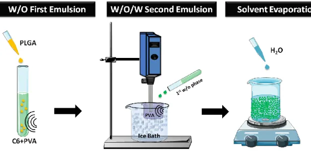
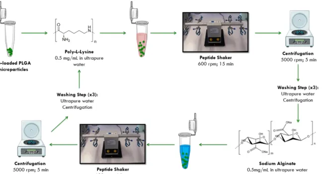
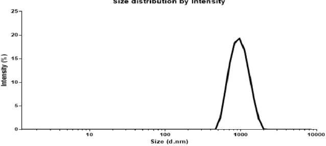
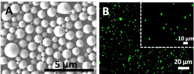
Figure 3.1.Schematic illustration of the synthesis of C6-loaded PLGA microparticles………40 Figure 3.2. Schematic illustration of the LbL assembly process towards the synthesis of unloaded and C6-loaded polyelectrolyte-coated PLGA microparticles……….41 Figure 4.1. Zeta-size distribution by intensity of the blank and uncoated PLGA microparticles………55 Figure 4.2. (A) SEM and (B) FL microscopy micrographs of the uncoated PLGA microparticles prepared at room temperature. (A) The SEM image shows the spherical shape
xviii
and nonporous surface morphology of the particles. (B) FL microscopy images of uncoated C6-loaded PLGA microparticles, which is fluorescence in green………56
Figure 4.3. Mean ζ-potential as a function of the polyelectrolytes layer number for the PLGA microparticles coated with alternating PLL and ALG layers. The odd layer numbers correspond to the PLL deposition and the even layer numbers to the ALG deposition. (n = 3; mean ± standard deviation (SD))………...…..59
Figure 4.4. (A,C,E) SEM and (B,D,F) FL microscopy micrographs of (PLL/ALG)2-, (PLL/ALG)4- and (PLL/ALG)6-coated C6-loaded PLGA microparticles prepared at room temperature. (A,C,E) SEM images show the spherical shape of the microparticles. (B,D,F) The FL microscopy images were acquired by the fluorescence feature of C6, which stains the microcores in green………...60 Figure 4.5. Cumulative release of C6 from the uncoated- (circles), (PLL/ALG)2- (triangles), (PLL/ALG)4- (squares) and (PLL/ALG)6-coated (rhomb) PLGA microparticles. The percentage of drug release was accessed for 21 days (A) and presented the burst of release during the first 30 min (B). C6 is a poorly-water soluble molecule, and thus present poor solubility in PBS. For that reason, Tween® 20 (0.5 % v/v) was added to PBS aqueous……….62 Scheme 5.1. Schematic illustration of the synthesis of (a) CaCO3 core microparticles, (b) core-shell microparticles with two CHT/ALG bilayers, (c) (CHT/ALG)2-based hollow multilayered microcapsules after CaCO3 core template dissolution with EDTA, and (d) well-dispersed (CHT/ALG)2-based hollow multilayered microcapsules after purification by dialysis……….65 Figure 5.1. (A,B) SEM, (C) TEM, (D) FL microscopy and (E,F) CLSM micrographs of the uncoated CaCO3 microparticles prepared at room temperature. (B) The SEM micrograph shows the high surface roughness and the porous structure of the microparticles. (D) FL microscopy and (E,F) 3D reconstructed CLSM micrographs were acquired by loading the CaCO3 core microparticles with FITC (green channel). F1 and F2 correspond to the upper (xy) and lateral (xz) views, respectively……….67 Figure 5.2. (A) Mean ζ-potential as a function of the polysaccharide layer number for the CaCO3 microparticles coated with alternating CHT and ALG layers. The odd layer numbers correspond to the CHT deposition and the even layer numbers to the ALG deposition. (B) TEM, (C) SEM and (D) FL microscopy micrographs of the core-shell CaCO3 microparticles after the deposition of two CHT/ALG bilayers. The insets in (B) and (C) show a higher magnification of the TEM and SEM images, respectively. The inset in (C) also shows the high surface roughness of the core-shell microparticles……… 70 Figure 5.3. (A,C) FL microscopy and (B,D) CLSM micrographs of (CHT/ALG)2-based hollow multilayered microcapsules before (A,B) and after (C,D) the dialysis purification step. Green channel: FITC-labeled CHT. The insets in B,D correspond to the middle plane of the microcapsules. (E,F) SEM micrographs of the (CHT/ALG)2-based hollow multilayered
xix
microcapsules after dialysis, showing folds and creases in their structure due to the drying procedure………...72
Figure 5.4. CLSM images of L929 cells upon contacting with well-dispersed or aggregated (FITC-CHT/ALG)2 hollow multilayered microcapsules. (A1–A4) Cells that were not in contact with microcapsules were used as controls. Dispersed (FITC-CHT/ALG)2 hollow multilayered microcapsules were internalized by cells (A5–A8). Contrarily, aggregated capsules were not incorporated and remained adsorbed on the cell surface (A9–A12). A zoomed-in region of A8 and A12 micrographs was done to distinguish the internalized dispersed capsules (B1–B1.2) from the ones adsorbed at the cell surface due to their higher aggregation degree (B2–B2.2). B1 and B2 correspond to 3D reconstructed z-stack micrographs as seen from top (view of the xy plane). B1.1–B2.1 and B1.2–B2.2 correspond to orthogonal projection images (view of the yz plane and view of the xz plane, respectively). Blue channel: DAPI nuclear probe; Red channel: rhodamine-labeled phalloidin; Green channel: (FITC-CHT/ALG)2-based hollow multilayered microcapsules. White circles indicate some examples of microcapsules internalized by L929 cells………..75
xxi
List of Tables
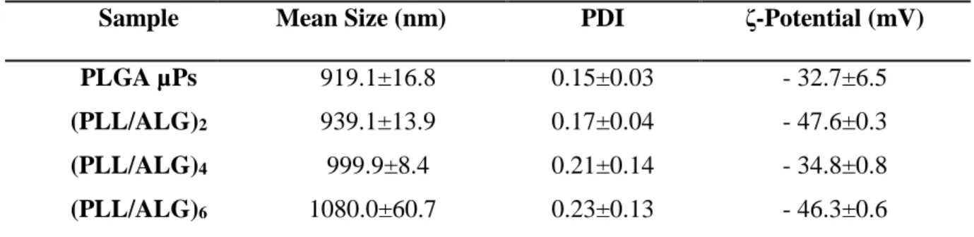
Table 1. Examples of synthetic polymers used in the DDS preparation………..21 Table 2. Mean size, PDI, and ζ-Potential of uncoated-, (PLL/ALG)2-, (PLL/ALG)4 and (PLL/ALG)6-coated C6-loaded PLGA microparticles (n = 3; mean ± standard deviation (SD))………..58
xxiii
List of Symbols and Abbreviations
ALG Sodium alginate
C6 Coumarin 6
CaCl2 Calcium chloride
CaCO3 Calcium Carbonate
CH3COOH Glacial acetic acid
CHT Chitosan
CLSM Confocal Laser Scanning Microscopy
Da Dalton
DAPI Rhodamine Phalloidin, 4’,6-Diamidino-2-phenylindole
DCM Dichloromethane
DDS Drug Delivery Systems
DI Deionized water
DLS Dynamic Light Scattering
DMEM Dulbecco’s Modified Eagle’s Medium
DOX Doxorubicin
DPBS Dulbecco’s phosphate-buffered saline
EDTA Ethylenediaminetetraacetic acid
EE Encapsulation Efficiency
ESE Emulsion-Solvent Extraction/Evaporation technique
xxiv
FBS Fetal Bovine Serum
FDA Food and Drug Administration
FITC Fluorescein 5(6)-isothiocyanate
FITC-CHT Isothiocyanate-labeled chitosan
FL Fluorescence Microscopy
GA Glycolic acid
HA Hyaluronic acid
HCl Hydrochloride acid
L929 Mouse Lung Fibroblast cell line
LA Lactic acid
LB Langmuir-Blodgett
LbL Layer-by-Layer
MPs Microparticles
NaCl Sodium Chloride
NaOH Sodium hydrochloride
NIR Near infrared
NPs Nanoparticles
PBS Phosphate-buffered saline
PDI Polydispersity index
PDLLA Poly(D,L-lactic acid)
PEG Polyethylene glycol
xxv
PLA Polylactic acid
PLGA Poly(lactic-co-glycolic acid)
PLL Poly-L-lysine
PLLA Poly(L-aspartic acid)
PMCs Polymeric microcapsules
PNIPAAM Poly(N-isopropylacrylamide)
PVA Polyvinyl alcohol
PVPON Poly(vinylpyrrolidone)
R2 Coefficient of determination
RES Reticuloendothelial System
SD Standard deviation
SEM Scanning Electron Microscope
TEM Transmission Electron Microscope
UV Ultraviolet
W/O Water-in-oil
PART A
Chapter 1 – General Introduction
3
Chapter 1 – General Introduction
1.1.
General
Concepts
in
Nanoscience
and
Nanotechnology
From the middle of twenty century to nowadays, the interdisciplinary field of nanoscience, with its promises of amazing nanotechnologies, has demonstrated to be one of the most challenging, attractive and widely explored fields of research among the scientific community with implications in the traditional areas of science, including chemistry, physics, biology and engineering, as well as in tackling societal challenges. Nanoscience and nanotechnology focus on the design, characterization, production, and application of structures, devices, and systems by controlling the size and shape at the nanoscale (from around one to hundreds of nanometers) (Drexler 1992). Briefly, the field is defined as the ability to build micro- and macro-materials with atomic precision through the manipulation of atoms and molecules at the nanoscale. The potential of nanotechnology to decisively contribute to influence the future of humanity and address major hurdles associated with human health led to the emergence of a novel field of research entitled nanomedicine, which has been and will continue to revolutionize disease diagnostics and treatment modalities (Farokhzad et al. 2006). The purpose may be broadly defined as the monitoring, control, repair, defense and improvement of all human biological systems, working from the molecular level using engineered devices and nanostructures, ultimately to achieve medical benefits (Albanese et al. 2012). These may be included in a microdevice that has a macrointerface in a biological environment. The focus, however, is always on nanointeractions within the framework of a larger device or directly within a subcellular system (Dowling et al. 2004).
A visionary in the field of quantum computing, the American and physicist Richard Feynman was a pioneer in the control and manipulation of matter at the atomic and molecular scale, launching the foundations of nanotechnology in his well-known and often-cited lecture entitled “There’s Plenty of Room at the Bottom” at the American Physical Society meeting in Caltech in 1959 (Feynman 1959). Due to his contributions and futuristic vision on the ability to manipulate atoms and molecules and on the intrinsic
4
impact of nanotechnology in a plethora of fields, Feynman revealed that there was no theoretical problem on the construction of small devices composed of very small elements, opening the doors for the development of even smaller and more powerful devices. He idealized the whole Encyclopedia Britannica fully written on the head of a pin and predicted the increasing ability to examine and control matter at the nanometer-scale level (Hess 2003). Later on, he noticed that nature also makes use of nanostructures in biological systems, for example the biological ion channels in the cell membrane (Feynman 1992).
The term nanotechnology started to be used worldwide in 1974 using the nanoscale science suggested by Feynman to point out some features between micrometric scale engineering and the new submicron-scale engineering field (Taniguchi 1974). Significant advances in nanotechnology were not noticed until the early 1980s, when the appearance of new nanomanipulation instruments enabled the measurement and manipulation of nanometer-scale materials (Ito 1998). Afterwards, in 1991, the Japanese scientist Sumio Iijima discovered the carbon nanotubes, which exhibit extremely high mechanical strength and unique properties for biomedical applications (Pautrat 2011; Iijima 1991). Hence, nanotechnology offers a new paradigm for the manufacture of materials using submicron scale manipulation with the aim of creating devices at the nanoscale, from fundamental structural units or building blocks, through the application of bottom-up or top-down methodologies (Augustine et al. 2009). The impact of nanotechnology is very penetrating in many areas including materials chemistry and engineering nanoelectronics and computer technology, medicine and health (Dowling et al. 2004). Parallel to the consolidation and expansion of nanotechnology, the developments in the areas of microelectronics (increasing the processing capacity of computers) and software (simulate the behavior of nanoscale materials), as well as in the biotechnology field are contributing for the development of new micro- and nano-sized systems (Lindquist et al. 2010). All this knowledge led to the flourishment of a new revolutionary area that helped to bring together science and technology for applications in human health, commonly called nanobiotechnology ( Ravichandran 2010, Pautrat 2011). Despite being a term mostly employed in academia, nanobiotechnology has a massive impact on diagnosing (Dowling et al. 2004), treating and preventing disease (Birch et al. 2007) and traumatic injury of relieving pain, thus extensively contributing for preserving and improving human health (Gaudana et al. 2009) through the use of molecular tools and knowledge of the human body (Moghimi et al. 2017). The developed
Chapter 1 – General Introduction
5 nanodevices are used in many areas of research related to human health, including in regenerative medicine (Walker et al. 2009) through the development of implanted devices (Wickline et al. 2006) or platforms for the controlled delivery of therapeutics (Lanza et al. 2002).
Classically, the delivery of therapeutics followed a systemic drug administration. However, this procedure is largely inefficient and entails a wide range of side effects that may ultimately lead to damage in healthy tissues and organs. Therefore, several research groups within academia and the pharmaceutical industry have been devoted to improve the drug administration strategy in order to hit the damaged targeted tissue and promote a sustained and prolonged release of the drug at the injured site (Smith et al. 1972).
Studies over the years, highlighted the successful encapsulation of drugs in carriers in an attempt to overcome the drawbacks associated to major conventional techniques (Saltzman 2001). Several progresses in polymeric nanoscience and nanotechnology brought a revolutionary chance to develop new sophisticated devices approaches for being used in the biomedical field (Gurwitz et al. 2006).
Drug delivery systems (DDS) offer a resource to encapsulate, protect and sustained deliver therapies directly and selectively at diseased cells or tissues, preventing the systemic diffusion and premature drug elimination, as well as supporting crossing of the in physiological barrier (Pardeike et al. 2009). Ultimately, it is intended to have a controlled drug release that target receptors inside cells with minimized biological risk (Wickline et al. 2006). This systems represent a new and innovative trend (Des Rieux et al. 2006) towards the development of novel and innovative therapeutic nanocarriers and diagnostics modalities for addressing many diseases, including cancer (Peer et al. 2007).
1.2. Motivation
In the biomedical field the term drug delivery refers to methods and approaches for administering drugs, preferentially selectively, to target sites at the proper dosage in order to accomplish their therapeutic action at the injured sited. Major hurdles associated to conventional techniques for drug administration include the premature interaction of drugs with the biological environment, their premature degradation, their release before reaching the diseased site or the burst release at the target site, which critically complicates the therapeutics action of the interest molecules. Having this is mind, the design of a new era of vehicles for drug delivery is needed to overcome these drawbacks
6
and improve the drug effectiveness, reduce unwanted side effects and avoid cellular drug resistance (Chertok et al. 2013).
1.3. Objectives
The main goal of this work is the development and in vitro validation of biomedical systems capable of either releasing model molecules in a sustained manner and at targeted sites and being internalized by cells. To achieve this goal, this work aimed to develop microparticles for (i) the encapsulation and sustained release of model compounds at the targeted sites aiming in order to overcome the conventional drug administration drawbacks, and (ii) for enhanced cellular uptake. The ideal microparticle should be biocompatible, biodegradable, and susceptible of carrying out molecules of interest and delivering it, with precision, to the therapeutic target sites and being internalized by living cells.
With this purpose in mind, two distinct microparticle templates were synthesized by resorting to calcium carbonate (CaCO3) and poly(lactic-co-glycolic acid) (PLGA) cores, and further surface-functionalized making use of the bottom-up Layer-by-Layer (LbL) assembly technology to obtain a core-shell multilayered system that was used as vehicle to evaluate the encapsulation and controlled release of model molecules coumarin 6 (C6) or as platform for enhanced cellular uptake. The CaCO3-based microparticles were surface functionalized with natural-origin polymers, namely marine biopolymers (oppositely charged chitosan (CHT) and alginate (ALG) biopolymers) to produce core-shell microparticles before the decomposition of the core to produce CHT/ALG hollow multilayered microcapsules and assess their cellular uptake. In the case of the PLGA-based microparticles encapsulating C6 as a model compound, their surface was functionalized with a hybrid system encompassing multilayers of oppositely charged natural (ALG) and synthetic poly-L-lysine (PLL) polymers and the encapsulation efficiency and release profile measured.
1.4. Organization
This dissertation is organized in six chapters. Chapter 1 intends to establish the scientific framework in which the present work is carried out. It also provides a brief context, motivation and organization of this work. Chapter 2 consists in a literature
Chapter 1 – General Introduction
7 review of the main subject of the dissertation. This chapter is mainly devoted to the state of the art of drug delivery systems and their relevance in biomedical applications. It includes a summary of different types of drug delivery systems, especially microparticles/capsules, different types of techniques to produce this microcarriers and a summary about Layer-by-Layer (LbL) technology as a way of coating the particles and processing microcapsules, using particles as template, and their biomedical applications. Chapter 3 details all information of the materials, sample preparation, methodologies and characterization techniques used to pursue this work. It aims to describe the materials, solutions/samples and methodologies employed throughout this thesis and theoretical background behind the main experimental techniques employed for the characterization of the sample formulations. Chapters 4 and 5 present the results and the discussion of the work developed concerning the development of microparticles for controlled drug released and in vitro cellular uptake. Chapter 4 entitled Preparation and Characterization of PLGA Microparticles aims to assess the influence of the number of PLL/ALG layers on the sustained release of C6 from the (PLL/ALG)n-coated PLGA microparticles. Chapter 5 entitled Preparation and Characterization of CaCO3 Microparticles and Hollow Microcapsules describes the synthesis of well-dispersed (CHT/ALG)n-coated CaCO3 microparticles and the subsequent preparation of (CHT/ALG)n-based hollow microcapsules, as well as their in vitro cellular uptake by mouse fibroblast cells (L929 cell line). Finally, Chapter 6 encloses the main conclusions regarding the work carried out during this MSc thesis. Besides, suggestions for future work in the field are also provided.
PART B
Chapter 2 – Drug Delivery Systems
11
Chapter 2 – Drug Delivery Systems
2.1. Introduction to Drug Delivery Systems
Over the last few decades, we have witnessed a tremendous evolution and impact of nanotechnology in several scientific areas (Rosen et al. 2005). The importance of nanotechnology and its application on the pharmaceutical and medical sciences, including human health, is clearly demonstrated by the exponential growth of the nanomedicine research field in the last few years (Doshi et al. 2009; Farokhzad et al. 2006; Moghimi et al. 2017). Several therapeutic strategies and diagnostic tools have been developed which have led to an unprecedented progress in the field of drug delivery and diagnostics aiming at fighting several well-known diseases, including cancer (Yih et al. 2006; Sinha et al. 2006).
The advent of new drug delivery systems (DDS) started in the mild 1900s (Smith 1999). Until then, most drugs were delivered through conventional methods such as intramuscular or subcutaneous injections (Shapiro et al. 2002; Barry 2001), oral delivery (Prisant et al. 2003; Maggi et al. 2000) or transdermal application (Viscusi et al. 2004; Rosen et al. 2005). Although effective, each of these methods has intrinsic disadvantages. The parenteral route is painful, invasive, and often requires administration by a trained person, resulting in reduced therapeutic efficacy (Edwards et al. 1998). Additionally, as the drug is often injected directly into the blood stream, the effect is somehow short lived, thereby reducing the therapeutic effect in the injured site (Allen et al. 2004). Oral delivery vehicles are associated with high patient compliance. However many pharmaceutical molecules cannot survive the harsh environment or be absorbed through the intestinal epithelial barrier (Sinha et al. 2003; Smith 1999). Topical delivery, again, generally improves patient compliance, but this method is limited to local delivery, as many therapeutics cannot diffuse through the protective epidermis layers (Malik et al. 2016).
Distinct types of nano- and micro-carriers have been proposed as DDS to effectively encapsulate, protect, transport and on-demand targeted release of well-defined amounts of therapeutic payloads in a controlled manner at the injured site, aiming to mitigate the limitations of the conventional delivery methodologies (Peer et al. 2007). In the late 1960s, Alejandro Zaffaroni, a pioneer in drug delivery research, designed the first transdermal controlled release system. His research is considered by many to have laid the foundation of the research in drug delivery (Peppas 2013). The family of drug delivery vehicles, schematically represented in
12
Figure 2.1, includes dendrimers (Caminade et al. 2014), lipid-based carriers, including micelles (Kataoka et al. 2012), polymersomes (Lee et al. 2012) and liposomes (Garg et al. 2014; Bochot et al. 2012), virus- or cell-based (Neu et al. 2001; Villa et al. 2016a; Fliervoet et al. 2016; Han et al. 2018; Ma et al. 2012; Yildiz et al. 2011) vehicles and inorganic and polymeric particles/capsules (Ariga et al. 2011; Becker et al. 2010; Costa et al. 2017; Cui et al. 2014; Kallinteri et al. 2007; Sukhorukov et al. 1998).
Virus-based vehicles are remarkable tools employed in the field of nanomedicine, which can provide many possible pathways to use different vehicles, including antimicrobial therapeutic materials and imaging markers (Ma et al. 2012). Currently nanotechnology advances have used viral particles as a tool to develop new multifunctional nanomaterials to be used in several applications, including in tissue engineering and drug delivery, or as semiconductor and energy-producing materials (Fliervoet et al. 2016). Virus-based nanofibrous tissue scaffold engineering allows efficient control of the chemical, physical and mechanical properties of the scaffolds. Although the use of virus-based nanostructures is leading to some safety concerns (Yildiz et al. 2011), the in vivo animal tests can help to better analyze the effectiveness and safety of phage-based matrices in tissue engineering applications (Farr et al. 2014).
Liposomal systems were developed in the 1960s for controlled release (Bochot et al. 2012). A liposome is an artificial vesicle made from two lipid bilayers, identical to cell membrane, resulting in a hydrophilic outer shell and an inner core, with a hydrophobic layer between them (Ariga et al. 2011). The dual attitude toward water allows the encapsulation of both hydrophobic and hydrophilic drug molecules within the carrier system. The physicochemical properties of the liposomal constituents, including the membrane fluidity, permeability or charge density have influence on the types of interactions between the liposomes and bloodstream or tissue elements (Yih et al. 2006). Current liposomal systems, including long-circulating liposomes, that can be formed by incorporating hydrophilic polymers into the lipid bilayer to create an aqueous coat on the surface, reduce the uptake by reticuloendothelial system (RES). An example of this modification is called PEGylation, where chains of polyethylene glycol (PEG) are attached to the particle surface (Lima et al. 2012). Active cationic liposomes have high affinity for cell membranes and to deliver materials to cells by fusing with cell membranes and depositing material into the cell (Bollhorst et al. 2017). Nucleic acids are the most common therapeutics form delivered within cationic liposomes (Lomas et al. 2007). As nucleic acids are negatively charged, stability is increased when
Chapter 2 – Drug Delivery Systems
13 compared with complexed positively charge liposomes, for drug delivery (Yildirim et al. 2017). It is also important to highlight that liposome-based medicines play a role in clinical trials, especially in drug administration or in food industry (Brandt et al. 2009).
Polymersomes are a class of artificial vesicles made from block copolymers (Bollhorst et al. 2017). Its ability to carry out biological elements has been highlighted in several applications in medicine, pharmacy and biotechnology, especially for drug delivery (Lee el al. 2012). The aqueous core may be used to encapsulate therapeutic molecules such as drugs, proteins, peptides and nucleic acids (Discher et al. 2006). Typically, the polymerosomes are hollow spheres with an aqueous central core surrounded by a two-layer membrane, which protects the nucleus from the external environment (Hu et al. 2017). Furthermore, to improve the therapeutic efficiency and prevent drug resistance, polymersomes must quickly release the encapsulated drug in a controlled manner when the target location is reached. Thus, several stimulus-responsive polymersomes, which aim to release the encapsulated molecules in response to various stimuli such as pH (Rofstad et al. 2006), redox (Napoli et al. 2004), temperature (Zhang et al. 2010), light (Tong et al. 2005), magnetic field (Krack et al. 2008) or ultrasound (Yu et al. 2006), have been developed by incorporating different responsive building blocks into polymers. Among all stimuli, the pH responsiveness is the most studied, due to pH gradients found in biological environments (Yildirim et al. 2017). In comparison to liposomes, these vehicles have thicker and more stable membranes and possess higher physical and chemical stability, largely due to the higher molecular weight of copolymers in comparison to lipids (Lima et al. 2016; Hu et al. 2017). This feature allows a high efficiency in the encapsulation by notably reducing the premature release, with applications in gene therapy or diagnostic imaging, thereby decreasing the biological risk. (Janapareddi et al. 2016; Discher et al. 2002).
Micelles are vehicles with small size and soluble features that can self-assemble by hydrophilic and hydrophobic segregation. In addition to hydrophobic and electrostatic interactions, metal complexation and hydrogen bonding may contribute to micelle formation (Oerlemans et al. 2010). A block copolymer, enclosing hydrophobic and hydrophilic segments, has the ability to self-assemble in an aqueous environment such that the hydrophobic segments form a core in the center and the hydrophilic segments form a shell in the exterior part, increasing the solubility of hydrophobic therapeutics (Kataoka et al. 2012). A popular choice for hydrophilic block copolymer sections is PEG, a widely used polymer in drug delivery applications and the gold standard of carrier systems, avoiding nonspecific uptake once
14
administered, and functions by creating an aqueous layer around the particle to avoid detection by the immune system (Torchilin 2007). Nonspecific uptake by the RES is a major obstacle to micelles and other small colloidal carrier systems. The RES is part of the body’s defense mechanism and includes cells, such as macrophages and monocytes. These cells are phagocytic in nature, and function by overcoming potential threatening substances, eventually accumulating in the liver or in spleen for degradation and elimination (Lima et al. 2012). This represents a major hurdle for drug carrier systems since the uptake by these cells will reduce the amount of therapeutic effect in the systemic circulation. Poly-L-amino acids, such as poly(D,L-lactic acid) (PDLLA), are frequently used as the hydrophobic portion of micellar block copolymers. For example, Genexol®-PM, a PEG–PDLLA copolymer micelle loaded with the chemotherapeutic paclitaxel, is currently approved in Korea for the treatment of breast cancer and is undergoing clinical trials in the United States for use in pancreatic cancer treatment (Shi et al. 2016). Micelle formation is influenced by factors such as the molecular weight of the copolymer, the ratio of copolymer blocks to each other, and the amount of polymer involved (Kataoka et al. 2012; Torchilin 2007). In short, micelles present high biocompatibility, can be easily prepared and loaded with drugs. Their nanosized dimension allow deep penetration into target cells or tissues and can successfully release the drug or imaging agents that is encapsulated in their hydrophobic cores or adsorbed to their hydrophilic surface, thus enhancing the therapeutic effect. (Steichen et al. 2013; Jones et al. 1999).
Dendrimers are highly branched, star-shaped macromolecular systems with a well-defined, homogeneous and highly monodisperse structure that typically comprise three core structures: a symmetric central core, an inner shell and an outer shell formed by dendritic structures containing functional groups (Boas et al. 2003). This vehicle is stable and has a surface that can be readily functionalized with targeting molecules and ligands (Caminade et al. 2014). Owing to its internal core and extensive branching structures in the inner shell, the dendrimers are highly suitable for drug encapsulation. They also present unique characteristics including a branched layered architecture, which confers them a globular structure and internal hollows that enhance sequestration (Lima et al. 2016).
Despite the huge research effort devoted to nanotechnology and its biomedical applications, the number of drug release systems approved for human use or in clinical trials is still very limited (Chen et al. 2010). Nowadays, one of the most explored vehicles in drug delivery research are nano (NPs)- and microparticles (MPs) (Couvreur 2013). Several studies have shown that drug encapsulation in micro- and nanoparticles results in an increase of drug
Chapter 2 – Drug Delivery Systems
15 bioavailability when compared to oral administration (Faraji et al. 2009). In addition, these carriers are able to transport both hydrophilic and hydrophobic drugs with a high loading efficiency and enhanced circulation time, making them excellent controlled release systems (Wang et al 2008).
Figure 2.1. Schematic representation of Drug Delivery Systems Family, (A) liposome, (B) micelle, (C) dendrimers, (D) polymer-based microparticle and (E) polymer-based microcapsule. Reprinted with permission from (Steichen et al. 2013).
Similarly, polymeric microcapsules (PMCs) show great potential in biomedical field, particularly in drug delivery, since they present low density, high specific surface area and large inner spaces for encapsulating guest molecules (Doshi et al. 2009; Javier et al. 2008). Due to their important features, the microparticles and microcapsules will be discussed in more detail in the Section 2.2. However, it is important to point out that, although each of the above DDS has its own advantages and drawbacks, depending on the envisioned application, they should
16
assure the encapsulation and protection of the drug to be administrated from premature degradation in order to secure a high accumulation and bioavailability of the drug at the pathological area, as well as prevent undesirable side effects in healthy tissues and/or organs (Fliervoet et al. 2016).
2.2.
Polymer-based
Microparticles/Microcapsules:
Applications in Drug Delivery and Cellular Uptake
The impact of drug delivery systems ranges from early developments in colloidal particles to a broad range of industrial and nanotechnology applications in physical, biological, and medical sciences. The design of novel drug delivery carriers has gained increasing attention during the past decade (Lima et al. 2016). Depending on the type of application there is the need for evaluating and gathering the physicochemical properties that best interact with biological systems (see Figure 2.2). In this concern, the shape, surface chemistry, charge and size were shown to have a major impact in in vivo experiments (Albanese et al. 2012).
A suitable DDS should be able to carry a specific therapeutic agent to the target locations without affecting healthy sites (Holzinger et al. 2014). With this is min, nano- and micro-sized polymer-based particles and capsules have attracted great interest as drug delivery systems and in the last few years we have witnessed that such interest has grown exponentially (Mitchell et al. 2017). After decades of research, some general guidelines have been established for the selection of the chemical properties including surface characteristics of the microparticles/capsules necessary to achieve good performances, minimizing side effects
(Albanese et al. 2012).
Most current microparticulate systems are spherical in shape, and extensive work has been devoted to the study of their biological behaviors both in vitro and in vivo (Schmid 2008). The particles’ shape plays a key in determining their intended biological functions. Recent data have shown that particle shape may have a deep effect on their biological properties (Hernández et al. 2010). For example, as demonstrated by Discher et al (Discher et al. 2002), cylindrically-shaped filomicelles can effectively evade non-specific uptake, allowing persistent circulation for up to one week after intravenous injection, which is ten times longer than the result found for spherical particles. Furthermore, the work on theoretical modeling has shown that non-spherical particles can significantly increase the particles adhesion to cellular receptors under flow conditions when compared with spherical particles. Experimentally, Muzykantov and
co-Chapter 2 – Drug Delivery Systems
17 workers (Villa et al. 2016) showed that disk-shaped nanoparticles increased intercellular targeting adhesion over spherical particles of similar size. Other non-spherical nanovehicles, including carbon nanotubes (Vashist et al. 2011) or worm shaped iron oxide-based nanoparticles (Arruebo et al. 2007), have also demonstrated considerably increased accumulation and retention in tumor tissues in vivo. All these information, together with the wide range of naturally occurring shape-specific nanoparticulate systems, like red blood cells (Han et al. 2018), are beginning to highlight the importance of controlling particle shape for biomedicine applications. Although shape plays an important role in various biological processes, it is important to understand it interdependence with other design parameters. This concept was proved by Gratton et al., who, in one of this works, reported the interdependence of shape, size, and surface chemistry on cellular uptake and internalization (Gratton et al. 2008). A top-down fabrication technique, PRINT® (Rolland et al. 2005) was applied to fabricate uniform populations of cationic, crosslinked PEG hydrogel based MPs/NPs with control over size, shape, and surface chemistry (Lewis et al. 2010).
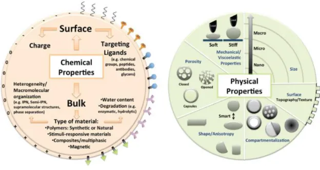
Figure 2.2. Schematic illustration of how chemical surface and the choice of bulk material as well as the physical features such as mechanical properties, size, porosity, surface topography/texture, shape and compartmentalization directly affect the design of particulate systems for biomedical applications. Reprinted with permission from (Lima et al. 2016).
18
Cellular internalization studies of different aspect ratios of cubic and cylindrical particles were performed, and it was found to have a strong dependence. Particle’ size,
geometry as well as surface curvature, directly affect the interactions between cell and particles and subsequently internalization kinetics and efficacy (Costa et al. 2014). It is generally accepted that if the surface properties of the particles are favorable, nanoparticles are internalized more efficiently by phagocytic cells (Radu et al. 2004). However, well-documented studies based on spherical and non-spherical particles indicate that MPs tend to bring more advantageous to deliver therapeutic agents to endothelial cells and tumor sites because these sizes are more likely to be taken up by cells (Bharali et al. 2005).
Additionally, the choice of bulk material that the particle is composed by is also a key point since it must fulfill a tailored biological behavior (bioactivity, biocompatibility, biodegradability), it must improve the payload capacity and be harmlessly eliminated from the body in a reasonable period after releasing the cargo and having carried out possible diagnostic function (Matricardi et al. 2013). For example, in non-permanent applications the materials used to obtain bare particles should degrade at adequate rates without releasing toxic degradation products. In the case of DDS, biological degradation affects directly the drug release profile (Edwards et al. 1998). Chemical surface and surface charge also influence the particles performance in biological environment. Several studies reported that positively charged MPs exhibit higher internalization by macrophages and dendritic cells than neutral or negatively charged particles (Gratton et al. 2008; Reajman et al. 2004).
Over the last decade, several studies have led to a host of new strategies being developed for the modification of the particle’s surface. In polymeric-based micro- and nano-composites fabrication, the particles are used as backbone to enhance the physiochemical features (Kwon et al. 2013; Costa et al. 2013) such as flexibility, smoothness, strength and stiffness, which are essential characteristics in biomedical applications (Janapareddi et al. 2016). The surface texture of the particles and their porosity, derived from the concentration or cross-linking of the material, are also relevant parameters to consider during the particles design, due to their influence on drug diffusion (Costa et al. 2013). Porous particles are particularly attractive as drug delivery systems due to their unique and superior properties (Volodkin et al. 2004). Particularly, CaCO3-based microparticles have received increasing interest as suitable templates for engineering prospective drug delivery vehicles for fulfilling biological and biomedical applications due to their easy and cost-effective synthesis, large surface to volume ratio, porous structure, high biocompatibility and mechanical stability, as well as mild core
Chapter 2 – Drug Delivery Systems
19 template decomposition upon core-shell formation (Wang et al. 2006; Petrov et al. 2005; Volodkin 2014). However, they show a great tendency to aggregate and have a high polydispersity index (Couvreur 2013). On the other hand, smooth or non-porous microparticles also show great potential in biomedical applications and present good stability in aqueous environment leading to formation of a monodisperse population of particles. (Parakhonskiy et al. 2010)Moreover, they show great potential application in medical diagnostics, biotechnology and pharmaceutical industry (Syamchand et al. 2013). When aiming for drug delivery purposes, smooth particles, such as silica or PLGA, suggest a great surface functionality and chemical stability that ensure the controlled release and target drug delivery of a variety of molecules (Lima et al. 2016; Doshi et al. 2009)
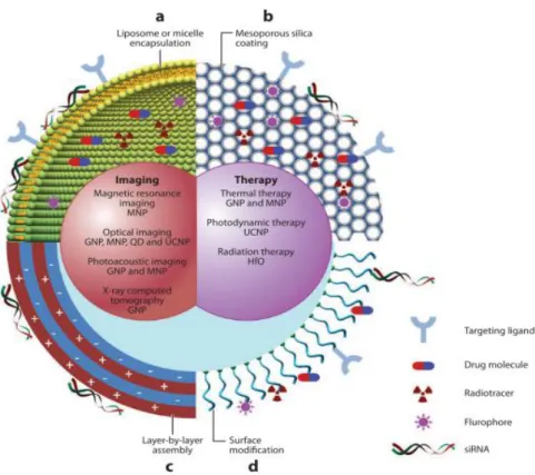
Figure 2.3. Schematic illustration of multifunctional MPs, which can be generated by either
combining nanocrystals with different functionalities or combining nanocrystals with functional small-molecule cargos through different surface engineering strategies: (a) liposome or micelle encapsulation, (b) mesoporous silica coating, (c) layer-by-layer assembly, and (d) surface conjugation. Reprinted with permission from (Syamchand et al. 2013).
20
All the aforementioned features of polymer-based particles are extremely important in determining the fate of the particles. Moreover, the particles can function as a template for the fabrication of other types of systems, including microcapsules, also important in biomedical applications, especially in drug delivery (see figure 2.3) (Syamchand et al. 2013). In today’s pharmaceutical industry, more efficient and effective ways of treating various diseases are needed. Currently, researchers are looking at hollow polymeric microcapsules as a new and innovative way for treating many diseases ranging from simple treatments such as vaccines to more complex treatments (Fliervoet et al. 2016b; Villa et al. 2016; Donath et al. 1998).
Polymeric micro- and nanosized capsules, obtained after core template dissolution, have been widely employed in the biomedical and biotechnological fields, owing to their versatility and finely-tuned physicochemical properties and functions. Specific examples of applications include drug delivery (De Cock et al. 2010), diagnostics (Kim et al. 2011), imaging (Chen et al. 2010; Wang et al. 2011), vaccination (Becker et al. 2009; Sexton et al. 2009), bio- and nanoreactors and immunomodulation (Lima et al. 2016). Polymeric microparticles consist of two parts: a core (inner part) and a shell (outer part). The shell protect the core material and the encapsulated drug and is usually made of natural or synthetic polymers or even a combination of both polymers (Urbas et al. 2017). Such structures have gained massive attention due to their intrinsic features, including low density, high specific surface area and large useful inner spaces for guest molecules. These structures can also encapsulate many types of biological molecules, such as drugs, nutrients, catalysts or fragrances (Szarpak et al. 2010). Polymer-based capsules usually exhibit porous shells which become advantageous since they establish more controlled interactions with the environment while simultaneously protecting the substances they carry on (Bollhorst et al. 2017). PMCs are produced by making use of sacrificial organic and inorganic templates (Urbas et al. 2017), as well as a variety of different engineering strategies including self-assembling Layer-by-Layer assembly technology (Caruso et al. 1998; Richardson et al. 2016), surface and interfacial polymerization, bioinspired assembly, and ultrasound assembly (Cui et al. 2014). Moreover, these systems might accomplish specific functions, must be selectively and specifically recognized by the target site and should retain the functional specificity of the surface ligands. Once the target cells recognize the carrier system, it must release the therapeutic drug moiety inside the targeted site (Richardson et al. 2015).
From simple to multifunctional systems, PMCs/MPs might target specific cells, improve the properties of therapeutic carriers and increase the circulation time, half-life, bioavailability, solubility and decrease toxicity (Wang et al. 2008). These carriers, with small and tunable size,
Chapter 2 – Drug Delivery Systems
21 have a large surface area capable of being functionalized with all sorts of molecules, including biological molecules such as proteins or nucleic acids (De Cock et al. 2010; Chassé et al. 2017). They have suitable stability, high encapsulation efficiency of hydrophobic and hydrophilic drugs, and are compatible with different routes of administration (De Cock et al. 2010; Chassé et al. 2017). By protecting the encapsulated drug, PMCs/MPs might increase the drug circulation time, improving the effect with smaller concentrations (Green et al. 2006; Bollhorst et al. 2017).
2.2.1.
Properties
of
Polymers
used
to
Produce
Microparticles/Microcapsules for Drug Delivery Applications
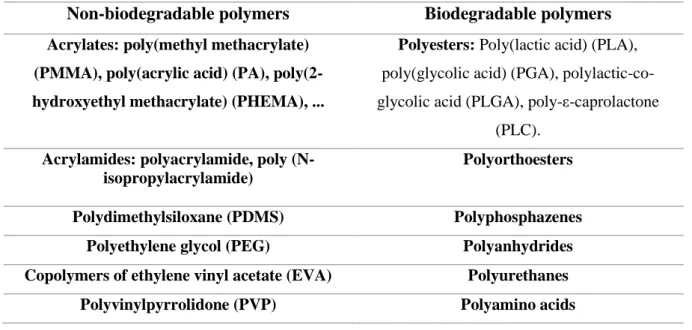
In which concerns the preparation of DDS, it’s important to highlight that a wide variety of biodegradable or non-biodegradable, synthetic or natural polymers can be used bearing in mind that these resulting DDS must be non-toxic, biocompatible and free of impurities (Chaun et al. 2015; Kumari et al. 2010). Table 1 lists some of the most widely explored biodegradable and non-biodegradable synthetic polymers used to develop DDS.
Table 1. Examples of synthetic polymers used in the preparation of DDS (Alvarez-Lorenzo et al. 2008; Saltzman 2001).
Non-biodegradable polymers Biodegradable polymers Acrylates: poly(methyl methacrylate)
(PMMA), poly(acrylic acid) (PA), poly(2-hydroxyethyl methacrylate) (PHEMA), ...
Polyesters: Poly(lactic acid) (PLA),
poly(glycolic acid) (PGA), polylactic-co-glycolic acid (PLGA), poly-ε-caprolactone
(PLC).
Acrylamides: polyacrylamide, poly (N-isopropylacrylamide)
Polyorthoesters
Polydimethylsiloxane (PDMS) Polyphosphazenes
Polyethylene glycol (PEG) Polyanhydrides
Copolymers of ethylene vinyl acetate (EVA) Polyurethanes
Polyvinylpyrrolidone (PVP) Polyamino acids
According to the Table 1, the DDS can be grouped depending on their composition. Within the synthetic polymers, it should be noted that the way they degrade (or not) in vivo determines the
22
viability of the developed systems. Thus, we can make a distinction between the polymers that are biodegradable and those which are non-biodegradable (Jain 2000). Synthetic polymers are composed mainly of organic compounds produced by humans through the polymerization process (Sharma et al. 2016).
A biodegradable polymer can be defined as a polymer which undergoes chemical degradation in vivo by hydrolysis or enzymatic action, resulting in non-toxic and biocompatible products capable of being metabolized and excreted by the normal physiological pathways (Luckachan et al. 2011). It is also characterized by extending the residence time when contact with mucous membrane due to its high degree of swelling property in aqueous medium. The rate and extent of drug release is controlled by the concentration of polymer and the release pattern (Luckachan et al. 2011). Polyesters, polyphosphazenes, polyorthoesters, polyamino acids, polyanhydrides, polyurethanes are some examples of biodegradable synthetic polymers used in the construction of delivery carriers.
PLGA is one of the most used synthetic polymer to produce microparticles. Although it is not a natural polymer, several studies have pointed out to its biocompatibility, biodegradability and low toxicity features (Martins et al. 2017). In addition, PLGA can improve drug solubility, efficacy, effectiveness, and safety through controlled release (Lu et al. 2009; Sharma et al. 2016). PLGA-based MPs/NPs present benefits related with their size. This crucial feature allows an enhancement of drug pharmacodynamics and bioavailability, since PLGA nanoparticles can penetrate into microcapillaries (Han et al. 2016; Sharma et al. 2016; Wischke et al. 2008).
Natural polymers, including proteins and polysaccharides are widely found in nature. Polysaccharides, including CHT and ALG are receiving great attention not only due to their broad availability, but also for their low cost, biodegradability and biocompatibility features (Shukla et al. 2013; Tønnesen et al. 2002). Moreover, we can obtain them in a wide range of molecular weights. In addition, due to the presence of various reactive groups in their structure, polysaccharides can be easily modified via chemical and biochemical process (Chai et al. 2017). In the nanomaterials field, CHT has attracted much attention owing to its mucoadhesive, antimicrobial, antitumor and anticoagulant properties (Sorlier et al. 2001; Berger et al. 2004). In addition, due to its ability to increase the diffusion of large molecules across mucosal surface, CHT has been widely used to produce microparticles (Xu et al. 2003). However, because of its insolubility in neutral and basic pH, its used is limited (Shukla et al. 2013). Gallo and co-workers were the first researchers to develop an approach using magnetic CHT microspheres,
Chapter 2 – Drug Delivery Systems
23 which are expected to be retained at the target site under the influence of an external magnetic field (Gallo et al. 1998).
The aforementioned properties have been extensively explored in the development of several systems for addressing multiple pharmaceutical and biomedical applications, including scaffolds for tissue engineering (Malafaya et al. 2007; Li et al. 2005), wound dressings (Wang et al. 2002; Öztürk et al. 2006) and controlled drug delivery systems (Bhardwaj et al. 2008). In the latter area of application, CHT and its derivatives have been processed in the form of gels, hydrogels, particles, nano- and microcapsules, films, sponges and tablets, encapsulating a wide variety of therapeutic agents (Wu et al. 2014). As drug carriers, these vehicles were investigated in vitro and in vivo by oral, ocular, nasal, subcutaneous and transdermal paths (Singla et al. 2001; Khor et al. 2003; Bhattarai et al. 2010). Similarly, ALG attracted a lot of attention in the biomedical field, especially in which concerns the biotechnological, cosmetic and pharmaceutical industries. This anionic polysaccharide is a versatile and useful tool to modify surfaces through biochemical functionality, imparting biocompatibility, biodegradability and antibacterial properties to the engineered systems (Dash et al. 2011; Muzzarelli et al. 2005; Fuente et al., 2010). In addition, ALG has a great potential in drug formulation due to its extensive application as food additive and non-toxicity (Tønnesen et al. 2002). Several studies related to CHT and ALG polysaccharides comprise the formation of controlled structures, for example spherical capsules or flat thin-films, through the deposition of each polymer onto template surfaces (Muzzarelli et al. 1989; Berger et al. 2004).
In order to better improve the mechanical properties, modify the hydrophobic/hydrophilic character and impart bioactivity of these two polysaccharides, their surfaces can be chemically functionalized through their functional groups. This physicochemical changes turn these two polysaccharides into highly promising biopolymers to be used in the fabrication of drug delivery systems (D’Ayala et al. 2008; Sashiwa et al. 2004; Jayakumar et al. 2010; Prabaharan et al. 2007).
2.3. Methods for the Fabrication of Polymer-based
Microparticles
Several methodologies have been commonly employed to fabricate polymeric microsystems. The chosen method should respect the nature of the polymer, the encapsulated