269
Revista da Sociedade Brasileira de Medicina Tropical 45(2):269-271, mar-abr, 2012
1. Departamento de Neurologia e Neurocirurgia, Complexo Hospitalar Santa Casa de Porto Alegre, Porto Alegre, RS.
Address to: Dra. Roberta Diehl Rodriguez.Rua Prof. Annes Dias 135, 90020-090 Porto Alegre, RS, Brasil.
Phone: 55 51 9209-1240; 55 51 3314-8636 e-mail: [email protected] Received in 02/12/2010 Accepted in 01/02/2011
Case Report/Relato de Caso
INTRODUCTION
CASE REPORT
Bruns’ syndrome and racemose neurocysticercosis: a case report
Síndrome de Bruns e neurocisticercose racemosa: relato de caso
Roberta Diehl Rodriquez
1, Denise Neme da Silva Crestani
1, José Otávio Dworzecki Soares
1, Paulo Roberto
Franceshini
1, Ronnie Petersen Alves
1, Ricardo Zimerman
1, Nelson Ferreira
1and Liselote Menke Barea
1ABSTACT
Cysticercosis is an infection caused by the larval stage of the tapeworm Taenia solium. he parasitemay infect the central nervous system, causing neurocysticercosis (NCC). he clinical manifestations depend on load, type, size, location, stage of development of the cysticerci, and the host’s immune response against the parasite. he racemose variety occurs in the ventricles or basal cisterns and is a malignant form. Mobile ventricular mass can produce episodic hydrocephalus on changing head posture with atacks of headache, vomiting, and vertigo, triggered by abrupt movement of the head, a phenomenon called Bruns’ syndrome (BS). We report a patient with racemose NCC and BS.
Keywords: Racemose neurocysticercosis. Cysticercotic meningitis. Bruns’ syndrome.
RESUMO
A infecção por cisticercose é causada pelo estágio larval da Taenia solium. O parasita pode infectar o sistema nervoso central, causando neurocisticercose (NCC). As manifestações clínicas dependem da quantidade, tipo, tamanho, local, estágio de desenvolvimento do cisticerco e resposta imune do hospedeiro contra o parasita. A variedade racemosa ocorre nas cisternas ventriculares ou basais e é considerada uma forma maligna. O cisticerco móvel no ventrículo pode produzir hidrocefalia episódica com ataques de cefaléia, vômitos e vertigem, provocados pelo movimento abrupto da cabeça, fenômeno chamado de síndrome de Bruns (SB). Relataremos o caso de uma paciente com NCC racemosa com SB.
Palavras-chaves: Neurocisticercose racemosa. Meningite cisticercótica. Síndrome de Bruns.
Cysticercosis is an infection caused by the larval stage of the tapeworm Taenia solium. he parasite may infect the central nervous
system, causing neurocysticercosis (NCC). NCC is the single most common cause of acquired epileptic seizures in the developing world1. Despite the advances in diagnosis and therapy, NCC remains
endemic in most low-income countries2. It is estimated that 50
million people in the world today are infected by the taeniasis-cysticercosis complex and 50,000 die every year3.
he clinical manifestations of NCC largely depend on load, type, size, location, and the stage of development of the cysticerci, as well as on the host’s immune response against the parasite4. here is neither
a pathognomonic feature nor a typical NCC syndrome.
he racemose variety occurs in the ventricles or basal cisterns and is characterized by abnormal growth of cystic membranes followed by degeneration of the scolex (parasite´s head). As ventricular and basal cisternal locations are considered malignant forms of NCC, when hydrocephalus secondary to cysticercotic meningitis is present, mortality rate is high (50%), and most patients die within 2 years of cerebrospinal luid (CSF) shunting5. Atacks of severe headache,
vomiting, and vertigo triggered by abrupt movement of the head due to a mobile ventricular mass producing episodic hydrocephalus on changing head posture happen. his unusual life-threatening phenomenon is called Bruns’ syndrome (BS)6-8.
In this paper, we report a patient with racemose NCC who presented symptoms compatible with Bruns’ syndrome and an atypical cyst location, whose diagnosis was only possible through magnetic resonance imaging (MRI).
A 43-year-old female maid from the urban region of the south of Brazil was admited to the Santa Casa emergency department in April 2008 presenting tonic-clonic seizure. She had no prior history of seizures. Over the previous 60 days, she had been presenting severe holocranial headaches with a tightening quality and progressive bilateral visual and hearing loss.
She denied any other signs or symptoms and reported no other possible trigger factor for a seizure than type II diabetes mellitus. he patient had a two-year history of direct contact with pulmonary tuberculosis at home.
270
Rodriquez RD et al - Intraventricular neurocysticercosis as cause of Brun´s syndrome
DISCUSSION
pointed to inlammatory granulomatosis and aracnoiditis and cystic lesions beside the pontine cistern, the interpeduncular cistern, and the bulbo-cerebellar cistern with hydrocephalus
(Figure 1A).
Her symptoms did not improve throughout the RHZ treatment, and on the 21st day of
hospitalization, a ventriculoperitoneal shunt (VPS) was performed. There was clinical improvement after the procedure. However, there were no changes in the CSF parameters.
Further investigation into the possible diagnosis of NCC was then initiated due to the indings on the MRI. A Weinberg reaction was, therefore, performed on the CSF, with low reactors result (1:2). he RHZ treatment was stopped and replaced by one with albendazole. he patient received albendazole for 15 days simultaneously with dexamethasone for 30 days.
Approx imately 2 weeks after being discharged, the patient returned to the hospital with symptoms suggestive of VPS infection. Antibiotic treatment was prescribed along with the removal of the VPS, followed by the endoscopic third ventriculostomy. Twenty four hours ater the procedure, the patient presented with intracranial hypertension. Subsequently, an external ventricular drainage was performed. A new VPS was placed later, and since then, the patient had been asymptomatic for 14 months.
In January 2010, the patient was admited because she had complained of sudden vertigo and vomiting, associated with head rotation and
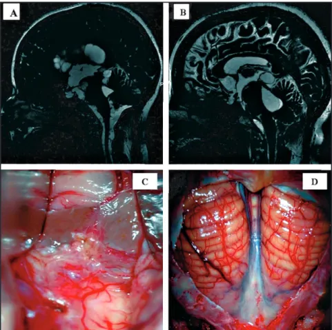
FIGURE 1: A - Sagital magnetic resonance Imaging (MRI) with a fast imaging employing steady state acquisition (FIESTA) acquisition demonstrating inlammatory granulomatosis, aracnoiditis and cystic lesions inside the lateral ventricles, third ventricle, pineal region and basal cisterns. B - T2 weighted sagital MRI showing a large cyst inside the fourth ventricle and in the pineal region and cysts in the basal cisterns. C, D - Surgical approach. In D the cerebellum with cisterna magna intact demonstrating aracnoiditis in this region. In C ater opening of the cistern magna and spatulation of the cerebellum hemispheres in the middle line also showing aracnoiditis. In C the surgeon is approaching the forth ventricle.
daily headaches in the past 30 days prior to her admission. She then underwent brain MRI that showed intra-fourth ventricular cystic lesion and cystic lesion of the pineal region (Figure 1B). The patient underwent surgery for removal of the cysticercus (Figures 1C
and 1D). Anatomopathological examination conirmed the
diagnosis of cysticercosis.
Extraparenchmal disease varies in its symptoms and prognosis according to the location of the parasites. The prognosis for intraventricular neurocysticercosis is worse than that for the intraparenchymal forms of the disease5. Intracranial hypertension
is a common manifestation and may be the result of a mass efect, distortion of the normal anatomy of CSF pathways, direct obstruction of the ventricular system by a cyst, or an inlammatory reaction in the meninges leading to arachnoiditis. Sometimes, like our patient, an intermittent or positional CSF obstruction with increasing intracranial pressure produces relapsing/remitting symptoms. he pathomechanism of the BS is not clear. Bruns believed the symptoms resulted from a change in the position of the cyst in the fourth ventricle with periodic blocking of the ventricular system on change of head posture and elevation of intracranial pressure due to a ball-valve mechanism6,8. he diagnosis of NCC is diicult
because clinical manifestations are nonspeciic. Most neuroimaging
indings are non-pathognomonic, while some serologic tests have low speciicity and sensitivity. A set of diagnostic criteria was proposed in 1996 and recently revisited to help with the diagnosis of NCC. Proper interpretation of these criteria permits two degrees of diagnostic certainty: deinitive or probable9. Magnetic resonance imaging (MRI)
is more accurate than CT for the diagnosis of most cases of NCC. herapeutic measures include antiparasitic drugs, surgery, and symptomatic medications. As inflammation is the conspicuous accompaniment in most forms of NCC, corticosteroids represent the primary form of therapy for meningitis, cysticercal encephalitis, and angiitis. Albendazole is considered the antiparasitic drug of choice for NCC because it has beter penetration into the CSF and because its concentration is not afected when administered along with steroids5,10.
Extra-parenchymal cysticercosis is associated with poor prognosis and requires a more aggressive approach. When feasible, complete surgical excision of lesions remains the deinitive therapy5-6.
271
Rev Soc Bras Med Trop 45(2):269-271, mar-abr, 2012
ACKNOWLEDGMENTS
REFERENCES
ventricular shunting to resolve the hydrocephalus. Hydrocephalus secondary to NCC is associated with high rates of shunt dysfunction.
Neurocysticercosis is a longstanding disease that remains endemic in most low-income countries and has increasingly afected high-income countries due to increased migration, tourism, and travel to endemic areas. We report this case because NCC is a highly prevalent infectious disease, but is potentially eradicable, and is a public health issue that entails many social and economic consequences. Control programs are urgently needed to reduce the burden of this disease.
he authors would like to thank the following: Dr. Rafael Ferreira, Dr. NeiGulcó, Dra. Cristina Fraga, Dra. Sabrina Sanvido, Dra. Marlise Ribeiro, Dr. César Aldabe, Dr. Vinicius de Oliveira, Dr. Luciano Carvalho, and Dr. Pablo Sebastian Velho.
1. Garcia HH, Evans CAW, Nash TE, Takayanagui OM, White AC, Botero D, et al. Current Consensus Guidelines for Treatment of Neurocysticercosis. Clin Microbiol Rev 2002; 18:747-756.
2. Garcia HH, Del Bruto OH. Neurocysticercosis: update concepts about an old disease. Lancet Neurol 2005; 4:653-661.
3. Takayanagui OM, Leite JP. Neurocysticercosis. Rev Soc Bras Med Trop 2001; 34:283-290.
4. Sotelo J, Del Brto OH. Brain Cysticercosis. Arch Med Res 2002; 31:3-14. 5. Takayanagui OM, Odashima NS. Clinical aspects of neurocysticercosis.
Parasitol Internat 2006; 55:111-115.
6. Torres-Corzo J, Rodriguez-Della Vecchia R, Rangel-Castilha L. Bruns Syndrome caused by intraventricular neurocysticercosis treated using lexible endoscopy. J Neurosurg 2006; 104: 746-748;
7. Das A, Kesavadas C, Radhakrishnan VV, Nair NS. Bruns Syndrome caused by intraventricular neurocysticercosis. Neurol 2009; 73:e34.
8. Krasnianski M, Muller T, Stock K, Zierz S. Bruns Syndrome caused by intraventricular tumor. Eur J Med Res 2008; 13:179-181;
9. Del Bruto OH, Rajshekhar V, White Jr AC. Proposed diagnostic criteria for neurocysticercosis. Neurol 2001; 57:177-183