ABSTRACT
ORIGINAL AR
João Pedro Marques-Pereira Andrés Delgado-Cañedo Nance Beyer Nardi
João Ricardo Michelin Sant’Anna Paulo Roberto Prates
Ivo Nesralla
by mini-thoracotomy in dilated
cardiomyopathy: technique and
early results
Instituto de Cardiologia do Rio Grande do Sul (IC-RS), Fundação
Universitária de Cardiologia (FUC), Porto Alegre, Rio Grande do Sul, Brazil
CONTEXT AND OBJECTIVES: There are few stud-ies concerning bone marrow mononuclear cell (BMMC) transplantation in cases of nonischemic dilated cardiomyopathy. This study describes a novel technique of BMMC transplantation and the results up to one year after the procedure. DESIGN AND SETTING: This was a case se-ries to evaluate the safety and viability of the procedure, at Instituto de Cardiologia do Rio Grande do Sul.
METHODS: Nine patients with symptomatic dilated cardiomyopathy, functional class III/IV and left ventricular ejection fraction (LVEF) < 35% received BMMC (9.6 ± 2.6 x 10⁷ cells) at 20 sites in the ventricular wall, by means of tho-racotomy of length 5 cm in the fi fth left intercostal space. Echocardiograms and nuclear magnetic resonance (NMR) were performed.
RESULTS: There were no major complications. The functional class results for the fi rst six pa-tients (preoperatively and at two, four, eight and twelve-month follow-ups, respectively) were: [IV-2, III-4] to [I-5, II-1] to [I-3, II-3] to [I-2, II-3] and [I-2, II-3]. Echocardiograms showed LVEF: 25.9 ± 8.2; 32.9 ± 10.4; 29.4 ± 7.2; 25.1 ± 7.9; 25.4 ± 6.8% (p = 0.023); and % left ventricular (LV) fi ber shortening: 12.6 ± 4.4; 16.4 ± 5.4; 14.3 ± 3.7; 12.1 ± 4.0; 12.2 ± 3.4% (p = 0.021). LV performance variation seen on NMR was non-signifi cant.
CONCLUSION: Intramyocardial transplantation of BMMC in dilated cardiomyopathy cases is feasible and safe. There were early improvements in symptoms and LV performance. Medium-term evaluation revealed regression of LV function, al-though maintaining improved functional class. KEY WORDS: Dilated cardiomyopathy. Stem cells. Heart surgery. Heart failure, congestive. Cell transplantation.
CLINICAL TRIAL REGISTRATION NUMBER: NCT00615394.
INTRODUCTION Cell transplantation is a promising al-ternative for promoting cardiac regeneration and improved function.1 In experimental
models, several types of cells have been used, such as: skeletal myoblasts,2 bone marrow
mononuclear cells (BMMCs),3 mesenchymal
stem cells4 and cardiomyocytes.5 So far, there
is no consensus about which type is most suitable. Because BMMCs are easy to obtain and manipulate, they are among the cell types most studied.
As these therapies begin to be trans-posed to clinical experiments, new inquiries are arising, such as the quantity of cells required, the ambient medium needed and the best administration route. With regard to the latter, several pathways have already been tested in clinical studies: intramyo-cardial transendointramyo-cardial, intramyointramyo-cardial transepicardial, intracoronary and periph-eral mobilization through the use of G-CSF (granulocyte colony-stimulating factor).1
An ideal approach would involve a mini-mally invasive method that would be capable of safely injecting the cells intramyocardially, in order to achieve greater local concentrations and fewer systemic effects.
OBJECTIVE In this article, our aim is to describe a technique for autologous transplantation of mononuclear bone marrow stem cells, man-aged by mini-thoracotomy.
METHODS Nine patients (six males) of mean age 57.5 ± 9.2 years with a previous diagnosis of nonischemic dilated cardiomyopathy were included. They fulfi lled the following selection criteria: (1) diagnosis made more than two years earlier and symptomatic in functional class III-IV according to the
New York Heart Association classifi cation (NYHA), despite optimal pharmacological therapy for at least six months prior to in-clusion; (2) left ventricular ejection fraction (LVEF) less than 35%, from echocardio-gram; (3) age less than 60 years; (4) absence of neoplasm; (5) absence of hematological disease or systemic disease; and (6) no pre-vious cardiac intervention. The exclusion criteria were: (1) episodes of tachycardia or ventricular fi brillation; (2) severe or moder-ate mitral insuffi ciency; and (3) any other valve diseases.
The preoperative evaluation included: Doppler echocardiogram, myocardial nuclear magnetic resonance, six-minute walk test, quality-of-life evaluation using the Minnesota Living With Heart Failure questionnaire, elec-trocardiogram, hemogram, electrolytes, chest x-ray and clinical evaluation. The patients included were selected for two studies: an initial pilot series of six cases to evaluate the safety and viability of the procedure, followed by a randomized clinical trial, which is now in progress, and in which three new cases have so far been included.
The project was approved by the local and the national ethics committees in the year 2004, under number 3549, and has been awarded a grant from FAPERGS (State of Rio Grande do Sul Research Foundation).
Stem cell preparation
mono-nuclear fraction was then collected, washed in a heparinized saline solution containing 5% autologous serum and filtered in order to remove the cellular adjunctive. The cells were again suspended in saline solution with autologous serum at 5% concentration, to make up an intramyocardial injection vol-ume of 5 ml. A small fraction was utilized for sterility tests, cell counting and viability tests. Viability greater than 90% was con-sidered acceptable. Another fraction of the cells was used for immunophenotyping using flow cytometry, in order to determine the subpopulations (CD 34+, CD 38+ and
sub-populations of lymphocytes and monocytes) and to carry out functional analysis.
Surgical technique
The surgical approach was through left mini-thoracotomy, consisting of an incision of approximately 5 cm in length, antero-laterally in the fifth left intercostal space
Figure 1. Left: cell suspension ready for use. Right: Intramyocardial injection of mono-nuclear cells.
0 1 2 3 4 5 6
5
4
2
1
3 3 3 3
2 2
Number of patients
I II III IV
I II III IV I II III IV I II III IV I II III IV
Baseline 2 months 4 months 8 months 12 months
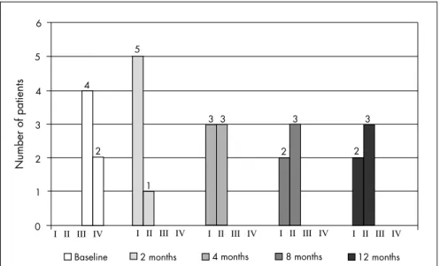
Figure 2. Patients’ functional class during the twelve months of follow-up.
(Figure 1), in order to expose the pericar-dium. A T-shaped pericardial incision was made and 2-0 polyester traction sutures were placed. Pericardial fluid was suctioned and the free wall of the left ventricle was exposed. Videothoracoscopy equipment for viewing was used only in the first patient. For subsequent patients, it was left avail-able in the operating room but not used, because it was considered unnecessary due to the proximity of the heart to the thoracic wall in these patients. Once the position of the coronary arteries had been defined, the injections of the cell suspension were made directly, through a 21F butterfly needle that was positioned in the myocardium and connected to an extension managed by the surgical assistant.
Twenty small injections in the myocar-dium were made, with a total volume of 5 ml or about 0.25 ml per injection site, in the anterior, lateral, posterior and apical faces of
the left ventricle, containing a mean total of 9.6 ± 2.6 x 10⁷ cells (Figure 2). After
review-ing the hemostasis, the pericardium was closed with a single suture of 2-0 polyester, the thoracic cavity was drained and the chest wall was closed. After the procedure, the patients were kept in the postoperative intensive care unit for a minimum period of 24 hours. They were released from the hospital after a time ranging from five to seven days.
RESULTS
Surgical procedure
No major complications were observed. During the procedure, one patient devel-oped ventricular arrhythmia because the heart was touched, which impeded adequate expose for injections. The arrhythmia ceased completely after endovenous lidocaine administration and the procedure was then completed. In another case, it was necessary to place hemostatic sutures at two sites in the ventricular wall where injections of the cell suspension were applied, due to slight but persistent bleeding that did not cease with prolonged compression.
The duration of the bone marrow col-lection procedure was 69 ± 31 minutes. The mean duration of the thoracotomy procedure was 105 ± 23 minutes. After the procedures, the patients remained under observation in the intensive care unit. There were no significant postoperative complications. There were no readmissions during the follow-up period due to heart failure.
Clinical results
Safety and hospital admission
One patient returned to the emer-gency room during the first postoperative month, vaguely complaining of malaise and anxiety. He was examined and did not present signs of cardiac failure, infection or postoperative complications. Seven months later, this patient presented a lower limb abscess and bacteremia that led to septic shock and death in an emergency room at another hospital. No postmortem examina-tion was carried out.
Cell analysis
Other cell fractions identified were: CD 45+ (74.6 ± 8.5%), CD 14+ (8.4 ± 4.7%), CD 34+ CD 38- (0.7 ± 0.5%) (Table 1).
Clinical results
All the patients presented clinical im-provement, as evaluated by the NYHA functional class (FC). At the baseline evalu-ation, four patients were in FC III and two were in FC IV. At the two-month follow-up, five patients were asymptomatic and one was in FC II. After one year of follow-up, two of the patients remained asymptomatic and three were in FC II (p = 0.001) (Figure 2). This clinical improvement was corroborated by the Minnesota Living with Heart Failure questionnaire, which showed significant improvements in quality of life, expressed as reductions in overall scores over the course of the twelve months of follow-up (p = 0.002) (Figure 3).
Functional capacity also showed a ten-dency towards improvement after four months of follow-up, and for the remaining of the fol-low-up period, as evaluated by the six-minute walk test, although not reaching statistical significance (Figure 4).
Left ventricular (LV) performance
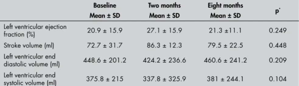
Nuclear magnetic resonance evaluation at the two-month follow-up showed reduc-tions in end systolic volume (ESV) and end diastolic volume (EDV), along with increased stroke volume (Table 2). As a result, the LVEF showed an early absolute increase of 6.2% and relative increase of 45.5% (20.9 o 15.9% to 27.1 o 15.9%; p = 0.249). At the eight-month follow-up, these parameters showed decreases towards baseline values, but a relative increase of 24.4% was still observed for LVEF. The ventricular mass was not modified (Table 2) (Figure 5).
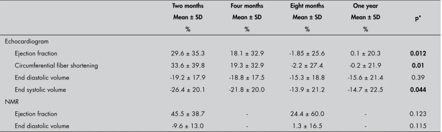
Improved ventricular function was shown by Doppler echocardiography (Table 3 and Figure 6). LVEF showed statistically signifi-cant differences, with an absolute increase of 6.93% and relative increase of 29.6% (25.9 o 8.2% x 32.9 o 10.4%; p = 0.023 and p = 0.012, respectively) at the two-month fol-low-up. A slight decrease in LVEF was observed at the four-month follow-up (29.4 o 7.2%), and the results at the eight and twelve-month follow-ups (25.4 o 6.8%) were similar to baseline values. Increased circumferential fiber shortening and reduced ESV and EDV were seen at the two-month follow-up; these values decreased over the next six months and showed a tendency towards stabilization at the eight and twelve-month postoperative assessments.
Table 1. Immunophenotyping of bone marrow mononuclear cells administered to patients (frequency of cells for each marker)
Patient CD45+* CD14+† CD3+ CD4+* CD3+ CD8+* CD34+† CD34+ CD38† Number of
cells injected
1 82.0 13.0 27.0 17.0 0.6 0.2 7.0 x 10⁷
2 81.0 14.0 27.0 10.0 0.9 0.3 1.3 x 10⁸
3 61.0 5.0 17.0 2.0 2.0 1.2 7.0 x 10⁷
4 77.0 6.0 19.0 3.0 1.6 ND 1.0 x 10⁸
5 72.0 4.0 24.0 9.0 2.6 1.1 1.1 x 10⁸
Mean 74.6 8.4 22.8 8.2 1.5 0.7 9.6 x 10⁷
SD 8.6 4.7 4.6 6.1 0.8 0.52 2.6 x 10⁷
*Percentage of cells in relation to total number of viable cells; †Percentage of cells in relation to CD45+ cells.
SD = standard deviation.
Table 2. Absolute values for evaluation of left ventricular performance by means of nuclear magnetic resonance
Baseline Mean ± SD
Two months Mean ± SD
Eight months
Mean ± SD p
*
Left ventricular ejection
fraction (%) 20.9 ± 15.9 27.1 ± 15.9 21.3 ±11.1 0.249
Stroke volume (ml) 72.7 ± 31.7 86.3 ± 12.3 79.5 ± 22.5 0.448
Left ventricular end
diastolic volume (ml) 448.6 ± 201.2 424.2 ± 236.6 460.6 ± 241.2 0.209
Left ventricular end
systolic volume (ml) 375.8 ± 215 337.8 ± 325.9 381 ± 244.1 0.104
*p = for repeated-measurement analysis of variance (ANOVA) adjusted according to Bonferroni; SD = standard deviation.
Figure 3. Quality of life of the patients during the 12 months of follow-up, from Min-nesota Living With Heart Failure questionnaire.
80
70
60 50
40
30 20
10
0
Baseline 2 months 4 months 8 months 12 months
p = 0.002
Score
Figure 4. Six-minute walk test on the patients during the twelve months of follow-up.
500
400
300
200
100
0
Baseline 2 months 4 months 8 months 12 months
p = 0.054
The echo imaging also showed a reduction in ventricular mass (226.2 ± 41.0 to 214.05 ± 54.9 g after twelve months), which did not reach significance level (p = 0.44).
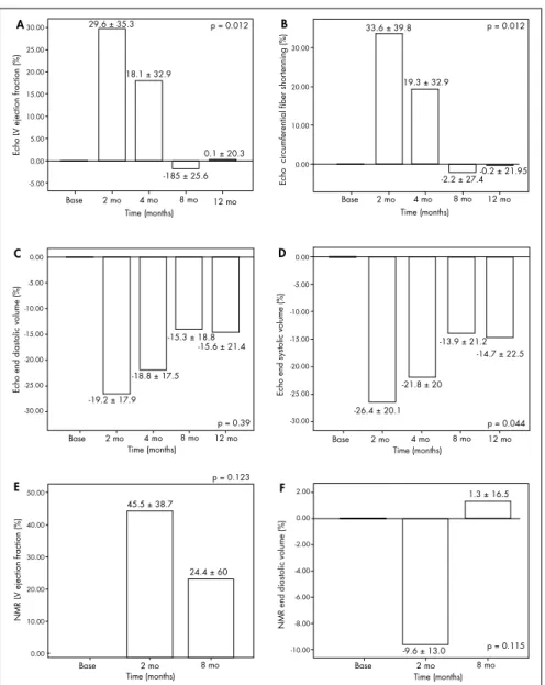
Figure 7 presents the variation (increases or decreases) in LV performance assessment values from echocardiograms and nuclear magnetic resonance, expressed as percent-ages of the baseline (preoperative) levels. The most significant findings were a relative and temporary improvement of up to 29.6% in LVEF on echocardiogram at the two-month follow-up (A); a 33.6% improvement in circumferential fiber shortening at the same time (B) and a similar relative increase in LVEF from NMR evaluation (45.5%) (E). Table 4 shows the percentage increases or decreases in left ventricular echocardiogram and NMR values, demonstrating the varia-tions relative to preoperative baseline mea-surements (Table 4).
DISCUSSION Our experience indicated that intramyo-cardial stem cell transplantation in patients with nonischemic dilated cardiomyopathy, by means of mini-thoracotomy, was feasible, safe, well-tolerated and associated with initial clinical improvement, as expressed by better
Table 3. Absolute values for left ventricular performance, by means of echocardiogram
Baseline Mean ± SD
Two months Mean ± SD
Four months Mean ± SD
Eight months Mean ± SD
One year
Mean ± SD p*
Left ventricular ejection fraction (%) 25.9 ± 8.2 32.9 ± 10.4 29.4 ± 7.2 25.6 ± 7.9 25.4 ± 6.8 0.023
Left ventricular circumferential fiber shortening (%) 12.6 ± 4.4 16.4 ± 5.4 14.3 ± 3.7 12.1 ± 4.0 12.2 ± 3.4 0.021
End diastolic volume (ml) 235.6 ± 103 185.3 ± 74.7 185 ± 70.9 192.4 ± 71.8 192.2 ± 77.7 0.163
End systolic volume (ml) 179 ± 92.7 130.7 ± 73.2 134.2 ± 65.2 148.3 ± 69.1 146.9 ± 69.9 0.156
*p = for repeated-measurement analysis of variance (ANOVA) adjusted according to Bonferroni; SD = standard deviation.
20.9 ± 15.9
27.1 ± 15.9
21.3 ± 11.1
0 10 20 30 40
Base 2 mo 8 mo
LV ejection fraction (%)
Time (months)
p = 0.240
72.7 ± 31.7
86.3 ± 12.3 79.5 ± 22.5
110 100 90 80 70 60 50 40
8 mo 2 mo
Base
Time (months)
p = 0.448
Stroke volume (ml)
448.6 ± 201.2 424.2 ± 236.6 460.6 ± 241.2
800
600
400
200
0
End diastolic volume (ml)
p = 0.209
Base 2 mo 8 mo
Time (months)
Base 2 mo 8 mo
Time (months) 375.8 ± 215
337.8 ± 235.9
381 ± 244.1 p = 0.104
End systolic volume (ml)
700
600
500
400
300
200
100
Figure 5. Nuclear magnetic resonance results for left ventricular functional parameters.
Table 4. Percentage increase or decrease in left ventricular echocardiogram and NMR values, demonstrating the variations relative to preoperative baseline measurements
Two months Four months Eight months One year
p*
Mean ± SD Mean ± SD Mean ± SD Mean ± SD
% % % %
Echocardiogram
Ejection fraction 29.6 ± 35.3 18.1 ± 32.9 -1.85 ± 25.6 0.1 ± 20.3 0.012
Circumferential fiber shortening 33.6 ± 39.8 19.3 ± 32.9 -2.2 ± 27.4 -0.2 ± 21.9 0.01
End diastolic volume -19.2 ± 17.9 -18.8 ± 17.5 -15.3 ± 18.8 -15.6 ± 21.4 0.39
End systolic volume -26.4 ± 20.1 -21.8 ± 20.0 -13.9 ± 21.2 -14.7 ± 22.5 0.044
NMR
Ejection fraction 45.5 ± 38.7 - 24.4 ± 60.0 - 0.123
End diastolic volume -9.6 ± 13.0 - 1.3 ± 16.5 - 0.115
functional capacity and ventricular func-tion in the immediate postoperative period. However, this improvement in ventricular function was lost after four months of fol-low-up, with progressive deterioration of some parameters, which returned to levels similar to the preoperative values. No major procedure-related complications occurred. There was a relatively prolonged length of hospital stay, until the patients achieved thorough recovery after the procedure. This was certainly longer than hospital stays relat-ing to less invasive procedures. Nevertheless, early hospital discharge was not of concern in this initial series.
This method of surgical transplantation of bone marrow mononuclear stem cells has potential advantages: (1) application in the myocardium under direct viewing, which allows precise and safe injection; (2) low complexity and low cost, making it avail-able to almost all cardiac surgical centers; (3) minimal dispersion of cell solution, thus minimizing systemic effects. The potential drawbacks would be: (1) the need for tho-racotomy, with its related morbidity; (2) using small incisions, especially in patients with significant cardiac dilatation, it may be difficult to reach the entire free wall of the LV; (3) the ventricular septal myocardium cannot be directly approached.
Clinically, the types of therapy1 that
bone marrow stem cells have most been used for are intracoronary infusion6,
transepicar-dial intramyocartransepicar-dial infusion during cardiac surgery7, transendocardial intramyocardial
infusion in patients for whom revasculariza-tion using the NOGA System was not pos-sible8 and peripheral mobilization by means
of G-CSF.9 Generally speaking, the
intra-coronary route would be beneficial, since it is less invasive and less expensive, although it would have the drawback of not apply-ing the cells directly to the lesioned tissue and would therefore depend on a homing process. One other disadvantage of intra-coronary injection would be the fact that cells tend to migrate to systemic circulation, which could theoretically accelerate diseases dependant on neovascularization, such as diabetic retinopathy and neoplasms.
In patients with chronic ischemia, the transendocardial intramyocardial approach by catheterization has been preferred, using the NOGA navigation system.6 This method
not only enables intramyocardial application less invasively, but also allows mapping of myocardial areas such that injection becomes more viable. Its drawbacks are that it presents
little system availability and high cost. In specific clinical experiments, its use has been associated with clinical symptomatic and LV function improvements that were attribut-able to better myocardial perfusion.
Peripheral mobilization of bone marrow cells using G-CSF has not been associated with favorable results in several randomized clini-cal trials among patients with heart failure, or has only been associated with very small benefits. Different results had been obtained in experimental studies. Thus, this approach seems to be less effective.
There are no published complete original studies using bone marrow cells in patients with nonischemic dilated cardiomyopathy. A successful clinical series of patients with Chagas disease on whom intracoronary infu-sion was used has recently been published.10
Trainini et al.11 evaluated the effects in eight
cases of nonischemic dilated cardiomyopathy. No procedure-related complications occurred. Over a mean follow-up period of 295 ± 37 days, they found a significant improvement in NYHA functional class (2.5 ± 0.8 to 1.5 ± 0.5) and LVEF (18.3 ± 7% to 26.4 ± 10%).11
In our experience, we found it unneces-sary to use thoracoscopy equipment, because the proximity between the heart (especially when dilated) and the chest wall made the
procedure completely achievable, in safety, through a small incision that was similar to the one utilized for epicardial electrode implants for pacemakers.
The main limitations of this study were: (1) the relatively small sample size: (2) the uncontrolled phase I study design, such that the improvements observed could be at least partly due to better adherence by the patients to the treatment; and (3) although the fol-low-up period used in the present study was one of the longest among cell therapy clinical trials for nonischemic dilated cardiomyopathy, it did not allow conclusions regarding the long-term effects.
Progressive improvement in heart function was observed for two months, with a slight decline during the following 10 months of follow-up. The clinical situa-tion as a whole showed a slight late worsen-ing (Figure 2), but some degree of recovery was maintained and none of the patients returned to functional class III or IV during the twelve months of follow-up. Major re-sponses to the therapy were seen in ejection fraction and end systolic volume.
It is possible that the temporary nature of the improvement in ventricular function observed in the present study occurs more generally and was not reported by others be-p = 0.023
LV ejection fraction (%)
25.9 ± 8.2 32.9 ± 10.4
29.4 ± 7.2
25 ± 7.9 25.4 ± 6.8
Base 2 mo 8 mo
Time (months)
4 mo 12 mo
12.5 ± 4.4 16.4 ± 5.4 45
40
35
30
25
20
15
14.3 ± 3.7
12.1 ± 4 12.2 ± 3.4 21
18
15
12
9
p = 0.021
% Circumferential fiber shor
tening (CFS)
Base 2 mo 8 mo
Time (months)
4 mo 12 mo
Base 2 mo 8 mo
Time (months)
4 mo 12 mo
End diastolic volume (ml)
p = 0.16
350
300
250
200
150
100 235.6 ± 103
185.3 ± 74.7
185 ± 70.9192.4 ± 71.8
192.2 ± 77.7
p = 0.156
Base 2 mo 8 mo
Time (months)
4 mo 12 mo
End systolic volume (ml)
300
250
200
150
100
50 179 ± 92.7
130.7 ± 73.2 134.2 ± 65.2
148.3 ± 69.1 146.9 ± 69.9
cause most investigations have been restricted to short and medium-term follow-up of a few weeks. In some studies, such as the one reported by Perin et al.8, however, sustained
positive results from cell therapy for ischemic heart failure were reported after six and twelve-month follow-up periods. In a randomized study with transplantation of BMMCs in cases of acute infarction, Meyer et al.12 observed
improvements six months after treatment but, at the 18-month follow-up, the conditions of the treated and control patients did not show any significant differences.
All of our patients, even those for whom the objective evaluation did not show improved ven-tricular function, reported subjective improve-ment following cell therapy. The placebo effect cannot be ignored in this kind of approach, and it may result in better adherence to treatment. Even without a control group, however, the re-sults in general show significant improvement in heart function, particularly considering that the inclusion criteria involved at least two years since diagnosis and complete treatment for at least six months, conditions under which progressive clinical deterioration would be expected.
CONCLUSIONS In conclusion, the described technique of mini-thoracotomy in the fifth left in-tercostal space, even without the aid of videothoracoscopy, is feasible and safe, with low surgical risks. Therefore, this may be a suitable approach for accessing the free wall of the LV to inject planned quantities of bone marrow stem cells, in cases of nonisch-emic dilated cardiomyopathy. There were early improvements in symptoms, quality of life and LV performance. Medium-term evaluation revealed regression of LV func-tion nearly to preoperative levels, although improved functional class and quality of life were maintained.
Base 2 mo 8 mo
Time (months)
4 mo 12 mo
Echo L
V ejection fraction (%)
A30.00
25.00 20.00 15.00 10.00 5.00 0.00 -5.00
p = 0.012 29.6 ± 35.3
18.1 ± 32.9
0.1 ± 20.3
-185 ± 25.6
Echo circumferential fiber shor
tenning (%)
B
30.00
20.00
10.00
0.00
Base 2 mo 8 mo
Time (months)
4 mo 12 mo
p = 0.012 33.6 ± 39.8
19.3 ± 32.9
-2.2 ± 27.4-0.2 ± 21.95
Echo end diastolic volume (%)
C 0.00
-5.00
-10.00
-15.00
-20.00
-25.00
-30.00
Base 2 mo 8 mo
Time (months)
4 mo 12 mo
p = 0.39 -19.2 ± 17.9
-18.8 ± 17.5 -15.3 ± 18.8
-15.6 ± 21.4
NMR L
V ejection fraction (%)
E
50.00
40.00
30.00
20.00
10.00
0.00
Base 2 mo 8 mo
Time (months)
p = 0.123
45.5 ± 38.7
24.4 ± 60
Echo end systolic volume (%)
D 0.00
-5.00
-10.00
-15.00
-20.00
-25.00
-30.00 p = 0.044
Base 2 mo 8 mo
Time (months)
4 mo 12 mo
-26.4 ± 20.1 -21.8 ± 20
-13.9 ± 21.2 -14.7 ± 22.5
NMR end diastolic volume (%)
F 2.00
0.00
-2.00
-4.00
-6.00
-8.00
-10.00
Base 2 mo 8 mo
Time (months)
p = 0.115 -9.6 ± 13.0
1.3 ± 16.5
AUTHOR INFORMATION
Renato Abdala Karam Kalil, MD, PhD.Professor at Instituto de Cardiologia do Rio Grande do Sul (IC-RS), Fundação Universitária de Cardiologia (FUC), Fundação Faculdade Federal de Ciências Médicas de Porto Alegre (FFFCMPA), Porto Alegre, Rio Grande do Sul, Brazil.
Daniele Ott, MD.Postgraduate student at Fundação Faculdade Federal de Ciências Médicas de Porto Alegre, Porto Alegre, Rio Grande do Sul, Brazil.
Roberto Sant’Anna, MD.Medical doctor at Instituto de Cardiologia do Rio Grande do Sul (IC-RS), Fundação Universitária de Cardiologia (FUC), Porto Alegre, Rio Grande do Sul, Brazil.
Eduardo Dias, MD. Postgraduate student at Fundação Facul-dade Federal de Ciências Médicas de Porto Alegre, Porto Alegre, Rio Grande do Sul, Brazil.
João Pedro Marques-Pereira, MD, PhD.Professor at Fundação Faculdade Federal de Ciências Médicas de Porto Alegre, Porto Alegre, Rio Grande do Sul, Brazil.
Andrés Delgado-Cañedo, PhD.Geneticist and Researcher at Instituto de Cardiologia do Rio Grande do Sul (IC-RS), Fundação Universitária de Cardiologia (FUC), Porto Alegre, Rio Grande do Sul, Brazil.
Nance Beyer Nardi, BSc, PhD.Professor at Instituto de Cardio-logia do Rio Grande do Sul (IC-RS), Fundação Universitária de Cardiologia (FUC), Porto Alegre; and Researcher in De-partment of Genetics, Universidade Federal do Rio Grande do Sul, Porto Alegre, Rio Grande do Sul, Brazil.
João Ricardo Michelin Sant’Anna, MD, PhD. Professor at Instituto de Cardiologia do Rio Grande do Sul (IC-RS), Fundação Universitária de Cardiologia (FUC), Porto Alegre, Rio Grande do Sul, Brazil.
Paulo Roberto Prates, MD.Cardiovascular surgeon at Instituto de Cardiologia do Rio Grande do Sul (IC-RS), Fundação Universitária de Cardiologia (FUC), Porto Alegre, Rio Grande do Sul, Brazil.
Ivo Nesralla, MD, PhD.Professor at Instituto de Cardiologia do Rio Grande do Sul (IC-RS), Fundação Universitária de Cardio-logia (FUC), Porto Alegre, Rio Grande do Sul, Brazil.
Address for correspondence:
Renato Abdala Karam Kalil
Unidade de Pesquisa, Instituto de Cardiologia do Rio Grande do Sul/Fundação Universitária de Cardiologia (IC/FUC)
Av. Princesa Isabel, 370 — Santana Porto Alegre (RS) — Brasil — CEP 90620-001 Tel/Fax. (+ 55 51) –3219-2802 Extensions 22, 23, 24 E-mail: [email protected] E-mail: [email protected]
Copyright © 2008, Associação Paulista de Medicina
RESUMO
Transplante autólogo de células-tronco de medula óssea por mini-toracotomia — técnica e resultados iniciais
CONTEXTO E OBJETIVO: Há pouco estudos avaliando o transplante de células mononucleares da medula óssea (CMMO) na miocardiopatia dilatada não-isquêmica. O presente estudo descreve uma técnica de im-plante intramiocárdico de CMMO por mini-toracomia e resultados com até um ano de acompanhamento. TIPO DE ESTUDO E LOCAL: Série casos para avaliar segurança e viabilidade do procedimento, no Instituto de Cardiologia do Rio Grande do Sul.
MÉTODOS: Nove pacientes com miocardiopatia dilatada, em classe funcional III/IV e fração de ejeção do ventrículo esquerdo (FEVE) < 35% receberam CMMO (média 9,6 ± 2,6 x 10⁷ células) em 20 pontos da parede livre do ventrículo esquerdo, através de toracotomia de 5 cm no quinto espaço intercostal esquerdo. Foram realizados ecocardiograma e ressonância nuclear magnética (RNM).
RESULTADOS: Não ocorreram complicações maiores. Os resultados pré-operatórios aos 2, 4, 8 e 12 meses de acompanhamento dos seis primeiros pacientes são: classe funcional: IV-2, III-4 para I-5, II-1 para I-3, II-3 para I-2, II-3 e I-2, II-3. Ecocardiograma: FEVE = 25.9 ± 8.2, 32.9 ± 10.4, 29.4 ± 7.2, 25.1 ± 7.9, 25.4 ± 6.8% (p = 0.023); Fração de encurtamento do ventrículo esquerdo (VE) = 12.6 ± 4.4, 16.4 ± 5.4, 14.3 ± 3.7, 12.1 ± 4.0, 12.2 ± 3.4% (p = 0.021). RNM demonstrou diferenças discretas, não significativas. CONCLUSÕES: O implante intramiocárdico de células-tronco na miocardiopatia dilatada é viável e seguro. Houve melhora precoce nos sintomas e na performance de VE. Avaliação a médio prazo demonstrou regressão da função VE, mantendo, contudo, a melhora na qualidade de vida e na classe funcional. PALAVRAS-CHAVE: Cardiomiopatia dilatada. Células-tronco. Cirurgia torácica. Insuficiência cardíaca congestiva. Transplante celular.
NÚMERO DE REGISTRO DO ENSAIO CLÍNICO: NCT00615394.
1. Dimmeler S, Zeiher AM, Schneider MD. Unchain my heart: the scientific foundations of cardiac repair. J Clin Invest. 2005;115(3):572-83
2. Scorsin M, Hagège A, Vilquin JT, et al. Comparison of the effects of fetal cardiomyocyte and skeletal myoblast transplantation on postinfarction left ventricular function. J Thorac Cardiovas Surg. 2000;119(6):1169-75.
3. Tomita S, Li RK, Weisel RD, et al. Autologous transplanta-tion of bone marrow cells improves damaged heart functransplanta-tion. Circulation. 1999;100(19 Suppl):II247-56.
4. Nagaya N, Kangawa K, Itoh T, et al. Transplantation of mesen-chymal stem cells improves cardiac function in a rat model of dilated cardiomyopathy. Circulation. 2005;112(8):1128-35. 5. Yoo KJ, Li RK, Weisel RD, Mickle DA, Li G, Yau TM.
Autolo-gous smooth muscle cell transplantation improved heart function in dilated cardiomyopathy. Ann Thorac Surg. 2000;70(3):859-65. 6. Strauer BE, Brehm M, Zeus T, et al. Repair of infarcted
myocardium by autologous intracoronary mononuclear bone marrow cell transplantation in humans. Circulation. 2002;106(15):1913-8.
7. Stamm C, Westphal B, Kleine HD, et al. Autologous bone-marrow stem-cell transplantation for myocardial regeneration. Lancet. 2003;361(9351):45-6.
8. Perin EC, Dohmann HF, Borojevic R, et al. Improved exercise capacity and ischemia 6 and 12 months after transendocardial injection of autologous bone marrow mononuclear cells for ischemic cardiomyopathy. Circulation. 2004;110(11 Suppl 1): II213-8.
9. Joseph J, Rimawi A, Mehta P, et al. Safety and effectiveness of granulocyte-colony stimulating factor in mobilizing stem cells and improving cytokine profile in advanced chronic heart failure. Am J Cardiol. 2006;97(5):681-4.
10. Vilas-Boas F, Feitosa GS, Soares MB, et al. [Early results of bone marrow cell transplantation to the myocardium of patients with heart failure due to Chagas disease]. Arq Bras Cardiol. 2006;87(2):159-66.
11. Trainini JC, Lago NE, Alvarez M, et al. Mononuclear bone marrow stem cells implant as an alternative treatment in non-ischemic dilated cardiomyopathy. J Am Coll Cardiol. 2006;47(4 Suppl A):A80. [abstract].
12. Meyer GP, Wollert KC, Lotz J, et al. Intracoronary bone marrow cell transfer after myocardial infarction: eighteen months’ fol-low-up data from the randomized, controlled BOOST (BOne marrOw transfer to enhance ST-elevation infarct regeneration) trial. Circulation. 2006;113(10):1287-94.
Sources of funding: Fundação de Amparo à Pesquisa do Estado do Rio Grande do Sul (Fapergs), grant number 3549. Conselho Nacional de Desenvolvimento Científico e Tecnológico (CNPq) and Ministry of Health of Brazil.
Conflict of interest: None
Date of first submission: December 27, 2006
Last received: March 7, 2008
Accepted: March 7, 2008