DNA extraction and quantification from
touch and scrape preparations obtained
from autopsy liver cells
Departamentos de 1Patologia and 2Genética,
Faculdade de Medicina de Ribeirão Preto,
Universidade de São Paulo, Ribeirão Preto, SP, Brasil C.N.M. Ribeiro1,
L.C. Peres1 and
J.M. Pina-Neto2
Abstract
The objective of the present study was to develop a simplified low cost method for the collection and fixation of pediatric autopsy cells and to determine the quantitative and qualitative adequacy of extracted DNA. Touch and scrape preparations of pediatric liver cells were obtained from 15 cadavers at autopsy and fixed in 95% ethanol or 3:1 methanol:acetic acid. Material prepared by each fixation procedure was submitted to DNA extraction with the Wizard® genomic DNA
purification kit for DNA quantification and five of the preparations were amplified by multiplex PCR (azoospermia factor genes). The amount of DNA extracted varied from 20 to 8,640 µg, with significant differences between fixation methods. Scrape preparation fixed in 95% ethanol provided larger amount of extracted DNA. However, the mean for all groups was higher than the quantity needed for PCR (50 ng) or Southern blot (500 ng). There were no qualitative differences among the different material and fixatives. The same results were also obtained for glass slides stored at room temperature for 6, 12, 18 and 24 months. We conclude that touch and scrape preparations fixed in 95% ethanol are a good source of DNA and present fewer limitations than cell culture, tissue paraffin embedding or freezing that require sterile material, culture medium, laboratory equipment and trained technicians. In addition, they are more practical and less labor inten-sive and can be obtained and stored for a long time at low cost.
Correspondence
L.C. Peres
Departamento de Patologia FMRP, USP
Av. Bandeirantes, 3900 14049-900 Ribeirão Preto, SP Brasil
Fax: +55-16-633-1068 E-mail: [email protected]
Research supported by CAPES (No. DS103/004).
Publication supported by FAPESP.
Received February 19, 2003 Accepted December 8, 2003
Key words
•Autopsy
•Congenital anomalies •DNA
•Touch preparative •Scrape preparative •Polymerase chain reaction
Introduction
Congenital anomalies are structural, func-tional, biochemical or behavioral alterations observable at birth. They involve about 3% of liveborns in the general population (1) and represent the major cause of post-neonatal death in the city of Ribeirão Preto (2), corre-sponding to about 25% of the cases of pedi-atric autopsy at the University Hospital of
the Faculty of Medicine of Ribeirão Preto, University of São Paulo (3).
those presenting multiple defects, but re-quires the presence of viable cells and an appropriately equipped laboratory. Its disad-vantages are a high cost, time-consuming procedures (4) and limitation by lack of growth or by contamination with bacteria and fungi (5). In addition, non-numerical chromosome alterations or alterations below the level of optical resolution (microdele-tions) cannot be identified by this method but require molecular techniques.
DNA can be extracted from different tis-sues, fixed or not. The choice of a method should consider not only the quality and quantity of extracted DNA, but also the costs of collection, preservation and storage. Touch and scrape preparatives are easy to obtain at autopsy and can be obtained from different tissues, providing a large number of samples (6-8). They do not require complicated tech-nical training and require only new and clean glass slides (9,10) and a fixative fluid, mate-rials of low cost. Fixation can be performed with ethanol, acetone, methanol or by air drying, among other techniques, with care taken to always preserve DNA integrity (11,12). The slides thus obtained can be stored for long periods of time at room tem-perature (13) and also provide a large num-ber of whole cells with intact nuclei, which can be recovered for DNA extraction for different purposes.
Touch and scrape preparatives are useful means of obtaining material for molecular studies and can be easily obtained from pa-tients, for example, by washes or scrape preparatives of the oral cavity (14-16). How-ever, this cell source is not adequate at au-topsy since the oral cavity presents marked contamination with cells and food debris and biological agents such as bacteria and fungi that may impair certain analyses. On the other hand, at autopsy it is possible to obtain better preserved cells, with a lower risk of tissue contamination with foreign elements. Thus, liver touch and scrape pre-paratives represent an excellent cell source.
The objective of the present study was to define and validate a method for the collec-tion and fixacollec-tion of liver cells obtained by touch and scrape preparatives at autopsy for DNA extraction and amplification for the purpose of diagnosing congenital anomalies and other genetic diseases.
Material and Methods
The touch and scrape preparatives used in the present study were obtained from 15 children of both sexes ranging in age from 25 min to four years who presented some type of congenital anomaly, and submitted to au-topsy at the Pathology Service of the Univer-sity Hospital, Faculty of Medicine of Ribeirão Preto (HCFMRP-USP), from July to Decem-ber 2001. The project was approved by the Research Ethics Committee of HCFMRP-USP.
A fragment of the left liver lobe measur-ing approximately 4 x 3 x 0.5 cm was re-moved from each liver and its surface was used for the collection of touch (group TP) and scrape preparatives (group SP). The frag-ments were first washed in running water to remove excess blood and 4 touch prepara-tives were then performed by simply press-ing new and clean microscope slides against the surface of the fragment. Scrape prepara-tives were then obtained with a new and clean surgical knife blade and deposited on three new and clean microscope slides.
Immediately after collection, the slides were placed in appropriate plastic tubes con-taining 95% ethanol for 10 min (groups TP95 and SP95) or 3:1 methanol:glacial acetic acid solution for 45 s (groups TPMA and SPMA). After fixation, the slides were air dried, wrapped in polypropylene film and stored in cardboard boxes at room tempera-ture until the next stage of the experiment.
or scrape preparative slide with the aid of a new sterile surgical knife blade and the mate-rial obtained was placed in new clean 1.0-ml Eppendorf microtube to which 300 µl cell lysis solution, 100 µl nucleus lysis solution and 20 µl proteinase K (20 mg/ml, Promega) were added. The mixture was incubated over-night in a water bath at 50ºC, and 35 µl of the protein precipitation solution of the kit (not specified by the manufacturer) was then added and the sample kept in an ice bath for 5 min. Next, the mixture was centrifuged at 15,600 g
for 4 min, the supernatant was transferred to a microtube containing 300 µl 100% isopro-panol, mixed by inversion, and again centri-fuged for 1 min and the supernatant was dis-carded. The pellet was resuspended in 300 µl 70% ethanol and centrifuged at 15,600 g for 1 min, and the supernatant was discarded. The pellet was air dried and then hydrated with 20 µl hydration solution. Finally, the extracted DNA was stored at -20ºC.
DNA was quantified at 1:100 dilution with a GeneQuant II RNA/DNA calculator spectrophotometer (Pharmacia Biotech, Cambridge, UK) at 260 nm wavelength for determination of double-stranded DNA.
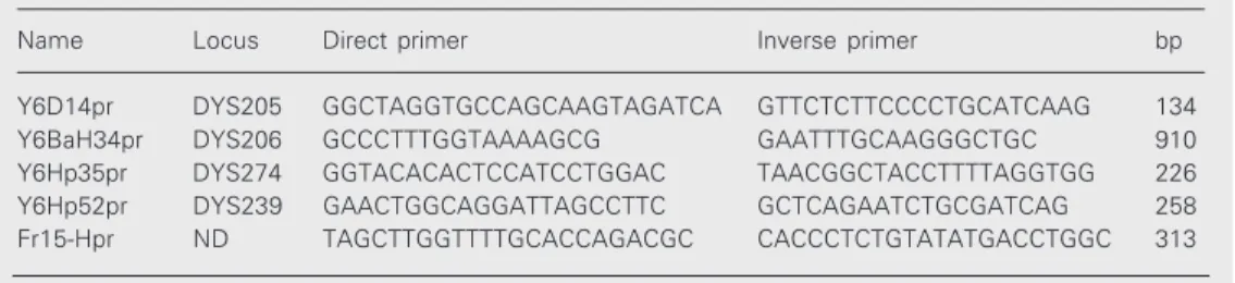
DNA was amplified by PCR in five cases from each group in order to determine the quality of extracted DNA. A multiplex PCR system with a primer mix for the study of azoospermia factor - AZF (Yq11) was used (Table 1) and for this reason all subjects studied were males.
PCR was performed using 3 µl DNA, 0.8 µl Taq (Thermus aquaticus) DNA
Poly-merase (Gibco-BRL, Gaithersburg, MD, USA), 10.45 µl ddp water, 5 µl Taq DNA polymerase buffer (Gibco), 2.5 µl 2 mM deoxyribonucleotide 5' triphosphate, 1.25 µl dimethylsulfoxide, and 2 µl primer mix, placed in a PTC-100 thermocycler (MJ Re-search Inc., Watertown, MA, USA). The following cycles were used: 94ºC/5 min, 94ºC/30 s, 52ºC/45 s, and 65ºC/2 min, with 44 repetitions starting from the second step, and a final cycle at 65ºC/5 min.
After amplification, 5 µl of the material was stained with Orange G and submitted to 1.5% agarose gel electrophoresis (Gibco) in Tris borate EDTA with 5 µl ethidium bro-mide at 80 V for a maximum of 1 h using a 100-bp ladder as a molecular weight marker. The result was photographed with a Kodak digital camera.
To test if long-term storage affected the amount and quality of the extracted DNA, one slide each of touch and scrape prepara-tives with both fixaprepara-tives, stored for 6, 12, 18 and 24 months on cardboard at room temper-ature, was submitted to the procedures de-scribed above.
Statistical analysis
Data were analyzed statistically using the GMC software. When testing for nor-mality we observed that the experimental data were not normally distributed and there-fore we opted for the Kruskal-Wallis test for analysis of variance of multiple independent samples.
Table 1. Primers used in multiplex PCR.
Name Locus Direct primer Inverse primer bp
Y6D14pr DYS205 GGCTAGGTGCCAGCAAGTAGATCA GTTCTCTTCCCCTGCATCAAG 134 Y6BaH34pr DYS206 GCCCTTTGGTAAAAGCG GAATTTGCAAGGGCTGC 910 Y6Hp35pr DYS274 GGTACACACTCCATCCTGGAC TAACGGCTACCTTTTAGGTGG 226 Y6Hp52pr DYS239 GAACTGGCAGGATTAGCCTTC GCTCAGAATCTGCGATCAG 258 Fr15-Hpr ND TAGCTTGGTTTTGCACCAGACGC CACCCTCTGTATATGACCTGGC 313
Results
Touch or scrape preparatives were easily obtained in all cases, usually with good qual-ity material. However, some of the slides showed large amounts of blood and in many cases, after fixation, they exhibited a turbid coloration of the fixative, indicating that part of the material had remained diluted in the latter. In other cases there was little material adhered to the slides even before they were placed in the fixative.
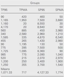
DNA extraction by the methodology used was efficient, as can be seen in Table 2, which shows the results of quantification for the 15 samples studied in each group. There was wide variation among touch and scrape preparative samples and between fixatives from 20 to 8,640 ng/µl, with a mean of 1,071.33 ng/µl for TP95, 717 ng/µl for TPMA, 4,127.33 ng/µl for SP95, and 1,774 ng/µl for SPMA.
Pair-wise comparison of the samples (Table 3)showed that there was a significant difference between TP95 and SP95, TPMA and SP95 and SP95 and SPMA, indicating that 95% ethanol is a better fixative than methanol:acetic acid and that scrape preparatives are better than touch preparatives. Comparison of TP95 and TPMA, TP95 and SPMA and TPMA and SPMA showed no significant differences at the 5% level.
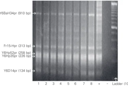
With respect to the quality of the extract-ed DNA, PCR amplification with the prim-ers used showed that there were no differ-ences between fixatives and materials, as illustrated in Figure 1.
Table 4 shows the results of quantifica-tion of DNA extracted from long-term stor-age slides. The amount was similar to what was obtained with shorter storage time.
PCR amplification done on the DNA extracted from long-term storage at room temperature showed no difference when compared to short-term storage (data not shown).
Table 2. Concentration in ng/µl of DNA extracted from the different samples (total volume of each, 20 µl).
Groups
TP95 TPMA SP95 SPMA
90 420 460 50
1,165 1,950 7,500 3,680
1,160 20 8,550 30
4,170 20 8,640 50
560 885 450 3,980
340 2,580 2,960 1,210
1,585 335 4,670 6,540
455 265 1,430 2,980
90 1,195 130 50
775 395 7,500 500
1,725 1,095 8,360 90
985 775 2,090 1,040
520 215 2,070 2,970
1,200 250 3,400 1,900
1,250 355 3,700 1,540
Means
1,071.33 717 4,127.33 1,774
TP = touch preparatives; SP = scrape prepara-tives; 95 = 95% ethanol; MA = methanol:glacial acetic acid (3:1).
Table 4. DNA extracted from different samples stored for different times.
Samples Storage time in months
6 12 18 24
TPMA 4,630 - - 2,320
SP95 5,000 - - 5,160
SPMA - 4,670 2,390
-TP95 - 6,540 2,330
-TP = touch preparatives; SP = scrape prepara-tives; 95 = 95% ethanol; MA = methanol:glacial acetic acid (3:1).
Table 3. Kruskal-Wallis test for the comparison of sample rank means.
Samples Differences Significance compared between means
TP95 x TPMA 7.3333 ns TP95 x SP95 16.5000 0.01 TP95 x SPMA 1.7667 ns TPMA x SP5 23.8333 0.001 TPMA x SPMA 9.1000 ns SP95 x SPMA 14.7333 0.05
Discussion
Congenital anomalies require attention and more in-depth studies since they are appearing at increasing frequency, not only in our region (2), but also in general in the Brazilian Southeast and in developed coun-tries (17). This frequency tends to increase as avoidable causes of death such as prema-turity, infections and malnutrition are better controlled, and also with better pre- and postnatal care. Thus, only causes of death of more difficult control persist; particularly important among them are congenital anoma-lies (3,17).
Paraffin-embedded tissue can be used as a source of cells for DNA extraction, al-though not all of them are susceptible to PCR and some fail to yield detectable prod-ucts (18,19). The explanation for this failure is unknown, but may be due to differences in fixation. The use of 10% buffered formalin for more than 3 days reduces amplification (18). Shorter times of fixation can reduce the quantity of amplified material (19). The amount of DNA decreased progressively when non-fixed frozen sections, frozen sec-tions fixed with 10% formalin for 10 min and paraffin-embedded sections were com-pared in terms of DNA extraction (19). The deleterious action of formalin is due to DNA cross-linking with proteins (20,21), which increases with time of fixation (11), as also observed for RNA (22). The quality of DNA extracted from paraffin-embedded tissue may also be inadequate due to the presence of phenols, salts, EDTA (23), paraffin, forma-lin (19,23), and substances used for staining (19). Thus, the degraded DNA generates an unknown amount of amplified products (23), usually smaller than that obtained with etha-nol (24). Amplified products of up to 250 bp can be obtained from histological sections of tissues previously fixed in formalin and em-bedded in paraffin (25).
Cell culture, which would guarantee excel-lent material for molecular studies, is of
lim-ited application because it is usually employed only in specific cases such as multiple con-genital anomalies. It is an expensive and time-consuming procedure which may also result in lack of growth and in contamination (1,3,5).
Another adequate source both in terms of quantity and quality is tissue fragments which are frozen in liquid nitrogen and stored fro-zen at very low temperatures (26-29), a pro-cedure that involves relatively high costs.
The present results show that the use of liver touch and scrape preparatives can be a good alternative because of the simple meth-odology involved, the amount and quality of the material obtained and the low cost of obtaining and storing the material, which is not impaired by long-term storage at room temperature. Touch and scrape preparatives vary widely in the amount of DNA extracted, possibly as a function of the presence of blood or even of inadequate adherence of the material to the slide (30), with the material being lost when the slide is placed in the fixative, with consequent turbidity of the fluid. To avoid this problem, the fragment could be washed in saline to remove most of the blood, and the surface to be used should
Y6BaH34pr (910 bp)
Fr15-Hpr (313 bp)
Y6Hp52pr (258 bp) Y6Hp35pr (226 bp)
Y6D14pr (134 bp)
Ladder (100 bp) 1 2 3 4 5 6 7 8 +
be dried before doing the touch or scrape preparative procedure. To guarantee cell ad-hesion to the glass, it is advisable to wait 30 s before placing the slides in the fixative. The preservation of the morphological and chemical characteristics of the tissue with this procedure would not be impaired since the material will not dry up. In any case, the quality of amplification is guaranteed, as shown in studies that demonstrated the use of material obtained from the skin of human patients with leishmaniasis that were first air dried and stained with Giemsa and then stored for up to 4 years without a coverslip (31). DNA extraction and amplification of Leish-mania genes were possible even in cases in which cytologic analysis failed (31). Good results were also obtained with the amplifi-cation of Mycobacterium tuberculosis DNA obtained from air-dried sputum slides (13). The use of adhesive substances such as albu-min and gelatin (30), silane, etc. is inad-equate since the presence of any contaminat-ing material should be avoided as much as possible when the objective is DNA extrac-tion and amplificaextrac-tion (23).
Logically, scrape preparatives would be expected to contain more cells and therefore to be more appropriate than touch prepara-tives, as actually indicated by our results. This agrees with literature data showing that fresh tissue scrape preparatives are more efficient than simple touch preparatives when evaluated by cytological methods for preop-erative diagnosis (10,23). The amount of DNA which can be extracted must therefore be larger in this type of material. However, despite the difference, the mean quantity of DNA extracted from all groups was much more than needed for PCR, which usually requires 50 to 100 ng, with the possibility of using only 5 ng (32,33), or even than needed for Southern blotting, for which about 500 to 1000 ng is necessary (32). This means that any of these forms of obtaining tissue samples can be utilized. Literature data agree with these results since epidemiological studies
involving DNA extraction, quantification and amplification using oral mucosa washes or scrape preparatives (16) have demonstrated that the amount of cells, and consequently of DNA, obtained permits the identification of different genes amplified by PCR. Tumor touch preparatives have also been utilized, with or without fixation to obtain DNA and to amplify specific genes such as p53 (34,35). As determined here, these cytologic meth-ods are of low cost, easy to set up and execute, and can be utilized on a large-scale basis (16). These advantages are also taken into account when cells are to be obtained for cytologic analysis (10,36) or even for fluorescent in situ hybridization (FISH).
Not only the method for material collec-tion, but also the fixative is of fundamental importance. The present results show that 95% ethanol is superior to methanol:acetic acid. Ethanol at 14% concentration, i.e., a concentration much lower than the one used here, has been used in studies of DNA ex-traction from oral mucosa cells obtained by oral brushing or washing and the results obtained have shown that DNA is of suffi-cient quality and quantity for the determina-tion of gene sequences of different sizes (16). Ethanol is also the fixative used for cytologic analysis (10,36) and even for FISH (37,38), showing potential for adequate pres-ervation not only of the chemical character-istics of cell components, but also of their morphological aspect. Cells obtained in this manner can therefore be utilized for differ-ent types of analysis, a fact supporting the importance of the definition of a protocol such as the one used here.
well-de-fined, strong bands positioned at the ex-pected points for the amplified genes. In addition, there was no dispersal or migration of the material beyond or short of the ex-pected points.
In conclusion, the proposed method of obtaining cells from autopsy cases fulfills the criteria for low cost, practicity and easy storage for a long time making it a good
option for the analysis of congenital disor-ders mainly in places with few resources.
Acknowledgments
We wish to thank the Department of Ge-netics, FMRP-USP, and the residents of the Department de Pathology, HCFMRP-USP.
References
1. Keeling JW & Boyd P (1993). Congenital malformation, prenatal diagnosis and fetal examination. In: Keeling JW (Editor), Fetal and Neonatal Pathology. 2nd edn. Springer Verlag, London, UK. 2. Maria e Silva J & Manço AMRX (1998). Aspectos epidemiológicos
da mortalidade infantil em Ribeirão Preto: Informativo Epidemioló-gico de Ribeirão Preto. Ano II, No. 24.
3. Peres LC (2000). Criação de um banco de dados de necropsias pediátricas realizadas no Hospital das Clínicas da Faculdade de Medicina de Ribeirão Preto da Universidade de São Paulo no período de abril de 1993 a abril de 1999. “Livre-Docente” thesis, Departa-mento de Patologia, Faculdade de Medicina de Ribeirão Preto, Universidade de São Paulo, Ribeirão Preto, SP, Brazil.
4. Pirmez C, Trajano VS, Neto MPO, Cruz AM, Costa SCG, Catanho M, Degrave W & Fernandes O (1999). Use of PCR in diagnosis of human American tegumentary leishmaniasis in Rio de Janeiro, Bra-zil. Journal of Clinical Microbiology, 37: 1819-1823.
5. Ortolan D (2001). Estudo citogenético em crianças com anomalias congênitas submetidas a necropsia no Hospital das Clínicas da Faculdade de Medicina de Ribeirão Preto da Universidade de São Paulo, no período de novembro de 1996 a novembro de 1999. Master’s thesis, Departamento de Genética e Matemática Aplicada à Biologia, Faculdade de Medicina de Ribeirão Preto, Ribeirão Preto, SP, Brazil.
6. de Launoit Y, Kiss R & Danguy A (1990). Influence of smear prepara-tion and fixatives on the ploidy and the morphonuclear features of the MXT-mammary tumor and normal tissues in the mouse. Cytom-etry, 11: 691-699.
7. Hutchinson ML, Agarwall P, Denault T, Berger B & Cibas ES (1992). A new look at cervical cytology. Thinprep multicenter trial results. Acta Cytologica, 36: 499-504.
8. Geyer JW, Hancock F, Carrico C & Kirkpatrick M (1993). Preliminary evaluation of Cyto-Rich: an improved automated cytology prepara-tion. Diagnostic Cytopathology, 9: 417-422.
9. Avidor B, Varon M, Marmor S, Lifschitz-Mercer B, Kletter Y, Ephros M & Giladi M (2001). DNA amplification for the diagnosis of cat-scratch disease in small quantity clinical specimens. American Jour-nal of Clinical Pathology, 115: 900-909.
10. Blumenfeld W, Hashmi N & Sagerman P (1998). Comparison of aspiration, touch and scrape preparations simultaneously obtained from surgically excised specimens - Effect of different methods of smear preparation on interpretative cytologic features. Acta Cytolo-gica, 42: 1414-1418.
11. Greer CE, Lund JK & Manos MM (1991). PCR amplification from paraffin embedded tissues: recommendations on fixatives for
long-term storage and prospective studies. PCR Methods and Applica-tions, 1: 46-50.
12. Burton MP, Schneider BG, Brown R, Escamilla-Ponce N & Gulley ML (1998). Comparison of histologic stains for use in PCR analysis of microdissected, paraffin embedded tissues. Biotechniques, 24: 86-92.
13. Patnaik M, Liegmann K & Peter JB (2001). Rapid detection of smear-negative Mycobacterium tuberculosis by PCR and sequenc-ing for rifampin resistance with DNA extracted directly from slides. Journal of Clinical Microbiology, 39: 51-52.
14. Mollaoglu N, Wilson MJ & Cowpe JG (2001). Extraction of DNA from oral cytological samples by scrape preparatives and smear method suitable for restriction site mutation analysis: A pilot study. Diagnostic Cytopathology, 25: 83-85.
15. Tanigawara Y, Kita T, Hirono M, Sakaeda T, Komada F & Okumura K (2001). Identification of N-acetyltransferase 2 and CYP2C19 geno-types for hair, buccal cell swabs, or fingernails compared with blood. Therapeutic Drug Monitoring, 23: 341-346.
16. Garcia-Closas M, Egan KM, Abruzzo J et al. (2001). Collection of genomic DNA from adults in epidemiological studies by cytobrush and mouthwash. Cancer Epidemiology, Biomarkers and Prevention, 10: 687-696.
17. Dastigiri S, Stone DH, Le-Ha C & Gilmour WH (2002). Prevalence and secular trend of congenital anomalies in Glasgow, UK. Archives of Diseases in Childhood,86: 257-263.
18. Shibata D, Kurosu M & Noguchi TT (1991). Fixed human tissues: a resource for the identification of individuals. Journal of Forensic Sciences, 36: 1204-1212.
19. Serth J, Kuczyk MA, Paeslack U, Lichtinghagen R & Jonas U (2000). Quantitation of DNA extracted after micropreparation of cells from frozen and formalin-fixed tissue sections. American Journal of Pa-thology, 156: 1189-1196.
20. Kuykendall JR & Bogdanffy MS (1992). Efficiency of DNA-histone crosslinking induced by saturated and unsaturated aldehydes in vitro. Mutation Research, 283: 131-136.
21. Mao Y, Wei W, Zhang J, Zhang S & Rao X (2001). Real-time monitor-ing of formaldehyde-induced DNA-lysozyme cross-linkmonitor-ing with pi-ezoelectric quartz crystal impedance analysis. Analyst, 126: 1568-1572.
22. Goldsworthy SM, Stockton PS, Trempus CS, Foley JF & Maronpot RR (1999). Effects of fixation on RNA extraction and amplification from laser capture microdissected tissue. Molecular Carcinogen-esis, 25: 86-91.
fixation on the amplification of nucleic acids from paraffin-embed-ded material by polymerase chain reaction. Journal of Histochemis-try and CytochemisHistochemis-try, 39: 351-354.
24. Li J, Liao X & Yang H (1999). Molecular characterization of a parasite tapeworm (Lígula) based on DNA sequences from formalin-fixed specimens. Biochemical Genetics, 38: 309-322.
25. Cawkwell L & Quirke P (2000). Direct multiplex amplification of DNA from a formalin fixed, paraffin wax embedded tissue section. Journal of Clinical and Molecular Pathology, 53: 51-52.
26. Ingjer F (1977). A method of correlating ultrastructural and his-tochemical data from individual muscle fibers. Histochemistry, 54: 169-172.
27. Van Noorden CJ & Fredericks WM (1992). Enzyme Histochemistry: a Laboratory Manual of Current Methods. Oxford University Press, Oxford, UK.
28. Vogels IMC & Van Noorden CJ (1993). Use of frozen biologic mate-rial for combined light and electron microscopy. Ultrastructural Pa-thology, 17: 537-546.
29. Prento P (1997). The effects of freezing, storage and thawing on cell compartment integrity and ultrastructure. Histochemistry and Cell Biology, 108: 543-547.
30. Polack JM & McGee JO (Editors) (1992). In Situ Hybridization: Principles and Practice. 2nd edn. Oxford University Press, New York.
31. Motazedian H, Karamian M, Noyes HA & Ardehali S (2002). DNA extraction and amplification of Leishmania from archived, Giemsa-stained slides, for the diagnosis of cutaneous leishmaniasis by PCR.
Annals of Tropical Medicine and Parasitology, 96: 31-34.
32. Thompson MW, McInnes RR & Willard HF (1993). Thompson & Thompson. Genética Médica. 5th edn. Guanabara Koogan, Rio de Janeiro, RJ, Brazil.
33. McArthur M, Gerum S & Stamatoyannopoulos G (2001). Quantifica-tion of DNAseI-sensitivity by real-time PCR: quantitative analysis of DNAseI-hypersensitivity of the mouse ß-globin LCR. Journal of Molecular Biology, 13: 327-334.
34. Kovach JS, McGovern RM, Cassady JD, Swanson SK, Wold LE, Volgestein B & Sommer SS (1991). Direct sequencing from touch preparations of human carcinomas: analysis of p53 mutations in breast carcinomas. Journal of the National Cancer Institute, 83: 1004-1009.
35. Ricevuto E, Ficorella C, Fusco C et al. (1996). Molecular diagnosis of p53 mutation in gastric carcinoma by touch preparation. American Journal of Pathology, 148: 405-413.
36. Shidham VB, Dravid NV, Grover S & Kher AV (1984). Role of scrape cytology in rapid intraoperative diagnosis. Value and limitations. Acta Cytologica, 28: 477-482.
37. Huang SF, Hsu HC & Fletcher JA (1999). Investigation of chromoso-mal aberrations in hepatocellular carcinoma by fluorescence in situ hybridization. Cancer Genetics and Cytogenetics, 111: 21-27. 38. Oliveira K, Haase G, Kurtzman C, Hyldig-Nielsen JJ & Stender H