A method for multiple sequential
analyses of macrophage functions
using a small single cell sample
Departamento de Imunologia, Instituto de Ciências Biomédicas, Universidade de São Paulo, São Paulo, SP, Brasil
F.R.F. Nascimento*, D. Rodríguez*, E. Gomes, E.C. Fernvik and M. Russo
Abstract
Microbial pathogens such as bacillus Calmette-Guérin (BCG) induce the activation of macrophages. Activated macrophages can be charac-terized by the increased production of reactive oxygen and nitrogen metabolites, generated via NADPH oxidase and inducible nitric oxide synthase, respectively, and by the increased expression of major histocompatibility complex class II molecules (MHC II). Multiple microassays have been developed to measure these parameters. Usu-ally each assay requires 2-5 x 10⁵ cells per well. In some experimental conditions the number of cells is the limiting factor for the phenotypic characterization of macrophages. Here we describe a method whereby this limitation can be circumvented. Using a single 96-well microassay and a very small number of peritoneal cells obtained from C3H/HePas mice, containing as little as ≤2 x 10⁵ macrophages per well, we determined sequentially the oxidative burst (H2O2), nitric oxide pro-duction and MHC II (IAk) expression of BCG-activated macrophages. More specifically, with 100 µl of cell suspension it was possible to quantify H2O2 release and nitric oxide production after 1 and 48 h, respectively, and IAk expression after 48 h of cell culture. In addition, this microassay is easy to perform, highly reproducible and more economical.
Correspondence
M. Russo
Departamento de Imunologia ICB IV, USP
Av. Prof. Lineu Prestes, 1730 05508-900 São Paulo, SP Brasil
Fax: +55-11-3091-7377 E-mail: [email protected] Research supported by FAPESP and CNPq.
*Both authors contributed equally to the work.
Received November 13, 2002 Accepted May 23, 2003
Key words
•Macrophages •Nitric oxide •Hydrogen peroxide •MHC II
•Method
Introduction
Activated macrophages play an essential role in innate and T cell-mediated immunity. In the process of activation, macrophages undergo profound functional changes that result in enhanced antimicrobial and anti-tumor activities and an increased capacity to present antigens to T cells. The microbicidal and tumoricidal activities are associated with the generation of highly toxic molecules
de-rived from reduction of oxygen or oxidation of nitrogen, referred to as reactive oxygen intermediates and reactive nitrogen interme-diates (1,2). The increased capacity to pres-ent antigens is associated with an increased expression of major histocompatibility com-plex (MHC) molecules (3).
the limiting factor usually is the number of cells available the drawback of these meth-ods is the amount of cells required for meas-uring these three macrophage functions, i.e., roughly one million cells. However, in some experimental conditions the number of avail-able cells is less than one million. Keeping this in mind, we developed a 96-well microassay in which multiple macrophage functional parameters could be determined sequentially with a small number of cells
(2 x 10⁵ macrophages per well). The results
were compared to those obtained with mul-tiple samples. The parameters analyzed were
macrophage hydrogen peroxide (H2O2)
re-lease, nitric oxide (NO) production and the
expression of MHC class II antigens (IAk).
We show that the results obtained with a
single macrophage sample (2 x 10⁵ cells) are
similar to those obtained with multiple samples of the same macrophage popula-tion. Moreover, the microassay permits the measurement of more than three macrophage
functions with only 2 x 10⁵ cells and it is easy
to perform, highly reproducible and eco-nomical.
Material and Methods
Mice
C3H/HePas mice were originally obtained as breeding units from Institut Pasteur (Paris, France) and have been maintained for many generations in our own Animal Breeding Unit (Biotério de Camundongos Isogênicos, ICB, USP, São Paulo, SP, Brazil) under stan-dard pathogen-free conditions. Animals were age and sex matched, fed sterilized food and acidified water and treated according to ICB-USP Animal Welfare guidelines.
Bacillus Calmette-Guérin treatment
C3H/HePas mice received two
intraperito-neal (ip) injections of 2 mg bacillus
Calmette-Guérin (BCG, ONCO oral BCG, 500 mg;
Instituto Butantan, São Paulo, SP, Brazil) on day 0 and day 14. Control animals received
two ip injections with phosphate-buffered
sa-line (PBS). The peritoneal cells were har-vested four days after the last injection.
Peritoneal cell harvesting
Mice were killed by CO2 asphyxia and
the peritoneal cells were aseptically collected by washing the peritoneal cavity with 5 ml sterile ice-cold PBS devoid of calcium and magnesium ions. Total cell counts were per-formed using a bright-line hemacytometer (Sigma, St. Louis, MO, USA).
Single analysis of macrophage functions using multiple macrophage samples
Two million peritoneal cells from
indi-vidual mice were centrifuged at 160 g for 10
min at 4ºC and suspended in 1 ml phenol red solution, or complete RPMI 1640 medium or PBS with 5% fetal calf serum (FCS) for
determination of H2O2 release, NO
produc-tion and IAk expression as described
else-where (4-7). In all microassays 100 µl of the cell suspensions was plated onto each well of a 96-well flat-bottomed tissue culture plate (Corning, New York, NY, USA).
Hydrogen peroxide assay
H2O2 release was measured using the
horseradish peroxidase-dependent phenol red oxidation microassay (6). The phenol red solution was freshly prepared and consisted of ice-cold Dulbecco’s PBS containing 5.5 mM dextrose, 0.56 mM phenol red (Sigma) and 8.5 U/ml horseradish peroxidase type II (Sigma). Peritoneal cells suspended in fresh phenol red solution were incubated for 1 h at 37ºC in a humid atmosphere containing 5%
CO2-95% air. The reaction was stopped with
USA). Conversion of absorbance to µM H2O2 was deduced from a standard curve obtained
with known concentrations of H2O2 (5 to 40
µM) as described by Pick and Keisari (4).
Nitric oxide assay
For NO determinations, peritoneal cells were suspended in RPMI 1640 medium supplemented with 10 mM HEPES, 11 mM sodium bicarbonate, 100 U/ml penicillin, 100 µg/ml streptomycin, 2 mM L-glutamine, 23 mM L-asparagine, 1 mM folic acid, 0.1 mM pyruvic acid, and 5% FCS. After plat-ing, the cells were incubated for 48 h at 37ºC
in a humid atmosphere containing 5% CO2
-95% air. The accumulation of nitrite (a stable end product of NO) in supernatants was determined with the standard Griess reagent (5). Briefly, 50 µl of the supernatants was incubated with an equal volume of Griess reagent (1% sulfanilamide/0.1% naphthalene
diamine dihydrochloride/2.5% H3PO4) at room
temperature for 10 min and absorbance at 550 nm was determined. Conversion of absorb-ance to µM NO was deduced from a standard curve using known concentration (5-60 µM) of sodium nitrite diluted in RPMI medium.
IAk expression
IAk expression by the peritoneal
macro-phages was determined by cell-ELISA using
a mouse H-39.487.7 (IgG2a) anti-IAk
biotin-ylated monoclonal antibody as described previously (7-9). Briefly, 100 µl of perito-neal cells was suspended in PBS-10% FCS and incubated for 1 h at 37ºC in a humid
atmosphere containing 5% CO2-95% air. The
nonadherent cells were removed by gentle washing of the wells with PBS-5% FCS three times. The remaining adherent cells were fixed with 1% paraformaldehyde dis-solved in PBS for 15 min, washed twice with PBS-0.05% Tween 20 (PBS-T20), and
incu-bated for 30 min with 1 µg/ml of the anti-IAk
biotinylated monoclonal antibody. The plates
were washed five times with PBS-T20 and 25 µl of ExtrAvidin peroxidase conjugate (Sigma) diluted 1:10,000 in PBS was added to each well. After incubation for 15 min at room temperature, the wells were rinsed five times with PBS-T20. Five milligrams ortho-phenylenediamine (Sigma) was diluted in 10 ml sodium citrate buffer (0.1 M sodium cit-rate plus 0.1 M citric acid, pH 5.0)
contain-ing 5 µl H2O2 (30%).
Ortho-phenylenedia-mine was added to each well (100 µl) and the plate was incubated at room temperature in the dark for 15 min. The reaction was stopped with 50 µl 4 N sulfuric acid per well and absorbance at 492 nm was determined.
Sequential analyses of macrophage functions using a single sample of macrophages
In a single macrophage sample we
se-quentially determined H2O2 release, NO
pro-duction and IAk expression using the same
standardized microassays as described above. Macrophages were harvested and two million peritoneal cells were suspended in 1 ml freshly prepared phenol red solution. One hundred microliters of the cell suspension (60-80% macrophages) was plated onto each well and incubated for 1 h at 37ºC in a humid
atmosphere containing 5% CO2-95% air. The
plates were centrifuged once at 150 g for 3
min and the supernatants were collected for
H2O2 determination as described above. Next,
the wells were washed three times with PBS and the remaining adherent cells were cul-tured in complete RPMI 1640 medium (100 µl/well) for 48 h. The supernatants were collected and NO production was quantified as described above. Finally, the plates were washed again with PBS and fixed with 25 µl 1% paraformaldehyde for 15 min. After washing three times with PBS the plates were incubated for 15 min in the presence of
PBS containing 0.045% H2O2 (100 µl/well)
Statistical analysis
Statistical significance was determined by ANOVA, with the level of significance set at P < 0.05. All analyses were carried out using the Instant Program (Graph PAD Soft-ware, Inc., San Diego, CA, USA). All deter-minations were performed in quadruplicate and the results are reported as means ± SEM.
Results and Discussion
Comparison between the multiple analysis and single analysis methods
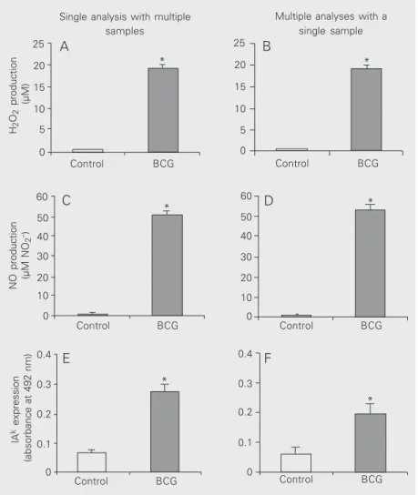
We first determined the macrophage func-tions by standard methods that use single analysis and multiple samples. We observed that BCG-activated macrophages release and
produce large amounts of H2O2 and NO and
express high levels of IAk molecules as
com-pared to nonactivated control macrophages (Figure 1A,C and E). These results demon-strate that our protocol of BCG administra-tion is highly effective in inducing macro-phage activation. Next, we determined mac-rophage functions by the multiple analysis method using a single cell sample. For this
procedure we sequentially determined H2O2
release, NO production and IAk expression.
The results obtained were statistically equiva-lent to those obtained by the standard meth-ods employing single analysis of macrophage functions using multiple cell samples (Fig-ure 1B,D and F). Although not statistically
significant, the results of IAk expression
ob-tained were always lower for the multiple analysis method compared to the single
anal-ysis method. The lower IAk expression might
be due to the time of macrophage culture.
Using single analysis, IAk expression is
meas-ured immediately after cell harvesting, while in multiple analyses it is measured after 48 h. Nevertheless, our results clearly show that when using a single macrophage sample it is possible to determine at least three macro-phage functions as accurately as when using standard microassays with multiple macro-phage samples. Actually, with the multiple analysis method it is possible to measure more than three functions. For instance, it is possible to analyze macrophage spreading
after performing the H2O2 analysis. For this,
the number of spread macrophages can be quantified under an inverted phase contrast microscope as described by Rabinovitch et al. (10). Moreover, it is possible to measure
Figure 1. Comparison of H2O2 release, NO production and MHC class II (IAk) expression by
bacillus Calmette-Guérin (BCG)-activated peritoneal macrophages. C3H/HePas mice were injected ip with 2 mg BCG or 0.2 ml PBS at days 0 and 14 and the macrophage functions determined 4 days later. H2O2 release, NO production and MHC II expression were
determined using multiple cell samples (left) or a single cell sample (right). H2O2 release (A
and B) was quantified 1 h after cell culture. NO production (C and D) was determined 48 h after cell culture. IAk expression was evaluated after 1 h using multiple cell samples (E) or
48 h after cell culture using a single cell sample (F). One representative experiment of three is shown. Results are reported as means ± SEM (N = 5). *P < 0.05 for BCG compared to control (ANOVA). There was no statistical difference between single and multiple analyses (ANOVA). MHC, major histocompatibility complex; NO, nitric oxide.
H2 O2 production (µM)
25 * 20 15 10 5 0 A Control BCG 25 20 15 10 5 0 B *
NO production (µM NO
2 -) 60 50 40 30 20 0 10 60 50 40 30 20 0 10 Control BCG
Control BCG Control BCG
Control BCG Control BCG
0.4 0.3 0.2 0.1 0 0.4 0.3 0.2 0.1 0 IA
k expression
(absorbance at 492 nm)
C D
E F
*
*
Single analysis with multiple samples
Multiple analyses with a single sample
*
different macrophage products such as TNF, GM-CSF, etc., by collecting the superna-tants (50 µl) from the wells at different time intervals (data not shown).
However, when using the multiple func-tion analysis method, it is fundamental to determine that the measurement of one mac-rophage function does not interfere with the macrophage response subsequently analyzed. For instance, phorbol myristate acetate (PMA) is often used to trigger the oxidative burst (11). Since PMA activates protein ki-nase C (12) and this enzyme is known to influence NO production (13), PMA should be avoided or a second macrophage sample without PMA run in parallel should be cluded. In our experiments we did not in-clude PMA because, as shown previously, freshly explanted BCG-activated macro-phages release spontaneously large amounts
of H2O2 (14). In contrast, concanavalin
A-stimulated macrophages only release
sig-nificant amounts of H2O2 when PMA is
pres-ent (15). Therefore, we suggest that for se-quential determination of reactive oxygen and nitrogen intermediate production, at least two macrophage samples (with and without PMA) should be included in the experiment.
References
1. Bogdan C, Rollinghoff M & Diefenbach A (2000). Reactive oxygen and reactive nitrogen intermediates in innate and specific immunity. Current Opinion in Immunology, 12: 64-76.
2. Nathan C & Shiloh MU (2000). Reactive oxygen and nitrogen inter-mediates in the relationship between mammalian hosts and micro-bial pathogens. Proceedings of the National Academy of Sciences, USA, 97: 8841-8848.
3. Long EO (1989). Intracellular traffic and antigen processing. Immu-nology Today, 10: 232-234.
4. Pick E & Keisari Y (1980). A simple colorimetric method for the measurement of hydrogen peroxide produced by cells in culture. Journal of Immunological Methods, 38: 161-170.
5. Ding AH, Nathan CF & Stuehr DJ (1988). Release of reactive nitro-gen intermediates and reactive oxynitro-gen intermediates from mouse peritoneal macrophages. Comparison of activating cytokines and evidence for independent production. Journal of Immunology, 141: 2407-2412.
6. Pick E & Mizel D (1981). Rapid microassays for the measurement of superoxide and hydrogen peroxide production by macrophages in
culture using an automatic enzyme immunoassay reader. Journal of Immunological Methods, 46: 211-226.
7. Warren MK & Vogel SN (1985). Bone marrow-derived macrophages: development and regulation of differentiation markers by colony-stimulating factor and interferons. Journal of Immunology, 134: 982-989.
8. Pierres M, Kourilsky FM, Rebouah JP, Dosseto M & Caillol D (1980). Distinct epitopes of Ik gene products identified by monoclonal antibodies. European Journal of Immunology, 10: 950-957. 9. Nascimento FR, Calich VL, Rodriguez D & Russo M (2002). Dual role
for nitric oxide in paracoccidioidomycosis: essential for resistance, but overproduction associated with susceptibility. Journal of Immu-nology, 168: 4593-4600.
10. Rabinovitch M, Manejias RE, Russo M & Abbey EE (1977). In-creased spreading of macrophages from mice treated with interfer-on inducers. Cell Immunology, 29: 86-95.
11. DeChatelet LR, Shirley PS & Johnston Jr RB (1976). Effect of phorbol myristate acetate on the oxidative metabolism of human polymorphonuclear leukocytes. Blood, 47: 545-554.
Another relevant point regarding H2O2
de-termination in the peritoneal cell population is the number of polymorphonuclear cells present in the peritoneal exudates. In the BCG model, at the time of cell harvesting the number of polymorphonuclear cells was minimal and did not interfere with the assay (data not shown). However, if the percent-age of polymorphonuclear cells exceeds 5%, an adhesion step for selecting macrophages is required. Thus, peritoneal cells suspended
in Ca2+- and Mg2+-free PBS should be
al-lowed to adhere to a 96-well plate for 30 min and, after one PBS washing, the nonadherent cells (including polymorphonuclear cells) are easily removed.
Finally, we wish to emphasize that the major advantage of the multiple function analysis method is that it requires as few as 200,000 cells. Moreover, the method is easy to perform, reproducible and more economi-cal.
Acknowledgments
12. Castagna M, Takai Y, Kaibuchi K, Sano K, Kikkawa U & Nishizuka Y (1982). Direct activation of calcium-activated, phospholipid-depend-ent protein kinase by tumor-promoting phorbol esters. Journal of Biological Chemistry, 257: 7847-7851.
13. Severn A, Wakelam MJ & Liew FY (1992). The role of protein kinase C in the induction of nitric oxide synthesis by murine macrophages. Biochemical and Biophysical Research Communications, 188: 997-1002.
14. Russo M, Teixeira HC, Marcondes MC & Barbuto JA (1989).
Super-oxide-independent hydrogen peroxide release by activated macro-phages. Brazilian Journal of Medical and Biological Research, 22: 1271-1273.