LETTER TO THE EDITOR
Glioblastoma multiforme in childhood: a case report
Mauro Cruz Machado Borgo,IJulio Leonardo Barbosa Pereira,IFranklin Bernardes Faraj de Lima,IRafael Augusto Castro Santiago Branda˜o,IGerva´sio Teles C. de Carvalho,I,IIBruno Silva CostaI ISanta Casa de Belo Horizonte, Belo Horizonte, MG, Brazil.IIFaculty of Medical Sciences of Minas Gerais, Belo Horizonte, MG, Brazil.
Email: maurocmborgo@hotmail.com Tel.: 55 31 3238-8896
INTRODUCTION
Among high-grade gliomas, childhood glioblastoma multiforme (GBM) is particularly challenging in terms of therapeutic treatment.1-5 Cerebral tumors are the most frequent childhood solid neoplastic disorders and are the primary cancer-related cause of death among children.1,3 Gliomas constitute approximately 60% of all cerebral tumors, and approximately half of them are considered to be high-grade malignant tumors.1 The prognosis for recovery is conservative, and 5-year survival rates range from 5% to 15%.2,6,7This case report documents a GBM that was located deep in the right cerebral hemisphere of a 9-year-old child. Because this is a rare illness for a patient of this age, we also provide a brief literature review to supplement this case report.
CASE REPORT
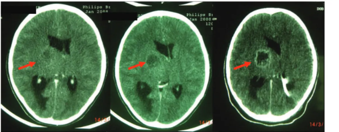
A previously healthy 9-year-old girl with adequate neuropsychomotor development was admitted to the hospital with a 15-day-long hemiparesis and disorientation. A skull computerized tomography (CT) scan showed a deep-seated, irregularly shaped expansive lesion on the right of brain, with peripheral contrast uptake that was
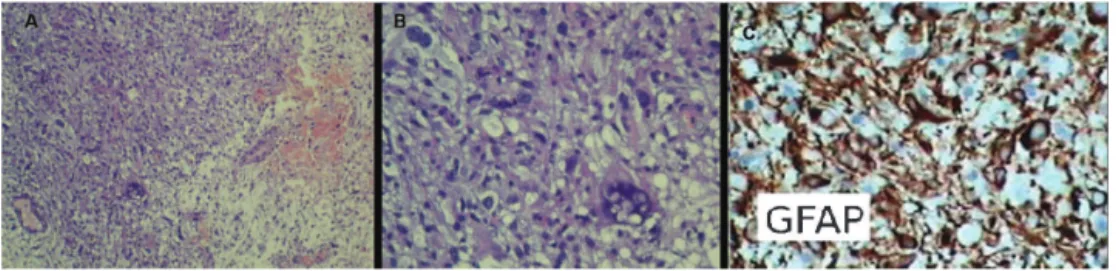
impinging on and obstructing the cerebrospinal fluid (CSF) pathways (see Figure 1). We performed a ventriculoper-itoneal shunting and stereoctatic biopsy of the lesion. The histopathology showed a pleomorphic neoplasia, which was associated with vascular neoformation, and necrosis with elongated cells and hyperchromatic and pleomorphic nuclei that had atypia and mitotic figures. The immunohistochem-istry (see Table 1) was positive for the glial fibrillary acidic protein (GFAP), Ki-67 proliferation antigen, and S-100 protein. These findings, along with the morphological features and the presence of necrosis, confirmed the diagnosis of GBM (see Figure 2). The patient, whose level of consciousness improved with steroids but whose motor functioning remained impaired, was referred to radio-therapy. After 40 days, there was a significant neurological worsening with hemiplegia and a fluctuating level of consciousness. Skull CT and MRI results showed an increase of the lesion size (see Figure 3). A craniotomy with a partial tumor removal was performed to reduce the
Copyrightß2010CLINICS– This is an Open Access article distributed under the terms of the Creative Commons Attribution Non-Commercial License (http:// creativecommons.org/licenses/by-nc/3.0/) which permits unrestricted non-commercial use, distribution, and reproduction in any medium, provided the original work is properly cited.
Figure 1 -Skull CT: Deep-seated cerebral lesion with an irregular outline, mass-effect, peripheral contrast uptake, and central necrosis.
Table 1 -Antigens that were used for the immunohistochemistry.
Antigens Clone Result
Glial fibrillary acidic protein (GFAP) Policlonal Positive Ki-67 cellular proliferation antigen M1B1 Positive S-100 protein Policlonal Positive
Neu-N MAB377 Negative
CD45RB – leukocyte-common antigen (pan-hematopoietic)
PD7/26/16&2B11 Negative
CLINICS 2010;65(9):923-925 DOI:10.1590/S1807-59322010000900016
intracranial hypertension. The patient died ten days after the procedure.
DISCUSSION
Malignant gliomas are rare in childhood, comprising approximately 6.5% of all intracranial neoplastic disorders in the pediatric population. Although these gliomas may occur in any anatomical site within the central nervous system, they are most frequently located in the supratentor-ial site.1,3,5 Males are slightly more affected than females
(male:female ratio = 1.5:1). In terms of histology, anaplastic astrocytomas are characterized by hypercellularity, nuclear atypia, mytotic figures, nuclear pleomorphism and vascular proliferation; GBM also has associated necrosis.2,6
Children with high-grade gliomas present with a variety of signs and symptoms that chiefly depend on their age and the tumor localization. The rate of neurological impairment is characteristically quick and may range from months to
days. Seizures may herald the onset, especially when tumors are close to the cerebral cortex. Other common clinical manifestations include hemiparesis, visual deficit, headache, and, in some cases, signs of intracranial hyper-tension due to an obstruction of the CSF pathways.2-8In our case, the first clinical manifestation was hemiparesis and clouding of the consciousness, with rapidly evolving intracranial hypertension despite partial removal of the tumor.
A brain MRI is the investigational tool of choice for deter-mining a GBM diagnosis.1-3,8 In this case, there was an irregularly outlined, deep-seated cerebral lesion with a mass effect, peripheral contrast uptake and central necro-sis. Such findings are consistent with the literature.1,3
The treatment of malignant gliomas is still a challenge, particularly in children. Chemotherapy and radiotherapy, far from being satisfactory treatment options, are associated with a significant rate of morbidity.9-12 Present day treatment includes tumor resection, local radiotherapy,
Figure 2 -A, B: Histological sections of cerebral tissue showing areas of elongated cells with pleomorphic and hyperchromatic nuclei that are associated with vascular neoformation and extensive hemorrhage and necrosis.
C: Immunohistochemistry positive for GFAP.
Figure 3 -CT (above) and MTI (below): A large lesion impinging on the thalamus and basal ganglia with peripheral contrast uptake and central necrosis.
Glioblastoma multiforme in childhood
Borgo MCM et al. CLINICS 2010;65(9):923-925
and chemotherapy, which are approaches that promote an improvement in the length of survival but do not seem to change the inexorable course of the disease.1,3-5,13,14
Our patient developed obstructive hydrocephalus that demanded ventriculoperitoneal shunting. A subsequent stereotactic biopsy led to the histopathological confirmation of GBM. We chose to perform radiotherapy and chemother-apy. The quick, unfavorable evolution of the disease precluded the use of chemotherapy and led us to try a partial tumor resection for decompression, which was ultimately unsuccessful.
The role of adjuvant chemotherapy for the treatment of pediatric high grade gliomas (HGGs) was established in the 1980s,15 which is based on the results of a randomized Children’s Cancer Group study using lomustine and vincristine. Recently, studies have shown a small increase in survival rates using temozolomide and lomustine to treat pediatric HGGs.16 These studies have demonstrated that
surgery, chemotherapy, and radiotherapy were ineffective in achieving long-term survival. There have only been anecdotal reports of good results in the treatment of glioblastoma in children. Further research on this disease is needed so that better treatments may be developed to improve the quality of life and prognosis of these patients.
REFERENCES
1. Reddy AT, Wellons JC. Pediatric high-grade gliomas. The Cancer Journal 2003;9:107-12, doi: 10.1097/00130404-200303000-00006.
2. Rondinelli PIP, Martinez CAO. Meta´stases intrarraquidianas de glio-blastoma multiforme supratentorial da infaˆncia: relato de caso. Arq Neuro-Psiquiatr. 2002;60:643-6.
3. Tamber MS, Rutka JT. Pediatric supratentorial high-grade gliomas. Neurosurg Focus. 2003;14(2):e1, doi: 10.3171/foc.2003.14.2.2.
4. Pollack IF. The role of surgery in pediatric gliomas. J Neuro-Oncol 1999;42:271-88, doi: 10.1023/A:1006107227856.
5. Artico M, Cervoni L, Celli P, Salvati M, Palma L. Supratentorial glioblastoma in children: a series of 27 surgically treated cases. Childs Nerv Syst. 1993;9:7-9, doi: 10.1007/BF00301926.
6. Kleihues P, Cavenne W. Pathology and Genetics of Tumors of the Central Nervous System. Lyon, France: International Agency for Research on Cancer, 1997.
7. Wrensch M, Minn Y, Chew T, Bondy M, Berger MS. Epidemiology of primary brain tumors: current concepts and review of the literature. Neuro-Oncol. 2002;4:278-99.
8. Dropcho EJ, Wisoff JH, Walker RW, Allen JC. Supratentorial malignant gliomas in childhood: a review of fifty cases. Ann Neurol 1987;22:355-64, doi: 10.1002/ana.410220312.
9. Wisoff JH, Boyett JM, Berger MS, Brant C, Li H, Yates AJ, et al. Current neurosurgical management and the impact of the extent of resection in the treatment of malignant gliomas of childhood: a report of the Children’s Cancer Group trial no. CCG-945. J Neurosurg. 1998;89:52-9, doi: 10.3171/jns.1998.89.1.0052.
10. Kreth FW, Warnke PC, Scheremet R, Ostertag CB. Surgical resection and radiation therapy versus biopsy and radiation therapy in the treatment of glioblastoma multiforme. J Neurosurg. 1993;78:762-6, doi: 10.3171/jns. 1993.78.5.0762.
11. Phuphanich S, Edwards MS, Levin VA, Vestnys PS, Wara WM, Davis RL, et al. Supratentorial malignant gliomas of childhood. Results of treat-ment with radiation therapy and chemotherapy. J Neurosurg. 1984;60: 495-9, doi: 10.3171/jns.1984.60.3.0495.
12. Quigley MR, Maroon JC. The relationship between survival and the extent of the resection in patients with supratentorial malignant gliomas. Neurosurgery. 1991;29:385-9, doi: 10.1097/00006123-199109000-00008. 13. Hess KR. Extent of ressection as a prognostic variabble in the treatment of
gliomas. J Neuro-Oncol. 1999;42:227-31, doi: 10.1023/A:1006118018770. 14. Prados MD. Future directions in the treatment of malignant gliomas with
temozolomide. Semin Oncol. 2000;27(3 Suppl 6):41-6.
15. Sposto R, Ertel IJ, Jenkin RD, Boesel CP, Venes JL, Ortega JA, et al. The effectiveness of chemotherapy for treatment of high grade astrocytomas in children: results of a randomized trial. A report from the Childrens Cancer Study Group. J Neurooncol. 1989;7:165-77, doi: 10.1007/BF00165101. 16. Jakacki RI, Yates A, Blaney SM, Zhou T, Timmerman R, Ingle AM, et al.
A phase I trial of temozolomide and lomustine in newly diagnosed high-grade gliomas of childhood. Neuro Oncol. 2008;10:569-76, doi: 10.1215/ 15228517-2008-019.
CLINICS 2010;65(9):923-925 Glioblastoma multiforme in childhood
Borgo MCM et al.