44
CASE REPORTCutaneous epithelioid hemangioma
of atypical location: a case report
Hemangioma epitelioide cutâneo de localização atípica: relato de caso
Murilo O. L. Carapeba1; Benício R. Junior1; Mariana V. Cervelatti1; Fernanda A. L. Guaresemin1; Paula A. L. Guaresemin1; Marilda A. M. M. Abreu2
1. Universidade do Oeste Paulista (UNOESTE). 2. Hospital Regional de Presidente Prudente.
First submission on 14/04/14; last submission on 26/12/14; accepted for publication on 29/12/14; published on 20/02/15
ABSTRACT
The epithelioid hemangioma, also called angiolymphoid hyperplasia with eosinophilia, is a rare benign vascular tumor that manifests as nodules. Histopathological examination shows vascular spaces of various sizes lined by prominent endothelium, and inlammatory iniltrates composed of eosinophils, histiocytes, mast cells and lymphocytes. We report a case of epithelioid hemangioma characterized by skin lesion in the left upper limb. This is an unusual location, since the lesions occur most often in the head and neck.
Key words: angiolymphoid hyperplasia with eosinophilia; hemangioma; vascular diseases.
J Bras Patol Med Lab, v. 51, n. 1, p. 44-47, February 2015
INTRODUCTION
The epithelioid hemangioma, also called angiolymphoid hyperplasia with eosinophilia, due to its histopathological characteristics, is a rare benign vascular tumor, irst described by Wells and Whimster, in 1969(1). It is more common in adult
females over 30 years old(2). Until recently it was confused with
Kimura’s disease; their designations were even considered
synonymous(3). Both disorders cause nodular lesions in head and
neck, but in Kimura’s disease, there are subcutaneous nodules without skin color changes, and although there is also lymphoid and vascular proliferation with eosinophilia, it does not present epithelioid endothelial cells(4-6).
In spite the unknown pathogenesis of epithelioid hemangioma, reports suggest the involvement of arteriovenous shunts(7). There
are cases associated to traumas, infections, oral contraceptives or pregnancy. It remains uncertain whether the lesion is neoplastic or reactive in nature(8).
The cutaneous lesions consist of atypical and inlammatory vascular proliferations that affect the hypodermis or the dermis. They manifest clinically as papules or nodules, of angiomatoid aspect, with variable color, from red to brown, single or numerous, measuring around 1 cm, possibly reaching 5 cm to 10 cm when affecting the hypodermis. They are usually asymptomatic, but may be painful and pulsating. Spontaneous bleeding is reported in some cases. They can arise in any region, but are generally located in the head and neck (85% of the cases), principally in the frontal region, the scalp and around the ear, besides extremities, especially ingers. They are less frequently described in the trunk, vulva, and penis. The lesions may be associated with regional lymphadenopathy and peripheral eosinophilia (20% of the cases). The subcutaneous lesions resemble lymph nodes(2, 6-11).
The histopathological exam reveals vascular spaces of varying sizes, lined by endothelial cells, and a mixed inlammatory iniltrate, containing eosinophils(6, 8, 11, 12).
45
The treatment of choice is the surgical excision, which effects the complete cure in a large part of the cases. Other options are: intralesional corticosteroid, radiotherapy, laser treatment (argon, carbon dioxide [CO2] and neodymium-doped yttrium aluminum garnet [Nd:YAG]), cryotherapy and oral retinoids. Spontaneous regression is reported in some cases. Recurrence is common, in up to two-thirds of cases; there is no report of malignant transformation, and the evolution of the deep presentation is less predictable(7, 8, 13). The tumor may beseen in individuals infected by the human immunodeiciency virus (HIV)(14).
The present work reports the case of a patient who developed a cutaneous nodule on the upper left limb, which was diagnosed as hemangioma epithelioid at the histopathological exam. It also presents a literature review about the characteristics of this disease.
CASE REPORT
A 46-year-old white female presented with a one-year history of a mildly-painful slow-growing nodule on the upper left limb. In the dermatologic exam, a nodule of around 3 cm was observed, located in the dorsal surface of the upper left limb, in the transition zone between arm and forearm, slightly purple with a more violaceous center, soft in consistency and depressible (Figures 1 and 2). The clinical hypotheses of hemangioma, lipoma, angiolipoma and ruptured sebaceous cyst were formulated, and the surgical exploration was conducted, in which a bloody content was observed. Material was collected for histopathological examination, which grossly revealed an irregular fragment of cyst wall, measuring 1 × 0.5 cm, with a smooth surface and elastic consistency. Microscopic examination confirmed the presence of a hyperplastic lesion, composed of the proliferation of blood vessels lined by endothelial cells, delimited by histiocytic cell forms, sometimes with epithelioid aspect. The vessels contained red globules; some exhibited papillary hyperplasia in the lumen
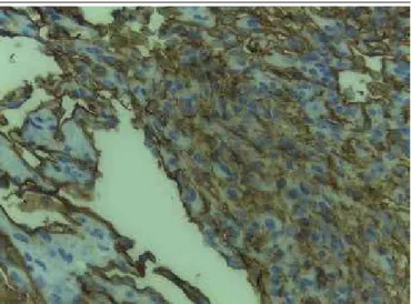
(Figures 3, 4, and 5). There were no signs of malignancy. Immunohistochemically the neoplasm was negative for pan-cytokeratins (AE1/AE2), but it was positive for vascular markers cluster of differentiation 31 (CD31) and cluster of differentiation 34 (CD34) (Figures 6 and 7), confirming the diagnosis of epithelioid hemangioma.
FIGURE 1 − Nodule of approximately 3 cm in diameter, in the transition zone between
arm and forearm, slightly purple with a more violaceous center
FIGURE 2 − Detail of the nodular lesion
FIGURE 3− Proliferation of epithelioid cells forming vascular spaces of varying sizes,
sometimes with papillary hyperplasia in the lumen (HE, 100×)
HE: hematoxylin and eosin.
46
FIGURE 4 − Blood vessels of varying sizes lined by endothelial cells with an epithelioid
pattern (HE, 400×)
HE: hematoxylin and eosin.
FIGURE 5 − Blood vessels containing erythrocytes: arrow in red; erythrocytes surrounded
by cells of epithelioid aspect: yellow arrow (HE, 400×) HE: hematoxylin and eosin.
FIGURE 6 − Immunohistochemistry showing lesion positive for CD31 (400×)
CD31: cluster of differentiation 31.
FIGURE 7 − Immunohistochemistry showing lesion positive for CD34 (400×)
CD34: cluster of differentiation 34.
DISCUSSION
The cutaneous epithelioid hemangioma is a benign vascular tumor, of mesenchymal origin, with no systemic involvement, which may be differentiated from lesions of similar aspects, such as lipoma, cylindroma, sebaceous and epidermoid cyst, lymphocytoma, insect bite reaction, vaccines, sarcoidosis, dermatoibroma, pyogenic granuloma, and Kimura’s disease, among others. The histopathological exam is the only one that can establish the deinite diagnosis of this condition. Currently, the epithelioid hemangioma is considered part of a spectrum of vascular lesions with epithelioid characteristics, including epithelioid hemangioendothelioma and epithelioid angiosarcoma(11-13).
Among its indings, it is interesting to highlight the identiication of two basic components: vascular and cellular. The vascular component is made of well-formed but generally immature dilated capillaries, lobular in appearance, with noticeable multiplication of the endothelial cells that present epithelioid aspect, that is, big cells with evident nucleoli and abundant eosinophilic cytoplasm. The cellular component, supericial and deep, is composed of iniltrate predominantly of lymphocytes, histiocytes and eosinophils, sometimes plasmocytes and mastocytes(6, 8-12).
In the immunohistochemical reaction, the epithelioid hemangioma is positive for CD31, CD34 and factor VIII-related antigen; and negative for keratins and epithelial membrane
antigens(15).
47
Murilo O. L. Carapeba; Benício R. Junior; Mariana V. Cervelatti; Fernanda A. L. Guaresemin; Paula A. L. Guaresemin; Marilda A. M. M. Abreu
RESUMO
O hemangioma epitelioide, também denominado hiperplasia angiolinfoide com eosinofilia, é um tumor vascular raro, de caráter benigno, que se manifesta como nódulos. O exame histopatológico evidencia espaços vasculares de diversos calibres, revestidos por endotélio proeminente e infiltrado inflamatório composto de eosinófilos, histiócitos, mastócitos e linfócitos. Relatamos um caso de hemangioma epitelioide caracterizado por lesão de pele no membro superior esquerdo. Essa é uma localização incomum, uma vez que as lesões ocorrem mais frequentemente na cabeça e no pescoço.
Unitermos: doenças vasculares; hemangioma; hiperplasia angiolinfoide com eosinofilia.
REFERENCES
1. Wells GC, Whimster IW. Subcutaneous angiolymphoid hyperplasia with eosinophilia. Br J Dermatol. 1969; 81: 1-5.
2. Rajendran R, Padmakumar SK, Kothawar S, et al. Angiolymphoid hyperplasia with eosinophilia (ALHE). J Oral Maxillofac Pathol. 2005; 9: 24-6.
3. Thompson JW, Colman M, Williamson C, et al. Angiolymphoid hyperplasia with eosinophilia of the external canal. Arch Otolaryngol. 1981; 107: 316-69.
4. Al-Muharraqi MA, Faqi MK, Uddin F, et al. Angiolymphoid hyperplasia with eosinophilia (epithelioid hemangioma) of the face: an unusual presentation. Int J Surg Case Rep. 2011; 2(8): 258-60.
5. Briggs PL. Doença de Kimura não é hiperplasia angiolinfoide com eosinoilia: correlação clinicopatológica com revisão da literatura e deinição de critérios diagnósticos. An Bras Dermatol. 2006; 2: 167-73. 6. Chagas SAP, Pinheiro SA, Mendes SC, et al. Hemangioma epitelioide vulvar (hiperplasia angiolinfoide com eosinoilia): relato de caso. Rev Méd Minas Gerais. 2010; 20: 419-21.
7. Kurihara Y, Inoue H, Kiryu H, et al. Hemangioma epiteloide con una distribución zosteriforme. Indian J Dermatol. 2012; 57: 401-3.
MAILING ADDRESS
Marilda Aparecida Milanez Morgado de Abreu
Rua São Paulo, 1949; Centro; CEP: 17900-000; Dracena-SP, Brazil; e-mail: [email protected].
8. Sánchez EL, Guerrero RC, Alfonso MCJ, et al. Hemangioma epitelioide cervical. ORL Aragon. 2014; 17(1): 47-8.
9. González AM, Ball E. Hemangiomas adquiridos inusuales. Estudio clínico-patológico. Hospital Universitario de Caracas, 2001-2011. Dermatol Venez. 2011; 49(3-4): 37-46.
10. Ismail M, Sesterhenn IA, Miettinen M, et al. Epithelioid hemangioma of the penis: case report and review of literature. J Med Case Reports. 2011; 5: 260.
11. Nogueira A, Accioly-Filho JW, Castro MCR, et al. Hiperplasia angiolinfoide com eosinoilia: relato de dois casos. An Bras Dermatol. 2003; 78(1): 79-85.
12. Villarinho JF, Boechem NT, Hadj LAE, et al. Hemangioma epitelioide de MAE: relato de um caso raro. Rev Bras Otorrinolaringol. 2008; 74(1): 49.
13. Tchernev G, Taneva T, Ananiev J, et al. Angiolymphoid hyperplasia with eosinophilia - an incidental inding after surgical excision. Wien Med Wochenschr. 2012; 162(19-20): 448-51.
14. D’Ofizi G, Ferrara R, Donati P, et al. Angiolymphoid hyperplasia with eosinophils in HIV infection. AIDS. 1995; 9(7): p. 813-4.
15. Gray HJ, Rendi MH, Holmes M. Painful clitoromegaly caused by rare epithelioid hemangioma. Gynecol Oncol Case Rep. 2013; 4: 60-2.
In this case, the uncommon location in the skin of the upper limb stands out, once the frequent involved sites are generally head and neck, what made it even more dificult to suspect epithelioid hemangioma.