0021-7557/$ - see front matter © 2013 Sociedade Brasileira de Pediatria. Published by Elsevier Editora Ltda. All rights reserved. http://dx.doi.org/10.1016/j.jped.2013.02.008
www.jped.com.br
☆Please, cite this article as: Moniz M, Silvestre C, Nunes P, Abadesso C, Matias E, Loureiro H, et al. High-frequency oscillatory ventilation in children: a 10-year experience.J Pediatr (Rio J). 2013;89:48-55.
* Corresponding author.
E-mail: [email protected] (M. Moniz).
ORIGINAL ARTICLE
High-frequency oscillatory ventilation in children:
a 10-year experience
☆Marta Moniz
a,*, Catarina Silvestre
b, Pedro Nunes
b, Clara Abadesso
b, Ester Matias
b,
Helena Loureiro
b, and Helena Almeida
ca Fellow Pediatrician, Pediatric Department, Hospital Professor Doutor Fernando Fonseca, Amadora, Portugal b MD. Pediatric Intensive Care Unit, Hospital Professor Doutor Fernando Fonseca, Amadora, Portugal
c MD. Chief, Pediatric Intensive Care Unit of Hospital Professor Doutor Fernando Fonseca, Amadora, Portugal
Received 22 May 2012; accepted 22 August 2012
KEYWORDS High frequency oscillatory ventilation; Child;
Acute respiratory failure;
Bronchiolitis; Pneumonia; Acute respiratory distress syndrome
Abstract
Objectives: The aim of the study was to describe the experience with high-frequency oscillatory ventilation (HFOV) in a Portuguese Pediatric Critical Care Unit, and to evaluate whether HFOV allowed improvement in oxygenation and ventilation.
Methods: This was a retrospective observational cohort study of children ventilated by HFOV between January, 2002 and December, 2011. The following parameters were recorded: demographic and clinical data, and blood gases and ventilatory parameters during the first 48 hours of HFOV.
Results: 80 children were included, with a median age of 1.5 months (min: one week; max: 36 months). Pneumonia (n = 50; 62.5%) and bronchiolitis (n = 18; 22.5%) were the main diagnoses. Approximately 40% (n = 32) of the patients developed acute respiratory distress syndrome (ARDS). Conventional mechanical ventilation was used in 68 (85%) of patients prior to HFOV. All patients who started HFOV had hypoxemia, and 56 (70%) also presented persistent hypercapnia. Two hours after starting HFOV, a significant improvement in SatO2/FiO2 ratio (128 ± 0.63 vs. 163 ± 0.72; p < 0.001) that was sustained up to 24 hours of HFOV and a decrease in FiO2 were observed. Since the beginning of HFOV, the mean PCO2 significantly decreased (87 ± 33 vs. 66 ± 25; p < 0.001), and the pH significantly improved (7.21 ± 0.17 vs. 7.32 ± 0.15; p < 0.001). Overall survival was 83.8%.
Conclusions: HFOV enabled an improvement in hypercapnia and oxygenation. It is a safe option for the treatment of ARDS and severe small airway diseases.
PALAVRAS-CHAVE Ventilação oscilatória de alta frequência; Criança;
Insuiciência respiratória aguda; Bronquiolite; Pneumonia;
Síndrome da angústia respiratória aguda
Ventilação oscilatória de alta frequência em crianças: uma experiência de 10 anos
Resumo
Objetivos: O objetivo do estudo foi descrever a experiência com ventilação oscilatória de frequência (VOAF) em uma unidade portuguesa de Cuidados Intensivos Neonatais e Pediátricos e avaliar se a VOAF permitiu uma melhoria na oxigenação e na ventilação.
Métodos: Estudo de coorte retrospectivo observacional em crianças submetidas À ven-tilação com VOAF entre janeiro de 2002 e dezembro de 2011. Os seguintes parâmetros foram registrados: dados demográficos e clínicos; gases sanguíneos; e parâmetros venti-latórios durante as primeiras 48 horas de VOAF.
Resultados: O estudo incluiu 80 crianças com uma idade média de 1,5 mês (mínima: uma semana; máxima: 36 meses). Pneumonia (n = 50; 62,5%) e bronquiolite (n = 18; 22,5%) foram os principais diagnósticos. Cerca de 40% (n = 32) dos pacientes desenvolveram a síndrome da angústia respiratória aguda (SARA). A ventilação mecânica convencional foi utilizada em 68 (85%) pacientes antes da VOAF. Todos os pacientes que começaram a VOAF tiveram hipoxemia, e 56 (70%) também apresentaram hipercapnia persistente. Duas horas após o início da VOAF, foi observada uma melhoria significativa na proporção SatO2/FiO2 (128 ± 0,63 em comparação a 163 ± 0,72; p < 0,001), que foi mantida durante as 24 horas de VOAF, e uma redução da FiO2. Desde o início da VOAF, a PCO2 média teve uma queda significativa (87 ± 33 em comparação a 66 ± 25; p < 0,001) e o pH aumentou significativamente (7,21 ± 0,17 em comparação a 7,32 ± 0,15; p < 0,001). A sobrevida geral foi de 83,8%.
Conclusões: A VOAF permitiu uma melhoria na hipercapnia e na oxigenação. Trata-se de uma opção segura no tratamento da SARA e de doenças graves das pequenas vias aéreas.
© 2013 Sociedade Brasileira de Pediatria. Publicado por Elsevier Editora Ltda. Todos os direitos reservados.
Introduction
Acute respiratory failure is a frequent problem in children admitted to pediatric intensive care units (PICUs). It is well known that mechanical ventilation is associated with barotrauma, volutrauma, atelectrauma, and biotrauma.1-4
Avoiding ventilator-induced lung injury has become a major concern when considering which ventilatory strategy to apply to patients with lung diseases. High frequency oscillatory ventilation (HFOV) is a lung-protective ventilatory mode that ensures alveolar recruitment and an optimal lung volume.5,6 During HFOV, very small tidal
volumes (1-2 mL/kg), high flow rates, and frequencies of 240-900 cycles per minute are used to open the lung, in order to avoid high peak airway pressures, alveolar over distension, and repeated cycles of recruitment.5 HFOV
has been most studied in the context of acute respiratory distress syndrome (ARDS), and several studies have demonstrated its safety as a lung volume recruitment strategy.7-9 However, there isn’t enough evidence to
support the use of HFOV over conventional mechanical ventilation (CMV). To a lesser extent, HFOV has been applied in children with air leak or small airway disease, such as bronchiolitis.10-14
This study aimed to describe the authors’ experience using HFOV in pediatric patients, and to evaluate its effect on oxygenation, ventilation, and associated complications. This study was based on retrospective analysis of the
patients treated at the PICU of Hospital Professor Doutor Fernando Fonseca in Amadora, Portugal.
Materials and methods
Design
This was an observational retrospective study from January, 2002 to December, 2011. The study was performed in an eleven-bed PICU of a Portuguese hospital with maximum capacity for six ventilated patients. The PICU is localized in the metropolitan area of Lisbon, which has 800,000 inhabitants, and admits approximately 500 children yearly. This study was approved by the institutional review board.
Patients and data collection
oxygen (FiO2), transcutaneous oxygen saturation (Sat. O2), and ventilator settings of HFOV (mean continuous distending airway pressure [Paw], amplitude [delta-p] and frequency [Hz]) were recorded immediately before and at 2, 6, 12, 24 and 48 hours of HFOV.
Maximal parameters during HFOV were considered as the highest ventilatory settings observed during six consecutive hours. Lung recruitment pressure was defined as the maximal mean airway pressure that allowed a stepwise decrease of FiO2 until reaching 0.6.
ARDS was defined according to the American-European Consensus Conference on ARDS.15 Airway obstructive
disease as bronchiolitis was defined as a respiratory disorder in children until two years of age with rhinitis, cough, tachypnea, wheezing, crackles, and use of accessory muscles, with or without fever and without consolidation on the X-ray. Pneumonia was defined as infiltrates on chest X-ray, in addition to one of the following: deterioration in pulmonary gas exchange, fever (temperature above 38 ºC), white blood cell count above 12.000/mm3, or a positive tracheal aspirate
culture. Sepsis was defined according to the guidelines of the Surviving Sepsis Campaign. Multiple organ failure was considered when at least two of the following organ dysfunctions occurred: central nervous system, cardiovascular, hepatic, respiratory, gastrointestinal, renal, or hematologic.
High frequency oscillatory ventilation protocol
The ventilatory modes used in CMV (Servoi®, Maquet Inc – Wayne, United States and Servo 300 Siemens Medical Systems – Solna, Sweden) were pressure control or pressure-regulated volume target. The authors opted to use CMV with non- aggressive settings (PIP < 30 cmH2O, tidal volume < 8 mL/kg, respiratory rate [RR] < 50/min). When refractory hypoxemia, defined as Sat.O2 below 90% with FiO2 1 or hypercapnia with severe acidosis (pH < 7.22 and pCO2 > 80 mmHg) occurred, patients were switched to HFOV.
Sensor-Medics 3100A® (Sensor Medics Corporation – Yorba Linda, CA, USA) was used for HFOV. An “open lung strategy” was adopted. The initial settings were FiO2 of 1.0, oscillation frequency of 10-12 Hz, a percent inspiratory time of 33%, and bias flow of 20-30 L/ min. The Paw was set 5 cmH2O above the mean airway pressure during CMV, and increments of 1 cmH2O were used until an optimal lung volume was reached, avoiding over distension and atelectasis. The oxygenation target was an adequate SatO2 (≥ 90%) with FiO2 ≤ 0.6. The delta-p was initially set to achieve chest wall vibration to the level of thigh in infants, or to the umbilical level in newborns. The frequency and delta-p were adjusted to obtain an adequate pCO2 and pH above 7.25. General supportive care included fluid restriction to 80% of daily needs, nutritional support, and antibiotics if needed. All patients were sedated with continuous infusion of an opioid (morphine), and a benzodiazepine (midazolam). Neuromuscular blockage agents were only used when there was a significant clinical deterioration related to spontaneous activity. Inotropic support, nitric oxide,
and chest tube drainage were used if necessary. The weaning process from HFOV was started when FiO2 was below 0.4. During this process, Paw was gradually decreased by 1-2 cmH2O until a value below 14 cmH2O was reached. After HFOV, patients started CMV (support mode), non-invasive ventilation, or nasal cannula oxygen.
Outcome measures
The primary outcome was to evaluate whether HFOV allowed improvement in oxygenation and ventilation. An improvement in oxygenation was considered if a decrease in FiO2 allowed Sat.O2 above 90%, and the ratio of Sat. O2/FiO2 increased along the study period.16 Evaluation
of ventilation was performed through changes of pH and pCO2 values obtained from capillary blood samples. HFOV success was defined as improvement in oxygenation and ventilation. HFOV failure was considered when death or intractable hypoxemia occurred.
The secondary outcomes were: a) to study complications related to HFOV and mortality rate. A new air leak, hypotension, hypoxemia or bradycardia during HFOV were considered as complications possibly related to HFOV; b) to compare clinical, blood gases, and therapeutic parameters in surviving and non-surviving patients; and c) to analyze predictive factors of mortality.
Statistical analysis
Quantitative variables were analyzed using measures of central location (mean and median) and dispersion (standard deviation). Qualitative or categorical variables were described as frequencies. To compare quantitative variables, Student’s t-test or Wilcoxin’s non-parametric test were used, depending on the distribution according to normality. Dichotomous variables were studied with the chi-squared test or Fisher’s exact tests. A multivariate analysis was performed using binary logistic regression through the Enter method. All tests were two-tailed, and a p-value < 0.05 was considered as significant. Statistical analyses were performed using the Statistical Package for Social Sciences (SPSS Inc. – Chicago, Illinois, USA) version 18.0 for Windows.
Results
Demographics
Ventilatory characteristics
CMV was performed in 68 (85%) of patients prior to HFOV, and non-invasive ventilation was the first option in 33 (44%) patients. The median time on CMV before HFOV was 12 hours (1-360 h). Maximum parameters during CMV were (mean±SD): PEEP 6±1 cmH2O, PIP 26±6 cmH2O, RR 52±8 cpm, and FiO2 0.70±0.25.
Immediately before starting HFOV, the median Sat. O2/FiO2 ratio was 156 (43-376) and the median value of PCO2 was 82.3 torr (31-200). Before HFOV, four patients presented a clinical deterioration due to pulmonary air leak (pneumothorax, n = 3; pulmonary interstitial emphysema, n = 1). HFOV was initiated based on hypercapnia in 56 (70%), hypoxemia with hypercapnia in 13 (16.3%), and hypoxemia without hypercapnia in 11 (13.8%) patients. HFOV was performed for a median time of 103 hours (12-576 h). The median of the highest parameters used during HFOV were: FiO2 0.8 (0.3-1), Paw 19 cmH2O (14-44 cmH2O), and delta-p 50 (30-93). The median frequency used was 10Hz (5-12 Hz). A median Paw of 19.5 cmH2O (15-44 cm2O) was used to perform lung recruitment. All patients were sedated with morphine and midazolam. Inotropic support was needed in 52 (65%) patients before starting HFOV. Dopamine was used alone in 29 (36%) patients, and two or more inotropics were needed in 18 (22.5%) cases. 22 (27.5%) patients were curarized for a median time of 60 hours (24-960 h). Nitric oxide was administered to 14 (17.5%) children.
Primary outcome
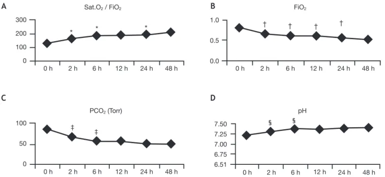
With HFOV, immediate and significant increase was achieved in the Sat.O2/FiO2 ratio (128±0.63 vs. 163±0.72; p < 0.001) that was sustained until 24 hours of HFOV. A FiO2
of approximately 0.6 was reached after 6 hours of HFOV. Immediately after starting HFOV, mean PCO2 significantly decreased (87±33 vs. 66±25; p < 0.001) and remained within target ranges during the entire study period. Also, pH significantly improved at the beginning of HFOV (7.21±0.17 vs. 7.32±0.15; p < 0.001), and remained within normal values during the 48 hours of the study. Figure 1 shows blood gases, FiO2, and Sat.O2/FiO2 ratios during the study period.
According to the protocol design, during the first two hours of HFOV an increment of Paw (17.8±3.5 vs. 18.0±3.4; p = 0.03) was observed to reach lung recruitment. After this time, significant oscillations in Paw were not registered. Significant variations of delta-p and frequency during the study were not observed. Table 2 shows ventilator settings and hemodynamic parameters during the 48 hours.
Mean arterial pressure did not show significant oscillations during the 48 hours studied. After 12-hours of HFOV, a significant decrease was observed in heart rate (149±21 vs. 141±23; p < 0.001). At 24 and 48-hours the heart rate was significantly lower (Table 2).
37 (46.3%) patients were successfully weaned from HFOV: 25 to non-invasive ventilation, and 12 directly to supplementary oxygen through face masks or nasal cannula.
Secondary outcome
During HFOV, seven patients presented with a new air leak related to central line insertion (two patients), to high-pressure hand-mask ventilation (two patients), and to fatal ARDS (three patients). HFOV did not cause worsening of pneumothorax in those patients with a diagnosis of air leak prior to HFOV. Endotracheal tube suction was associated with transitory hypoxemia and/ or bradycardia in 16 (20%) patients. During the course of HFOV, reintubation was needed in eight (10%) patients due to endotracheal tube obstruction or accidental extubation. Most patients (n = 51; 63.8%) did not present complications during HFOV.
Overall survival was 83.8% (67 patients). Five patients died from multi-organ failure, four from cardiac failure (hypoplastic left heart syndrome), and four from both septic shock and refractory respiratory failure. Children who died had a median age of 4 months (min = 1; max = 36 months), and were significantly older than those who survived, who presented a median age of 1 month (min = 9 days; max = 35 months) (p = 0.015). A relevant past medical history was more prevalent in the group of non-survivors (77% vs. 36%; p = 0.006). Also, in this group, mechanical ventilation before HFOV was performed for a significantly longer period when compared to survivors (72±106 vs. 24±39; p = 0.01). Differences in ventilator settings during HFOV between both groups are shown in Table 3. Non-survivors presented a lower Sat.O2/ FiO2 ratio (133±60 vs. 104±78; p = 0.006) since the beginning of HFOV when compared to survivors. This difference was observed during the 48 hours of the study period. Values of pH and PCO2 were similar between both groups up to 24 hours of HFOV. After that time, a significant improvement was observed only in the group of survivors
Table 1 Patient demographics, underlying diseases, and ARDS.
Variable
Weight (kg), mean±SD 4.1±2.1
Past medical history
Prematurity, n (%) 25 (31.3)
Cardiac disease, n (%) 7 (8.8)
Neurological disease, n (%) 7 (8.8)
AIDS, n (%) 1 (1.3)
Irrelevant, n (%) 40 (50)
Diagnosis
Pneumonia, n (%) 50 (62.5)
Bronquiolitis, n (%) 18 (22.5)
Pneumonia with sepsis, n (%) 10 (12.5) Cardiac insufficiency, n (%) 2 (2.5)
ARDS, n (%) 32 (40)
(7.40±0.10 vs. 7.30±0.08; p = 0.003). During HFOV, lower frequencies and higher Paw and delta-p were used in the group of patients who died (p < 0.05) (Table 3). In the univariate analysis, predictive factors of mortality were:
pH and PCO2 at 2 hours, pH at 24-hours, and Sat.O2/ FiO2 ratios at 2 hours, 6 hours, 12 hours, 24 hours, and 48 hours. In the multivariate analysis, these were not independent factors.
Table 2 Mean ventilator settings, gas exchange, blood pressure, and heart rate of the study population at multiple time intervals during HFOV.
Parameters At beginning 2 hours 6 hours 12 hours 24 hours 48 hours p-value
HFOV Mean±SD Mean±SD Mean±SD Mean±SD Mean±SD
Mean±SD Ventilator settings
Paw (cmH2O), 17.8±3.5 18.0±3.4 18.1±3.1 18.1±3.1 17.8±4.3 17.6±4.5 a
delta-p 45±12 45±12 45±12 46±13 44±15 45±17 ns
Frequency (Hz) 10±1 10±1 10±1 10±1 10±1 10±1 ns
Hemodynamic data
Mean arterial pressure 48±11 48±10 50±12 48±13 49±9 51±11 b
Heart rate (cpm) 145±23 145±22 149±21 141±23 136±18 129±17 c
delta-p, amplitude; ns, non-significant; HFOV, high-frequency oscillatory ventilation; paw, airway pressure; SD, standard deviation. aA significant increase was observed during the first 2 hours of the study (p = 0.03). During the remainder of the study, there were no significant variations.
bMean arterial pressure was stable during the entire study.
cHeart rate significantly decreased from the 12th hour of the study (p < 0.05 for 24-hour and 48-hour comparisons).
300
200
100
0
0 h 2 h 6 h 12 h 24 h 48 h
Sat.O2 / FiO2
0 h 2 h 6 h 12 h 24 h 48 h
1.0
0.5
0.0
100
50
0
0 h 2 h 6 h 12 h 24 h 48 h
PCO2 (Torr)
0 h 2 h 6 h 12 h 24 h 48 h
7.50
7.25 7.00
6.75
6.51
pH FiO2
*
‡ §
†
*
*
‡
§
† † †
Figure 1 Changes in mean Sat.O2/FiO2 ratio, FiO2, PCO2, and pH during the different periods of HFOV. (A) *p < 0.05, at initiation versus 2 hours; Sat.O2/FiO2 ratio became significantly higher 2 hours after starting HFOV, and a significant increase was observed until 24 hours of HFOV. (B) †p < 0.05 at initiation versus 2 hours, at 6 versus 2 hours, at 12 hours versus 6 hours, and at 24 hours
versus 12 hours; FiO2 was lower immediately after starting HFOV. Along the 24 hours of the study, significant lower levels of FiO2 were reached. (C) ‡p < 0.05 at initiation versus 2 hours, and at 6 hours versus 2 hours; PCO
2 significantly decreased at 2 hours and
6 hours of HFOV. Thereafter it remained within reference ranges. (D) §p < 0.05 at initiation versus 2 hours, and at 6 hours versus 2
hours; pH significantly rose during the first 6 hours of HFOV, when normal values were reached. FiO2, fraction of inspired oxygen; PCO2, partial pressure of carbon dioxide; Sat.O2, transcutaneous oxygen saturation.
A
C
B
Discussion
In this study, it is reported one of the largest single-center pediatric studies in which HFOV was a safe modality for ventilating young patients with acute lung disease associated with heterogeneous clinical diagnosis, such as pneumonia, bronchiolitis, ARDS, and sepsis. In all patients, a statistically significant improvement in pH and PCO2 values, together with a decrease in FiO2 and an increase of the Sat.O2/FiO2 ratio, demonstrated the efficacy of HFOV on ventilation and oxygenation. These effects were registered after 2 hours of HFOV. Similar results with HFOV have also been reported in pediatric and adult populations.8-9,17
Some of the patients who started HFOV presented small airway disease due to underlying bronchiolitis, in which PCO2 clearance is a main problem. In this subgroup of patients, when HFOV was started, not only was hypoxemia present but also hypercapnia was moderate or severe. In this study’s population, patients with bronchiolitis were mainly infants aged less than 3 months with minimal physiological time constant, so an approach of high volume and low respiratory rate was not used. HFOV proved to be very efficient in patients with diffuse alveolar disease or with increased airway resistance and hyperinflation. Slee-Wijffels et al. reported a single-center experience, which included 17 patients with small airway disease who were successfully ventilated on HFOV.10
In this study, maximum parameters in CMV were not aggressive, since HFOV was used as an early intervention strategy in the course of disease and when lung recruitment was needed. The optimal timing to initiate HFOV is not yet defined, and different approaches can be found in literature. Fedora et al. reported a survival benefit in children with ARDS ventilated in HFOV during the first 24 hours of mechanical ventilation.18 In their study, the mean length of
CMV prior to HFOV was 8.8 hours in the early intervention group, and 133.3 hours in the late intervention group.18 In
the study by Slee-Wijffels et al., the median length of CMV was 29.5 hours in the survivor group and 63 hours in the non-survivor group.10 In a multicenter experience reported
by Arnold JH et al., the duration of CMV before HFOV was
found to have a significant relationship with outcome.19 In
the present study, HFOV was the first invasive ventilation option in 12 patients, and in the others CMV was applied for a short period of time (median duration 12 hours).
Although several oxygenation strategies, including HFOV, have been studied in patients with acute lung injury, most published studies refer to the adult population; their extrapolation to pediatric patients is not always feasible. To date, there is not enough evidence to recommend one strategy over another.20,21 In PICU, only one prospective
study comparing HFOV and CMV was performed, and a statistically significant difference in mortality with HFOV could not be proved.8
In this study, only three cases of air leak occurred during HFOV. However, a direct relationship between air leaks and HFOV could not be established. All three patients had severe ARDS, which could also be responsible for the air leak. In the study by Ben Jaballah N et al., HFOV did not cause any new air leak syndrome; Arnold JH et al. found that HFOV was not associated with a higher number of air leaks when compared to CMV.8,22 Other problems found
during HFOV were transitory hypoxemia and bradycardia associated with endotracheal tube suction. During HFOV, increased secretions were produced. To avoid transitory hemodynamic instability and loss of alveolar recruitment, suction should be performed with closed loops and a slight increase in Paw (3-5 cmH2O). Also, attention to the vibration pattern may be important to detect endotracheal tube obstruction. This approach may have contributed to the low number of complications presented in this article.
During the study period, patients remained hemodynamically stable without significant oscillations in blood pressure and heart rate. Inotropics were prescribed in about 65% of the patients, which could be related to the severity of the underlying disease or possibly to HFOV. However, since an invasive hemodynamic study was not performed, a direct relation with HFOV could not be established. In the study by David M et al., transition to HFOV was related to a statistically significant increase in right atrial pressure and pulmonary occlusion pressure, together with a decrease in cardiac index.23 However, as
Table 3 Comparison of maximal ventilatory parameters during HFOV between the group of survivors and non-survivors.
Parameters Maximal HFOV settings
Survivors Non-survivors p-value
Maximal ventilator settings
MAP (cmH2O) 19.8±5 24.3±6 0.008
FiO2 0.7±0.2 0.9±0.2 0.023
delta-p 51±15 67±15 0.001
Frequency (Hz) 11±1 9.5±1 0.002
Minimal frequency (Hz) 10±1.4 8±1 < 0.001
in the present study, they did not observe hemodynamic instability in their patients, and after transition to HFOV changes in vasoactive or inotropic support were not performed.22
In the present study, the overall survival rate was 83.8%, higher than others reported in the literature (between 45% and 75%).21,24 This difference could be due
to the variety of underlying diseases that led to HFOV, and also probably due to the early institution of this ventilatory strategy.
Persistent hypoxemia at 48 hours of HFOV was associated with a poor prognosis. There were no other independent mortality factors.
The retrospective design and data collection were the major limitations of the present study. Patients were not randomly selected, and data were collected relying on nurses and medical records. Blood gases were mainly obtained from capillaries, thus oxygen partial pressure could not be calculated. A recently validated ratio in the pediatric population (Sat.O2/FiO2) was used to study improvements in oxygenation.16 This index is important
for monitoring oxygenation in situations where PaO2 is not obtained.
Conclusion
HFOV is an efficient ventilation strategy in a variety of clinical settings, and it is associated with a rapid improvement in oxygenation and ventilation. Lung injury associated with alveolar disease or with increased airway resistance was safely treated with HFOV. Minimal complications and high survival rates contributed to the successful use of HFOV.
It can be concluded that an approach using HFOV instead of aggressive CMV appears to be safe.
An adequate HFOV protocol remains important in order to avoid complications. Future randomized controlled trials comparing conventional ventilation and HFOV will be crucial to delineate the role of HFOV as an early strategy in lung recruitment.
Conlicts of interest
The authors have no conflicts of interest to declare.
References
1. Imai Y, Slutsky AS. High-frequency oscillatory ventilation and ventilator-induced lung injury. Crit Care Med. 2005;33: S129-34.
2. Dahlem P, van Aalderen WM, Bos AP. Pediatric acute lung injury. Paediatr Respir Rev. 2007;8:348-62.
3. Pipeling MR, Fan E. Therapies for refractory hypoxemia in acute respiratory distress syndrome. JAMA. 2010;304: 2521-7.
4. Duval EL, Markhorst DG, van Vught AJ. High frequency oscillatory ventilation in children: an overview. Respir Med CME. 2009;2:155-61.
5. López-Herce Cid J. Manual de ventilación mecánica en pediatría. Madrid: Publimed; 2003. 345p.
6. Martinón Torres F, Rodríguez Núñez A, Jaimovich DG, Martinón Sánchez JM. High-frequency oscillatory ventilation in pediatric patients. Protocol and preliminary results. An Esp Pediatr. 2000;53:305-13.
7. Courtney SE, Durand DJ, Asselin JM, Hudak ML, Aschner JL, Shoemaker CT, et al. High-frequency oscillatory ventilation versus conventional mechanical ventilation for very-low-birth-weight infants. N Engl J Med. 2002;347:643-52. 8. Arnold JH, Hanson JH, Toro-Figuero LO, Gutiérrez J, Berens
RJ, Anglin DL. Prospective, randomized comparison of high-frequency oscillatory ventilation and conventional mechanical ventilation in pediatric respiratory failure. Crit Care Med. 1994;22:1530-9.
9. Derdak S, Mehta S, Stewart TE, Smith T, Rogers M, Buchman TG, et al. High-frequency oscillatory ventilation for acute respiratory distress syndrome in adults: a randomized, controlled trial. Am J Respir Crit Care Med. 2002;166: 801-8.
10. Slee-Wijffels FY, van der Vaart KR, Twisk JW, Markhorst DG, Plötz FB. High-frequency oscillatory ventilation in children: a single-center experience of 53 cases. Crit Care. 2005;9: R274-9.
11. Duval EL, van Vught AJ. Status asthmaticus treated by high-frequency oscillatory ventilation. Pediatr Pulmonol. 2000;30:350-3.
12. Berner ME, Hanquinet S, Rimensberger PC. High frequency oscillatory ventilation for respiratory failure due to RSV bronchiolitis. Intensive Care Med. 2008;34:1698-702. 13. Medbø S, Finne PH, Hansen TW. Respiratory syncytial virus
pneumonia ventilated with high-frequency oscillatory ventilation. Acta Paediatr. 1997;86:766-8.
14. Duval EL, van Vaught AJ, Leroy PL, Gemke RJ. High frequency oscillatory ventilation (HFOV) in bronchiolitis patients. Meeting abstract. Crit Care. 1999;3:P034.
15. Bernard GR, Artigas A, Brigham KL, Carlet J, Falke K, Hudson L, et al. The American-European Consensus Conference on ARDS. Deinitions, mechanisms, relevant outcomes, and clinical trial coordination. Am J Respir Crit Care Med. 1994;149:818-24.
16. Khemani RG, Patel NR, Bart RD 3rd, Newth CJ. Comparison of the pulse oximetric saturation/fraction of inspired oxygen ratio and the PaO2/fraction of inspired oxygen ratio in children. Chest. 2009;135:662-8.
17. Mehta S, Granton J, MacDonald RJ, Bowman D, Matte-Martyn A, Bachman T, et al. High-frequency oscillatory ventilation in adults: the Toronto experience. Chest. 2004; 126:518-27.
18. Fedora M, Klimovic M, Seda M, Dominik P, Nekvasil R. The inluence of an early application of high-frequency oscillatory ventilation on the outcome in paediatric acute respiratory distress syndrome. Scr Med (Brno). 2001;74:233-44.
19. Arnold JH, Anas NG, Luckett P, Cheifetz IM, Reyes G, Newth CJ, et al. High-frequency oscillatory ventilation in pediatric respiratory failure: a multicenter experience. Crit Care Med. 2000;28:3913-9.
20. Matthews BD, Noviski N. Management of oxygenation in pediatric acute hypoxemic respiratory failure. Pediatr Pulmonol. 2001;32:459-70.
21. Diaz JV, Brower R, Calfee CS, Matthay MA. Therapeutic strategies for severe acute lung injury. Crit Care Med. 2010;38:1644-50.
23. David M, von Bardeleben RS, Weiler N, Markstaller K, Scholz A, Karmrodt J, et al. Cardiac function and haemodynamics during transition to high-frequency oscillatory ventilation. Eur J Anaesthesiol. 2004;21:944-52.