ISSN 0102-695X
DOI 10.1590/S0102-695X2013005000003 Received 20 Sep 2012
Accepted 9 Nov 2012 Available online 1 Feb 2013
effect of a sulfated-polysaccharide fraction
from the algae
Hypnea musciformis
against
ethanol-induced gastric damage in mice
Samara R. B. Damasceno,
1Jocélia C. Rodrigues,
1Renan O.
Silva,
1Lucas A. D. Nicolau,
1Luciano S. Chaves,
2Ana L. P.
Freitas,
2Marcellus H. L. P. Souza,
3André L. R. Barbosa,
1Jand-Venes R. Medeiros
*,11Núcleo de Pesquisa em Biodiversidade e Biotecnologia, Universidade Federal do
Piauí, Brazil,
2Departamento de Bioquímica e Biologia Molecular, Universidade Federal do Ceará,
Brazil,
3Departamento de Fisiologia e Farmacologia, Universidade Federal do Ceará,
Brazil.
Abstract:Seaweeds are the most abundant source of polysaccharides such as alginates and agar, as well as carrageenans. This study aimed to investigate the gastroprotective activity and the mechanism underlying this activity of a sulfated-polysaccharide fraction extracted from the algae Hypnea musciformis (Wulfen) J.V. Lamour. (Gigartinales– Rhodophyta). Mice were treated with sulfated-polysaccharide fraction (3, 10, 30, and 90 mg/kg, p.o.) and, after 30 min, they were administered 50% ethanol (0.5 mL/25 g, p.o.). After 1 h, gastric damage was measured using a planimeter. In addition, samples of the stomach tissue were obtained for histopathological examination and for assays to determine the glutathione and malondialdehyde levels. Other groups of mice were pretreated with NG
-nitro-L-arginine methyl ester (L-NAME, 10 mg/kg, i.p.), aminoguanidine (100 mg/kg, i.p.), or glibenclamide (10 mg/kg, i.p.). After 30 min to the aminoguanidine group and 1 h to the other groups, sulfated-polysaccharide fraction (30 mg/kg, p.o.) was administered and gastric damage was induced as described above. Sulfated-polysaccharide fraction prevented ethanol-induced gastric injury in a dose-dependent manner. However, treatment with L-NAME or glibenclamide reversed this gastroprotective effect. Administration of aminoguanidine did not inl uence the effect of sulfated-polysaccharide fraction. Our results suggest that sulfated-polysaccharide fraction exerts a protective effect against ethanol-induced gastric damage via activation of the NO/KATP pathway.
Keywords:
sulfated polysaccharide nitric oxide
gastric damage ethanol
Introduction
The search for natural products with pharmacological properties has significantly contributed to the discovery of substances with important applications (Sousa et al., 2008; Corrêa et al., 2008). Thus, marine algae are valuable sources of diverse structurally bioactive compounds such as carotenoids, pigments, polyphenols, enzymes, and diverse functional polysaccharides (Karnjanapratum & You , 2010; Wijesekara et al., 2011).
Sulfated polysaccharides derived from marine algae exhibit many biological and physiological activities (Rocha et al., 2007; Mayer et al., 2009; Machado et al., 2010). In recent years, polysaccharides
from seaweeds have shown to play an important role as free-radical scavengers and antioxidants for the prevention of oxidative damage in living organisms (Kim et al., 2007; Souza et al., 2007).
Ethanol increases the risk of ulcer formation, which causes gastric damage characterized by loss of epithelial cells, mucosal edema, and subepithelial hemorrhage (Li et al., 2008; Medeiros et al., 2008). Ethanol-induced gastric mucosal lesions are mainly mediated through the release of inflammatory mediators due to increased production of oxygen free radicals, which induce vasoconstriction/ischemia, oxidative stress, lipid peroxidation, and then cell death (Pan et al., 2008; Park & Oh, 2011).
Nitric oxide (NO) is a key mediator in gastric
defense mechanisms because it stimulates mucus production, inhibits neutrophil adherence to endothelial cells, and especially increases blood flow to the gastric mucosa (Coruzzi et al., 2000). However, few studies have shown the association between polysaccharides from marine seaweeds and NO in lesion models. Thus, we aimed to evaluate the protective effect of a sulfated-polysaccharide fraction extracted from the red algae
Hypnea musciformis against ethanol-induced gastric damage in mice and the involvement of the NO/KATP pathway in this effect.
Materials and Methods
Collection of alga and extraction of polysaccharide fraction
Specimens of Hypnea musciformis (Wulfen) J.V. Lamour. (Gigartinales–Rhodophyta) were collected in August 2008 from the Atlantic coast northeast of Brazil (Fleixeira Beach, Trairi-Ceará, latitude: 03°35′57′′ S, longitude: 39°31′37′′ W). The sample was identified with the help of a professor (Institute of Marine Sciences, Federal University of Ceará), and the specimen was deposited on Herbarium Ficológico do Instituto de Ciências do Mar at Federal University of Ceará- Fortaleza-CE, Brazil (exsicate no. 2165). The samples were cleaned of epiphytes, washed with distilled water, and stored at -20 °C. The sulfated polysaccharides were extracted according to the procedure described by Farias et al. (2000). Approximately 5 g of the dried tissue was milled and suspended in 250 mL of 0.1 M sodium acetate buffer (pH 6.0) containing 510 mg of papain (E. Merck), 5 mM ethylenediaminetetraacetic acid (EDTA), and 5 mM cysteine and was incubated at 60 °C for 12 h. The residue was removed by filtration and centrifugation (2700 × g for 25 min at 4 °C), and the sulfated polysaccharides were precipitated by addition of 48 mL of 10% cetylpyridinium chloride (CPC, Sigma Chemical). The mixture was centrifuged (2700 × g for 25 min at 4 °C), and the polysaccharides (kappa-carrageenan) in the pellet were washed with 200 mL of 0.05% CPC solution dissolved in 174 mL of a 2 M NaCl/ethanol (100:15, v/v) solution and was precipitated with 200 mL of 70% ethanol (v/v) for 12 h at 4 °C. After further centrifugation (2700 × g, 4°C for 25 min), the precipitate was washed twice with 200 mL of absolute ethanol and dried with acetone under hot air flow (60 °C). The sulfated-polysaccharide fraction thus derived is referred as “PLS” throughout this manuscript.
Drugs and reagents
NG-nitro-L-arginine methyl ester (L-NAME),
aminoguanidine (AG), and glibenclamide were purchased from Sigma-Aldrich (St. Louis, MO, U.S.A.). L-NAME and AG were dissolved in physiological saline. Glibenclamide was dissolved in 0.01 N sodium hydroxide (NaOH) containing 4% glucose.
Animals
We used male Swiss mice weighing 25-30 g. The animals were housed in temperature-controlled rooms and received food and water ad libitum; the animals were fasted for 18-24 h before experiments. All treatments and surgical procedures were performed in accordance with the Guide for Care and Use of Laboratory Animals (National Institute of Health, Bethesda, MD, USA) and were approved by the appropriate local ethics committee (protocol No. 0066/10).
Effect of PLS on ethanol-induced gastric damage
Initially, the mice were treated with PLS (3, 10, 30, and 90 mg/kg) by gavage. After 30 min, gastric damage was induced in the experimental groups by administration of ethanol (0.5 mL/25 g, p.o.), while the control groups were pretreated only with saline, which was administered 30 min after the administration of saline or ethanol. After 1 h, the animals were killed and their stomachs were immediately removed and opened via an incision along the greater curvature and pinned out on a wax block. Gastric damage (hemorrhagic or ulcerative lesions) was measured using a computer planimetry program (Image J). A sample of the corpus region of each stomach was fixed in 10% formalin immediately after removal for subsequent histopathological assessment. Further, gastric corpus samples were then weighed, frozen, and stored at -70 °C until they were assayed to determine glutathione (GSH) (Sedlak & Lindsay, 1968) and malondialdehyde (MDA) levels (Mihara & Uchiyama, 1978).
Role of NO in the gastroprotective effect of PLS
Role of KATP in the gastroprotective effect of PLS
The role of KATP in the gastric protection mediated by PLS was evaluated by pretreatment of mice with glibenclamide (10 mg/kg, i.p.), a drug that blocks KATP-dependent channels. The mice received PLS (30 mg/kg, p.o.) after 1 h. After 30 min, gastric damage was induced in experimental mice by intragastric instillation of ethanol 50% (0.5 mL/25 g, p.o.), while the mice in the control group received saline. After 1 h, gastric damage was determined as described above.
Histopathological analysis
For histopathological evaluation, stomach samples were ixed in 10% formalin solution, sectioned, and embedded in parafin. Four-micrometer-thick sections were deparafinized, stained with hematoxylin and eosin, and then examined under a microscope. We followed the procedure described by Laine & Weinstein (1988) for the analysis of samples. Briely, we examined 1 cm long sections for epithelial cell loss (a score of 0-3), edema in the upper mucosa (a score of 0-4), hemorrhagic lesion (a score of 0-4), and the presence of inlammatory cells (a score of 0-3). After this procedure, the sections were assessed in a “blind study” (without knowledge of the previous treatments) by an experienced pathologist.
Analysis of the GSH concentration
The concentration of glutathione (GSH) in the samples of the stomach tissue was estimated according to the method described by Sedlak & Lindsay (1968). A segment from each stomach was homogenized in 5 mL of cold 0.02 M EDTA solution (1 mL 100 mg/tissue). Aliquots (400 μL) of the tissue homogenate were mixed with 320 μL
of distilled water and 80 μL of 50% (w/v) trichloroacetic
acid in glass tubes and centrifuged at 3000 × g for 15 min. Next, 400 μL of each supernatant was mixed with 800 μL of Tris buffer (0.4 M, pH 8.9) and 20 μL of 0.01 M 5,5-dithio-bis (2-nitrobenzoic acid). Subsequently, the samples were stirred for 3 min and read on a spectrophotometer at 412 nm. GSH concentration was determined via a reduced GSH standard curve, which was generated in parallel. The results are expressed as micrograms of GSH per gram of tissue.
Determination of MDA levels
The malondialdehyde (MDA) levels in the homogenate from each group were measured using the method described by Mihara & Uchiyama (1978), which is based on a thiobarbituric acid reaction. Fragments of the gastric mucosa weighing between 100 and 150 mg were homogenized with cold 1.15% KCl to prepare 10%
homogenates. Briefly, 250 μL of each homogenate was added to 1.5 mL of 1% phosphoric acid (H3PO4) and 0.5 mL of 0.6% tert-butyl alcohol (aqueous solution). Then, this mixture was stirred and heated in a boiling water bath for 45 min. The mixture was then cooled immediately in an ice water bath followed by the addition of 4 mL of n-butanol. This mixture was shaken and the butanol layer was separated by centrifugation at 1200 × g for 10 min. Optical density was determined to be 535 and 520 nm, and the optical density difference between the two determinations was calculated as the tert-butyl alcohol value. MDA concentrations are expressed as millimoles per gram of tissue.
Gastric acid secretion in 4 h pylorus-ligated mice
We used the technique described by Shay et al. (1945) in the present study. Firstly, the pylorus was ligated under inhalation anesthesia. Then, saline and PLS (30 mg/kg) were injected intraperitoneally. In another group, we determined gastric acid secretion induced by intraperitoneal injection of histamine (5 mg/kg) or ranitidine (5 mg/kg) in pylorus-ligated mice. After 4 h, the animals were killed, their stomachs were opened, and the gastric contents were collected. The final volume and pH were directly determined after washing the mucosal side of the stomach with 2 mL of distilled water. Total acidity of the gastric juice was titrated with 0.01 N NaOH using 2% phenolphthalein as an indicator.
Statistical analysis
All values are expressed as means±SEM; analysis of variance (ANOVA) and Student–Newman-Keuls test were used to determine statistical significance of differences between groups. For analysis of results of histological assessment, the Kruskal-Wallis nonparametric test was used followed by Dunn’s test for multiple comparisons. The differences were considered statistically significant when p<0.05.
Results
Effect of PLS on ethanol-induced gastric damage in an animal model
In this study, we confirmed that ethanol induced macroscopic and microscopic gastric lesions (72.2±20.4 mm2). Treatment with PLS reversed the
gastric damage caused by ethanol in a dose-dependent manner, and a maximum effect was obtained with the dose of 30 mg/kg (3.1±0.9 mm2), which corresponded
best effect gastroprotective against ethanol-induced lesions, this dose was selected for studying the possible mechanisms of action involved in PLS-mediated gastroprotective effects.
Sal Sal 3 10 30 90
0 20 40 60 80 100
PLS (mg/kg)
50% Ethanol (0.5 mL/25 g)
*
#
*
*
Ma
cro
sco
p
ic
G
a
st
ri
c
L
e
si
o
n
(mm
2 )
Figure 1. The effect of sulfated-polysaccharide (PLS) on ethanol-induced gastric damage. Mice were treated by gavage with either saline or PLS (3, 10, 30, and 90 mg/kg). After 30 min, the mice in experimental groups were administered 50% ethanol (0.5 mL/25 g); the negative control group was administered saline (Sal). The total area of macroscopic gastric lesions was determined after 1 h. The results are expressed as mean±SEM of a minimum of five animals per group. #p<0.05 vs. saline group; *p<0.05 vs. ethanol group;
Analysis of variance (ANOVA) and Newman-Keuls test.
Histopathological analysis
Administration of ethanol disrupts the integrity of the gastric mucosa with excessive loss of epithelial cells, which causes a rupture in the surface
of the mucosa, besides accentuating hemorrhage (Figure 2). However, administration of 30 mg/kg of PLS maintained the integrity of the mucosa, which suggested that PLS exerts a potential gastroprotective effect on this lesion. Microscopic studies showed that PLS (30 mg/kg) decreased hemorrhagic damage, edema, and epithelial cell loss induced by ethanol (Table 1). Further, compared to control animals, animals treated with ethanol showed no increase in inflammatory cell infiltration after treatment with PLS; however, this is probably because the mice were sacrificed just 1 h after ethanol administration (Figure 2 and Table 1).
Analysis of the GSH concentration
Our results showed that the gastric GSH levels in mice treated with 50% ethanol (178.6±19.8 μg/g tissue) were lower than those in mice treated with saline (250.9±10.9 μg/g tissue, Figure 3). However, when animals were pretreated with PLS 30 mg/kg, a significant increase in the gastric GSH levels was observed (262.8±18.5 μg/g tissue), which reversed the decrease caused by ethanol (Figure 3).
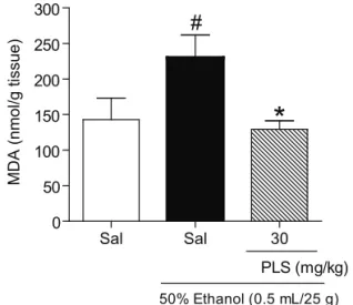
Determination of MDA levels
The gastric MDA levels in mice treated with ethanol (230.1±31.6 nmol/g of tissue) were significantly higher than those in mice treated with saline (142.8±30.5 nmol/g of tissue), and PLS (30 mg/ kg) pretreatment reversed this effect; the MDA level in the control group (129.4±11.9 nmol/g of tissue) was lower than that in the group treated with ethanol (Figure 4).
Sal Sal 30 0
50 100 150 200 250 300
PLS (mg/kg)
50% Ethanol (0.5 mL/25 g)
*
#
G
SH
µg/g
t
issu
e
Figure 3. The effect of sulfated-polysaccharide (PLS) on glutathione (GSH) levels in the gastric mucosa of mice treated with ethanol. Mice were treated by gavage with saline or PLS (30 mg/kg). After 30 min, 50% ethanol (0.5 mL/25 g) was administered to the experimental groups, while the control group was administered saline. Ethanol administration promoted a reduction in the gastric GSH levels. This effect was partially reversed when the animals were treated with PLS. The results are expressed as mean±SEM of cinco animals per group. #p<0.05 vs. saline group; *p<0.05 vs. ethanol group;
Analysis of variance (ANOVA) and Newman-Keuls test.
Role of NO and KATP channels on the gastroprotective effect of PLS
Our results show that in the animals treated with L-NAME (49.0±15.0 mm2, a non selective inhibitor of
NOS) or with glibenclamide (34.8±6.2 mm2, a drug that
blocks KATP-dependent channels), the gastroprotective effect of PLS (30 mg/kg) (3.1±0.9 mm2) was reversed.
However, the animals treated with AG (3.5±1.1 mm2, an inhibitor of iNOS) showed no significant changes (p<0.05, Figure 5).
Gastric acid secretion in 4-h pylorus-ligated mice
In this study, gastric juice obtained from pylorus-ligated mice was used to analyze the gastric biochemical parameters (Table 2). However, compared to the values obtained in the saline group, values of the animals pretreated with PLS showed no change in any biochemical parameter of the gastric juice such as the
volume, pH, or total acidity. In contrast, the volume and total acidity values in the group treated with ranitidine (a histamine [H2] antagonist) were decreased and increased by histamine compared to the corresponding values in the saline group.
Sal Sal 30
0 50 100 150 200 250 300
PLS (mg/kg)
50% Ethanol (0.5 mL/25 g)
*
#
MD
A
(n
mo
l/
g
t
issu
e
)
Figure 4. The effect of sulfated-polysaccharide (PLS) on malondialdehyde (MDA) concentration in the gastric mucosa of mice treated with ethanol. Mice were treated by gavage with PLS (30 mg/kg). After 30 min, mice in the experimental group were administered 50% ethanol (0.5 mL/25 g), and the control group was administered saline. The ethanol showed a marked increase in the gastric MDA levels. When the animals were pre-treated with PLS, this effect was reversed. The results are expressed as the means±SEM of five animals per group. #p<0.05 vs. saline group; *p<0.05 vs. ethanol group;
analysis of variance (ANOVA) and Newman-Keuls test. Discussion
Marine algae are important sources of new bioactive substances. Therefore, in the present study, we investigated the protective effect of a PLS fraction from the algae Hypnea musciformis (Wulfen) J.V. Lamour. (Gigartinales–Rhodophyta) against ethanol-induced gastric damage in mice and evaluated the role of NO/KATP channels in this effect.
Table 1. Effect of sulfated-polysaccharide (PLS) (30 mg/kg) on the microscopic gastric injury induced by ethanol.
Experimental group Hemorrhagic
damage (score, 0–4) Edema (score, 0-4)
Epithelial cell loss (score, 0-3)
Inlammatory cells (score, 0-3)
Total (score, 0-14)
Saline 0 (0–1) 0 (0–1) 0 (0–1) 0 0 (0–3)
Ethanol 3 (2–4) 2 (1–4) 2 (1–3) 0 (0–1) 7 (4–12)
Ethanol+PLS 30 mg/kg 1 (0–2)* 0 (0–1)* 1 (0–2) 0 2 (0–5)*
Sal Sal Sal L-NAME AG GLIB 0
25 50 75 100 125
PLS (30 mg/kg) 50% Ethanol (0.5 mL/25 g)
*
# #
Ma
cro
sco
p
ic
G
a
st
ri
c
L
e
si
o
n
(mm
2)
Figure 5. Evaluation of the effect of NG-nitro-L-arginine methyl ester (L-NAME), aminoguanidine (AG), and glibenclamide in sulfated-polysaccharide (PLS)-mediated gastroprotection against ethanol-induced lesion. The mice were pretreated with L-NAME (10 mg/kg, i.p.), AG (100 mg/ kg, i.p), or glibenclamide (GLIB: 10 mg/kg, i.p.). After 1 h, PLS (30 mg/kg, p.o.) was administered. After 30 min, 50% ethanol (0.5 mL/25 g) was administered to the experimental groups and saline was administered to the control group. After 1 h, the total macroscopic area of the gastric lesions was determined. The results are expressed as mean±SEM of a minimum of five animals per group. *p<0.05 vs. ethanol group; #p<0.05 vs. PLS+ethanol group; analysis of variance
(ANOVA) and Newman-Keuls test.
Table 2. The effects of sulfated-polysaccharide (PLS) on gastric acid secretion in 4 h pylorus-ligated mice.
Experimental
Group (n = 5) Volume (µL) pH
Total acid (mEq[H+]/L/4 h)
Saline 612.1±12.8 1.6±0.5 5.1±0.3
PLS (30 mg•kg-1) 598.5±70.2 1.7±0.3 5.1±0.7
Histamine 1009.1±75.3* 1.2±0.3 12.0±0.5*
Ranitidine 330.0±42.1# 2.9±0.4 2.3±0.4#
Data shown are expressed as mean±SEM (n=5). *p<0.05; #p<0.05,
vs. saline group; Analysis of variance (ANOVA) and Newman-Keuls test.
The pathogenesis of ethanol-induced gastric mucosal damage is a multifactorial process, which depends on the imbalance between the aggressive and protective factors that can occur by direct or indirect action through mediators such lipoxygenase and oxygen-derived free radicals (Abdel-Salam et al., 2001). Ethanol rapidly penetrates in the gastric mucosa, promotes injury characterized by membrane damage, erosive hemorrhage, and ulcer formation via destruction of the mucus barrier, and increases the vascular permeability and oxidative stress (Gazzieri et al., 2007; Li et al., 2008; Nassini et al., 2010). Thus, experimental models of ethanol-induced gastric ulcers have been widely used for evaluation of the gastroprotective activity of potentially active macromolecules extracted from
natural products.
Consistent with the previous findings, our results showed that administration of 50% ethanol caused macroscopic and microscopic gastric damage characterized submucosal edema, intense hemorrhage, and loss of epithelial cells (Medeiros et al., 2008; Abdon et al., 2012). However, pretreatment with PLS reversed the gastric damage and maintained the integrity of the gastric mucosa against the ethanol-induced gastropathy. Our results are consistent with those reported in previous studies on the gastroprotective effects of other sulfated-polysaccharides extracted from marine algae in experimental models of ethanol-induced gastric damage (Hwang et al., 2008; Silva et al., 2011).
The pathophysiology of ethanol-induced gastric damage in an experimental model is attributed to the creation of free radicals, increase in lipid peroxidation, and reduction in non-protein sulfhydryl (NP-SH) groups (Medeiros et al., 2009; Gomes et al., 2010). GSH is the major NP-SH of the gastric mucosa; therefore, GSH constitutes one of the main cytoprotective mechanisms against ulcer formation, acts as an important antioxidant in the maintenance of mucosal integrity (Cnubben et al., 2001; Chandranath et al., 2002). On the other hand, MDA is the inal product of lipid peroxidation (Dursun et al., 2009) and is considered as the major indicator of the lipid peroxidation process (Gawe et al., 2004).
We showed that ethanol administration decreased the gastric GSH levels and increased the MDA concentration compared to these values in the saline group. Our findings are consistent with those reported in previous studies in that oxygen free radicals play a major role in the pathogenesis of ethanol-induced gastric lesions, and reduction in oxidative stress is of primary importance in the gastroprotective effect (Pihan et al., 1987). On the other hand, PLS pretreatment reversed the decrease in the gastric GSH levels and increase in the MDA concentrations in ethanol-induced gastropathy. Several authors have shown that sulfated-polysaccharides from marine algae can act as free-radical scavengers and antioxidants; this mechanism plays an important role in preventing the free-radical-induced oxidative damage (Hu et al., 2001; Xue et al., 2001). These results suggest that the gastroprotective effect of PLS could be secondary to a decrease in the production of free radicals, which indicates possible antioxidant activity these marine algae. Therefore, sulfated-polysaccharides may be new therapeutic agents for inhibiting the damage caused by excessive free radicals.
which plays an important role in the modulation of gastric mucosal integrity via several mechanisms, including control of bicarbonate and mucus production, blood flow regulation, anti-inflammatory action, and promoting preservation and repair of gastrointestinal tract injuries (Allen et al., 1993; Andreo et al., 2006; Lanas, 2008). Endogenous NO has a dual action in the gastrointestinal tract: protective effects by constitutive NOS (cNOS or eNOS)/NO and proulcerogenic effects by iNOS/NO (Jimenez et al., 2002).
Several authors have shown that NO derived from eNOS is an important mediator that accelerates gastric ulcer healing and maintains the integrity of the gastric epithelium (Li et al., 2000). Our results showed that the gastroprotective effect of PLS were reversed using a nonselective NOS inhibitor, L-NAME. Other studies shows that L-NAME delays the healing of acute gastric injury produced by ethanol (Konturek et al., 1993) and increase the amount and intensity of lesion caused by alcohol in the stomach of rats (Nahavandi et al., 1999). Thus, we can infer that the protective effect of PLS was an NO-dependent process. In addition, we showed that iNOS is associated with severe inflammation in the ulcer tissue because high levels of NO, in special conditions, react with the superoxide anion (O2-), which lead to formation of peroxynitrite
(ONOO-) and tissue damage (Guo et al., 2003). In our experiments, we showed that pretreatment with AG, a selective iNOS inhibitor, did not alter the protective effect of PLS in ethanol-induced gastropathy. Thus, we suggest that the iNOS-derived NO associated with ethanol-induced gastric injury is not implicated in the protective effects of PLS.
Previous studies have shown that the NO plays a role in maintaining the integrity of the gastric mucosa through activation of the KATP channels (Medeiros et al., 2008; Chávez-Piña et al., 2011). In fact, NO can activate different types of K+ channels and induced mainly effects
via the opening of KATP channels (Aschcroft & Gribble, 2000). Many effects of NO, including vasodilation, inhibition of leukocyte adherence, inhibition of edema formation and gastroprotection, have been found to be inhibited by glibenclamide, a KATP channel antagonist (Daut et al., 1994; Toroudi et al., 1999; Medeiros et al., 2009). We performed pharmacological studies to show that glibenclamide significantly reversed the protective effects of PLS against ethanol-induced gastric damage, which showed the possible involvement of KATP channels in the gastroprotective effect of PLS. These results are consistent with those reported by other authors, which show that the KATP channels regulate gastric protection (Ockaili et al., 2002; Vale et al., 2007). Therefore, we suggest that the mechanism of the gastroprotective effect of PLS is dependent on the NO/KATP channels.
Another important factor that contributes
to the pathogenesis of gastric ulcers is an increase in gastric acid secretion (Goa & Monk, 1987). Studies have shown that molecules with the ability to reduce acid secretion can attenuate gastric mucosal damage induced by several aggressors of the mucosa (Patel et al., 2001; Takeuchi et al., 2003). The next step of this study was to evaluate the effect of PLS on gastric juice parameters to evaluate the possible anti-secretory action of the polysaccharide. Our results showed that PLS did not alter the gastric acid secretion. Therefore, we can infer that gastric secretion does not have any influence on the effect of PLS.
In summary, our results indicate that activation of the NO/KATP pathway plays an important role in the protective effect of the PLS against ethanol-induced gastric damage. The gastroprotective effect may also be mediated, in part, by a mechanism involving reduction of lipid peroxidation. Taken together, these data suggest that further studies should be performed to develop sulfated-polysaccharides as novel therapeutic strategies for gastropathy.
Acknowledgments
The authors gratefully acknowledge the financial support from the CNPq (Brazil).
Authors contributions
SRBD and JCR (undergraduate students) contributed in the execution of all biological experiments, running the laboratory work, analysis of the data and drafted the paper. ROS and LADN (undergraduate students) contributed to biochemical analysis. LSC (PhD student) and ALPF contributed to extraction and isolation of a sulfated-polysaccharide fraction from the algae Hypnea musciformis. ALRB contributed to histological analysis. JVRM designed the study, supervised the laboratory work and contributed to critical reading of the manuscript. All the authors have read the inal manuscript and approved the submission.
References
Abdel-Salam OME, Czimmer J, Debreceni A, Szolcs´anyi J, M´ozsik G 2001. Gastric mucosal integrity: gastric mucosal blood flow and microcirculation. Journal Physiology-Paris 95: 105-127.
Abdon APV, Souza GC, Souza LNC, Vasconcelos RP, Castro CA, Guedes MM, Júnior RCPL, Moreira RA, Monteiro-Moreira ACO, Campos AR 2012. Gastroprotective potential of frutalin, a D-galactose binding lectin, against ethanol-induced gastric lesions.
Fitoterapia 83: 604-608.
Gastroduodenal mucosal protection. Physiol Rev 73: 823-857.
Andreo MA, Ballesteros KV, Hiruma-Lima CA, Machado da Rocha LR, Souza BAR, Vilegas W 2006. Effect of Mouriri pusa extracts on experimentally induced gastric lesions in rodents: role of endogenous sulfhydryls compounds and nitric oxide in gastroprotection. J Ethnopharmacol 11: 431-441.
Aschcroft FM, Gribble FM 2000. New windows on the mechanism of action of KATP channel openers.
Trends Pharmacol Sci 21: 439-445.
Chandranath SI, Bastaki S, Singh JA 2002. Comparative study on the activity of lansoprazole, omeprazole and PD-136450 on acidified ethanol- and indomethacin-induced gastric lesions in the rat. Clin Exp Pharmacol Physiol 29: 173-180.
Chávez-Piña AE, Tapia-Álvarez GR, Reyes-Ramínrez A, Navarrete A 2011. Carbenoxolone gastroprotective mechanism: participation of nitric oxide/cGMP/KATP pathway in ethanol-induced gastric injury in the rat.
Fund Clin Pharmacol 25: 717-722.
Cnubben NHP, Rietjens IMCM, Wortelboer H, Van Zanden J, Van Bladeren PJ 2001. The interplay of glutathione-related process in antioxidant defense. Environ Toxicol Pharmacol 10: 141-152.
Corrêa MFP, Melo GO, Costa SS 2008. Natural products from plant origin potentially useful in the asthma therapy.
Rev Bras Farmacogn 18: 785-797.
Coruzzi G, Adami M, Morini G, Pozzoli C, Cena C, Bertinaria M 2000. Antisecretory and gastroprotective activities of compounds endowed with H2 antagonistic and nitric oxide (NO) donor properties. J Physiology-Paris 94: 5-10.
Daut J, Klieber HG, Cyrys S, Noack TK 1994. ATP channels and basal coronary vascular tone. Cardiovasc Res 28: 811-817.
Dursun H, Bilici M, Albayrak F, Ozturk C, Saglam MB, Alp HH, Suleyman H 2009. Antiulcer activity of fluvoxamine in rats and its effect on oxidant and antioxidant parameters in stomach tissue. BMC Gastroenterology 9: 36.
Farias WRL, Valente AP, Pereira MS, Mourão PAS 2000. Structure and anticoagulant activity of sulfated galactans. Isolation of a unique sulfated galactan from the red alga Botryocladia occidentalis and comparison of its anticoagulant action with that of sulfated galactans from invertebrates. J Biol Chem 275: 29299-29307.
Gawe S, Wardas M, Niedworok E 2004. Malondialdehyde (MDA) as a lipid peroxidation marker. Wiad Lek 57: 453-455.
Gazzieri D, Trevisani M, Springer J, Harrison S, Cottrell GS, Andre E, Nicoletti P, Massi D, Zecchi S, Nosi D, Santucci M, Gerard NP, Lucattelli M, Lungarella G, Fischer A, Grady EF, Bunnett NW, Geppetti P 2007.
Substance P released by TROV1-expressing neurons produces reactive oxygen species that mediate ethanol-induced gastric injury. Free Radical Biol Med 43: 581-589.
Goa KL, Monk JP 1987. Enprotil: a preliminary review of its pharmacodynamics and pharmacokinetic properties and therapeutic efficacy in the treatment of peptic ulcer diseases. Drugs 3: 539-559.
Gomes AS, Gadelha GG, Lima SJ, Garcia JA, Medeiros J-V.R., Havt A, Lima AA, Ribeiro RA, Brito GA, Cunha FQ, Souza MH 2010. Gastroprotective effect of heme-oxygenase 1/biliverdin/CO pathway in ethanol-induced gastric damage in mice. Eur J Pharmacol 642: 140-145.
Guo JS, Cho CH, Wang WH, Shen XZ, Cheng CL, Koo MW 2003. Expression and activity patterns of three inducible enzymes inthe healing of gastric ulcers in rats. Word J Gastroenterol 9: 1767-1771.
Hu JE, Geng MY, Zhabg JT, Jiang HD 2001. An in vitro study of the structure activity relations of sulfated polysaccharide from brown algae to its antioxidant effect. J Asian Nat Proc Rep 3: 353-358.
Hwang HJ, Kwon MJ, Kim IH, Nam TJ 2008. The effect of polysaccharide extracted from the marine alga Capsosiphon fulvescens on ethanol administration.
Food Chem Toxicol 46: 2653-2657.
Jimenez D, Martin MJ, Pozo D, Alarcon C, Md JE, Bruseghini L 2002. Mechanisms involved in protection afforded by L-argininein ibuprofen-induced gastric damage: role of nitric oxide andprostaglandins. Dig Dis Sci 47: 44-53.
Karnjanapratum S, You S 2010. Molecular characteristics of sulfated polysaccharides from Monostroma nitidum
and their in vitro anticancer and immunomodulatory activities. Int J Biol Macromol 48: 311-318.
Kim SH, Choi DS, Athukorala Y, Jeon YJ, Senevirathne M, Rha CK 2007. Antioxidant activity of sulfated polysaccharides isolated from Sargassum fulvellum. J Food Sci Nut 12: 65-73.
Konturek SJ, Brzozowski T, Majka J, Pytko-Polonczyk J, Stachura J 1993. Inhibition of nitric oxide synthase delays healing of chronic gastric ulcers. Eur J Pharmacol 239: 215-217.
Laine L, Weinstein WM 1988. Histology of alcoholic hemorrhagic gastritis: A prospective evaluation.
Gastroenterology 94: 1254-1262.
Lanas L 2008. Review: role of nitric oxide in the gastrointestinal tract. Arthritis Res Ther 10: 1-6. Li Y, Wang WP, Wang HY, Cho CH 2000. Intragastric
administrationof heparin enhances gastric ulcer healing through a nitric oxidedependentmechanism in rats. Eur J Pharmacol 399: 205-14.
injury. Can J Physiol Pharmacol 86: 675-681. Machado FLD, Kaiser CR, Costa SS Gestinari LM Soares AR
2010. Rev Bras Farmacogn 20: 441-452.
Mayer AMS, Rodriguez AD, Berlinck RGS, Hamann MT 2009. Marine pharmacology in 2005-6: Marine compounds with anthelmintic, antibacterial, anticoagulant, antifungal, anti-inflammatory, antimalarial, antiprotozoal, antituberculosis, and antiviral activities; affecting the cardiovascular, immune and nervous systems, and other miscellaneous mechanisms of action. Biochim Biophys Acta Gen Subj 1790: 283-308.
Medeiros J-VR, Gadelha GG, Lima SJ, Garcia JA, Soares PM, Santos AA, Brito GA, Ribeiro RA, Souza MH 2008. Role of the NO/cGMP/KATP pathway in the protective effects of sildenafil against ethanol-induced gastric damage in rats. Br J Pharmacol 153: 721-727.
Medeiros J-VR, Bezerra VH, Gomes AS, Barbosa ALR, Lima-Junior RCP, Soares PMG, Brito GAC, Ribeiro RA, Cunha FQ, Souza MHLP 2009.Hydrogen sulfide prevents ethanol-induced gastric damage in mice: role of ATP-sensitive potassium channels and capsaicin-sensitive primary afferent neurons. J Pharmacol Exp Ther 330: 764-770.
Mihara M, Uchiyama M 1978. Determination of malonaldehyde precursor in tissues by thiobarbituric acid test. Anal Biochem 86: 271-278.
Nahavandi A, Dehpour AR, Mani AR, Homayounfar H, Abdoli A 1999. NG-nitro-L-arginine methylester is protective against ethanol-induced gastric damage in cholestatic rats. Eur J Pharmacol 370: 283-286. Nassini R, Andre E, Gazzieri D, De Siena G, Zanasi A,
Geppetti P, MaterazziS 2010. A bicarbonate-alkaline mineral water protects from ethanol-induced hemorrhagic gastric lesions in mice. Biol Pharm Bull 33: 1319-1323.
Ockaili R, Salloum F, Hawkins J, Kukreja RC 2002. Sildenafil (Viagra) induces powerful cardioprotective effect via opening of mitochondrial KATP channels in rabbits. Am J Physiol Heart Circ Physiol 283: H1263-H1269. Pan JS, He SZ, Xu HZ, Zhan XJ, Yang XN, Xiao HM, Shi
HX, Ren JL 2008. Oxidative stress disturbs energy metabolism of mitochondria in ethanol-induced gastric mucosa injury. World J Gastroenterol 14: 5857-5867.
Park JG, Oh GT 2011. The role of peroxidases in the pathogenesis of atherosclerosis. BMB Rep 44: 497-505.
Patel HM, Santani DD, Goswami SG 2001. Evaluation of the effects of nicorandil on experimentally induced gastric ulcers. Pharmacology 63: 154-159.
Pihan G, Regillo C, Szabo S 1987. Free radicals and lipid peroxidation in ethanol- or aspirin-induced gastric mucosal injury. Dig Dis Sci 32: 1395-1401.
Rocha FD, Pereira RC, Kaplan MAC, Teixeira VL 2007. Natural products from marine seaweeds and their antioxidant potential. Rev Bras Farmacogn 17: 631-639.
Sedlak J, Lindsay RH 1968. Estimation of total, protein-bound, and nonprotein sulfhydryl groups in tissue with Ellman’s reagent. Anal Biochem 25: 1192-1205. Shay M, Kamarov SA, Fels D, Meranze D, Gruenstein H, Siplet
H 1945. A simple method for the uniform production of gastric ulceration in the rats. Gastroenterology 5: 43-61. Silva RO, Santos GMP, Nicolau LAD, Lucetti LT, Santana
APM, Chaves LS, Barros FCN, Freitas ALP, Souza MHL, Medeiros J-VR 2011. Sulfated-polysaccharide fraction from red algae Gracilaria caudata protects mice gut against ethanol-induced damage. Mar Drugs 9: 2188- 2200.
Sousa FCF, Melo CTV, Citó COM, Félix FHC,Vasconcelos SMM, Fonteles MMF, Barbosa Filho JM, Viana GSB 2008. Medicinal plants and their bioactive constituents: A scientific review of bioactivity and potential benefits in the anxiety disorders in animal models. Rev Bras Farmacogn 18: 642-654.
Souza MCR, Marques CT, Dore CMG, Silva FRF, Rocha HAO, Leite EL 2007. Antioxidant activities of sulfated polysaccharides from brown and red seaweeds. J Appl Phycol 19: 153-160.
Takeuchi Y, Kitano S, Bandoh T, Matsumoto T, Baatar D, Kai S 2003. Accelaration of gastric ulcer healing by omeprazole in portal hypertensive rats. Is its action mediated by gastrin release and the stimulation of epithelial proliferation? Eur Surg Res 35: 75-80. Toroudi HP, Rahgozar M, Bakhtiarian A, Djahanguiri B 1999.
Potassium channel modulators and indomethacin-induced gastric ulceration in rats. Scand J Gastroenterol 34: 962-966.
Vale ML, Rolim DE, Cavalcante IF, Ribeiro RA, Souza MH 2007. Role of NO/cGMP/KATP pathway in antinociceptive effect of sildenafil in zymosan writhing response in mice. Inflamm Res 56: 83-88. Wijesekara I, Pangestuti R, Kim SK 2011. Biological
activities and potential health benefits of sulfated polysaccharides derived from marine algae. Carbohyd Polym 84: 14-21.
Xue C, Fang Y, Lin H, Chen L, Li Z, Deng D, Lu CX 2001. Chemical characters and antioxidative properties of sulfated polysaccharides from Laminaria japonica. J Appl Phycol 13: 67-70.
*Correspondence
Jand-Venes R. Medeiros
LAFFEX, Universidade Federal do Piauí, Brazil
Av. São Sebastião, nº 2819, 64202-020 Parnaíba-PI, Brazil jandvenes@ufpi.edu.br