Brief communication
A handcrafted tissue microarray for a matrix arrangement of
tissue samples
João P.A. Sampaio
a, José R. Cavalcante
a, Francisco N.N. Furtado
a, Roberto C.P. Lima-Júnior
b,
Ronaldo A. Ribeiro
b,c, Paulo R.C. Almeida
a,⁎
aDepartment of Pathology and Forensic Medicine, Faculty of Medicine, Federal University of Ceará, Fortaleza, Ceará, Brazil bDepartment of Physiology and Pharmacology, Faculty of Medicine, Federal University of Ceará, Fortaleza, Ceará, Brazil cDepartment of Clinical Oncology, Haroldo Juaçaba Hospital, Cancer Institute of Ceará, Fortaleza, Ceará, Brazil
a b s t r a c t
a r t i c l e
i n f o
Article history:
Received 17 October 2013 Accepted 19 May 2014 Available online 28 May 2014
Keywords:
Tissue microarray Immunohistochemistry Diagnostic pathology Pathology
Introduction:Tissue microarray (TMA) wasfirst designed to enable more efficient immunohistochemical screening of antibodies and tissues. However, due to the high cost of commercial TMA builder instrument, such method is not affordable for many pathology laboratories. Then, methodological adaptations have been proposed in order to reduce TMA-associated cost.Methods:A manual leather puncher with an inner diameter of 2 mm was used to collect a tissue sample from the donor paraffin block. The conventional TMA method was adopted as a control group.Results:Empty paraffin recipient blocks were prepared and a standard 2-mm crochet needle was used to create 24 equidistant holes in the recipient block. Tissue cores obtained from the donor blocks were transferred to the holes in the recipient blocks and routine histopathological techniques were then per-formed.Discussion:In this study we proposed a new approach to produce TMA recipient blocks as an alternative to the conventional TMA.
© 2014 Elsevier Inc. All rights reserved.
1. Introduction
The tissue microarray (TMA) technique wasfirst described by Battifora in 1986 to enable more efficient immunohistochemical screening of antibodies and tissues (Battifora, 1986). Battifora developed the multitumor tissue block for use in clinical research on tumors. More recently, Kononen et al. developed a tissue array based on needle core sampling of donor blocks inserted into customized holes in recipient blocks (Kononen et al., 1998).
The conventional construction of a TMA block involves the use of a conventional TMA builder instrument to punch cores from donor blocks and the transference of these tissue cores to a recipient block. However, the conventional TMA builder instrument and its needles are not affordable for many pathology laboratories worldwide due to high cost. TMA technology allows the analysis of multiple biopsy specimens in a single paraffin block and saves time and antibodies. Certain low-cost methodological adaptations have been proposed (Chen & Foran, 2006; Gurgel, Dornelas, Lima-Júnior, Ribeiro, & Almeida, 2012; Miettinen, 2012; Pires, Andreiuollo, & Souza, 2006). This study was
performed to produce TMA blocks using a handcrafted TMA tech-nique as an alternative to the method proposed by our group (Gurgel et al., 2012).
2. Methods
Tissue cores were obtained from donor blocks using the puncher and technique described byGurgel et al. (2012). Briefly, a manual leather puncher (Graziano™, Brazil) with an inner diameter of 2 mm was used to collect a tissue sample from the donor paraffin block. While collecting the tissue sample, it is important to orient the 50 °C heated puncher vertically to the donor block to perform an adequate removal of the cylinder (Gurgel et al., 2012).
Empty paraffin blocks (recipient blocks) were prepared. To avoid trapping air bubbles, the cassettes were warmed to 62 °C beforefilling. After pouring, the paraffin blocks were cooled at room temperature to avoid cracks. The conventional TMA method was adopted as a control group.
3. Results
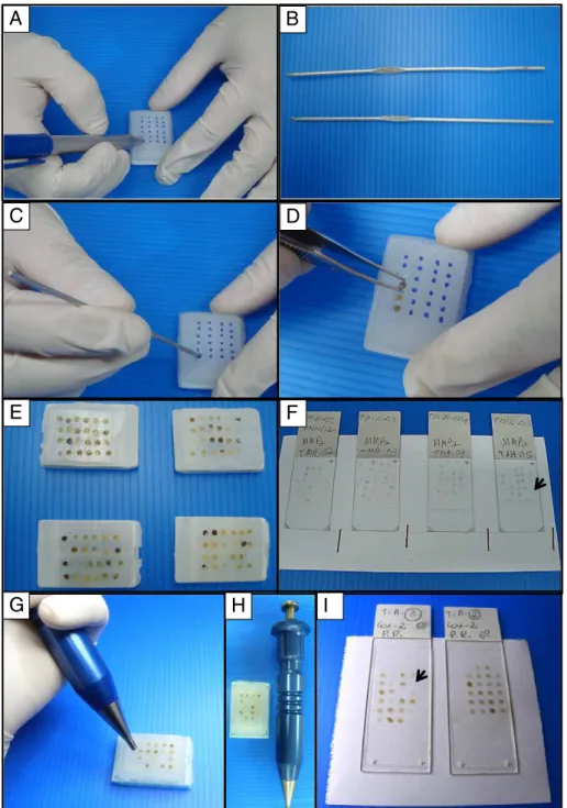
A pilot pen was used to mark 24 equidistant points (4 rows and 6 columns) on the paraffin recipient block (Fig. 1A). We then used a standard 2-mm crochet needle (Fig. 1B) to create 24 equidistant holes in the recipient block (Fig. 1C).
Journal of Pharmacological and Toxicological Methods 70 (2014) 70–72
⁎ Corresponding author at: Departamento de Patologia e Medicina Legal/FAMED/UFC, Rua Monsenhor Furtado S/N, Rodolfo Teófilo, 60.430-350 Fortaleza, CE, Brazil. Tel.: +55 85 3366 8307; fax: +55 85 3366 8316.
E-mail addresses:[email protected](R.C.P. Lima-Júnior),[email protected] (P.R.C. Almeida).
http://dx.doi.org/10.1016/j.vascn.2014.05.005 1056-8719/© 2014 Elsevier Inc. All rights reserved.
Contents lists available atScienceDirect
Journal of Pharmacological and Toxicological Methods
Tissue cores were obtained from the donor blocks (Gurgel et al., 2012) and transferred to the holes in the recipient blocks (Fig. 1D). The design of each block was detailed on a TMA map, indicating the position and identification of each core.
Once all cores were attached to the recipient paraffin blocks, melted paraffin was gently poured into the blocks to create and improve adher-ence between the tissue cores and the recipient block. The recipient blocks were incubated in an oven at 60 °C for 15 min and allowed to reach room temperature (Fig. 1E). Routine histopathological techniques were then performed to obtainfinal slides (Fig. 1F). The same procedure was performed using the conventional TMA method (Fig. 1G–I), which was adopted as a control group.
4. Discussion
In the present method, samples obtained from different donor blocks were allocated into holes previously opened in a receiver block with a standard 2-mm crochet needle. The use of a crochet needle is an alternative to bone marrow aspiration needles used in previously published TMA techniques (Hidalgo, Piña, Guerrero, Lazos, & Salcedo, 2003). By constructing holes in the recipient paraffin block, we avoided the loss of cylinder arrangement and incomplete paraffin homogenization.
Different TMA techniques have been described to facilitate the efficient immunohistochemical analysis of particular types of tumors.
A
B
C
D
E
F
G
H
I
Fig. 1.Adapted tissue microarray method. The alternative method is shown in Fig. 1A–F. The conventional method is shown in Fig. 1G–I. (A) Paraffin recipient block with 24 equidistant points. (B) A standard 2-mm crochet needle. (C) Holes being made in the recipient block. (D) Insertion of a cylinder core in the holes of recipient block. (E) Final recipient block with an array of cylinders. (F) Example of afinal immunohistochemistry slide produced by the alternative method. (G) Insertion of a cylinder core in the holes of recipient block using the conventional TMA. (H) Final recipient block and the conventional TMA builder. (I) Example of afinal immunohistochemistry slide produced by the conventional TMA method. Black arrows indicate the loss of tissue samples in both methods.
71
The method described here represents an alternative to the method de-scribed byGurgel et al. (2012)andPires et al. (2006).
However, the critical problem of the loss of tissue samples due to staining processing was observable on thefinal slide and was similar to loss using a conventional TMA, as indicated by the black arrows in Fig. 1F and I. In both conditions, about 8–15% of samples are lost while processing the slides, but specifically in the microtome step. We under-score that no additional difficulties were observable while comparing the conventional and the alternative method when a head to head assay was performed. Furthermore, the quality of slides obtained was quite similar when the methods were compared and the degree of reproducibility was noticeable considering the slides obtained inter-and/or intraassay. Thesefindings suggest that the conventional method can be replaced by the alternative method proposed in this manuscript without the loss of quality.
Many authors have tried to create handcrafted techniques that are more accessible and less expensive (Chen & Foran, 2006; Gurgel et al., 2012; Hidalgo et al., 2003; Miettinen, 2012; Pires et al., 2006). Patholo-gists are searching for new methods of constructing TMA blocks at low prices (Eguíluz, Viguera, Millán, & Pérez, 2006).
Our handcrafted TMA technique uses the puncher described in a previous study (Gurgel et al., 2012) to extract fragments from a donor block. Additionally, we use a standard 2 mm crochet needle to make holes in recipient blocks. These crochet needles can be purchased at a cost of approximately US$ 2.00. This adaptation preserves all advan-tages of machine-performed TMA, with easy handling and a low cost and requiring minimum skill and time. The method can be successfully performed in studies of solid tumors, inflammatory processes or any other indication for conventional TMA.
In summary, we described an alternative method for the construc-tion of TMA blocks that can be performed by any anatomic pathology laboratory at a low cost and that requires minimum skill and time.
Acknowledgments
We are grateful to the American Journal Experts for English edition. This work was also supported by the CNPq (Conselho Nacional de Desenvolvimento Científico e Tecnológico; Grant number: 308879/ 2009-0), CAPES (Fundação Coordenação de Aperfeiçoamento de Pessoal de Nível Superior) and FUNCAP (Fundação Cearense de Apoio ao Desenvolvimento Científico; Grant number: 11.01.00/88).
References
Battifora, H. (1986).The multitumor (sausage) block: Novel method for immunohisto-chemical antibody testing.Laboratory Investigation,55(2), 244–248.
Chen, W., & Foran, D. J. (2006).Advances in cancer tissue microarray technology: Towards improved understanding and diagnostics.Analytica Chimica Acta,564(1), 74–81. Eguíluz, C., Viguera, E., Millán, L., & Pérez, J. (2006).Multitissue array review: A
chronolog-ical description of tissue array techniques, applications and procedures.Pathology Research and Practice,202(8), 561–568.
Gurgel, D. C., Dornelas, C. A., Lima-Júnior, R. C. P., Ribeiro, R. A., & Almeida, P. R. C. (2012). An adapted tissue microarray for the development of a matrix arrangement of tissue samples.Pathology Research and Practice,208(3), 167–168.
Hidalgo, A., Piña, P., Guerrero, G., Lazos, M., & Salcedo, M. (2003).A simple method for the construction of small format tissue arrays.Journal of Clinical Pathology,56(2), 144–146.
Kononen, J., Bubendorf, L., Kallioniemi, A., Bärlund, M., Schraml, P., Leighton, S., et al. (1998).Tissue microarray for high-throughput molecular profiling of tumor speci-mens.Nature Medicine,4(7), 844–847.
Miettinen, M. (2012).A simple method for generating multitissue blocks without special equipment.Applied Immunohistochemistry & Molecular Morphology,20(4), 410–412. Pires, A.R. C., Andreiuollo, F. M., & Souza, S. R. (2006).TMA for all: A new method for the construction of tissue microarrays without recipient paraffin block using custom-built needles.Diagnostic Pathology,1, 14.