619
Brazilian Journalof otorhinolaryngology 75 (4) July/august 2009
http://www.bjorl.org / e-mail: revista@aborlccf.org.br
CASE REPORT
Rhino-orbito-cerebral
mucormycosis
Igor Teixeira Raymundo1, Beatriz Gonzalez De Araújo2,
Carina De Carvalho Costa3, Joana Pinho Tavares4,
Cleyverton Garcia Lima5, Luiz Augusto Nascimento6
INTRODUCTION
Mucormycosis is the most lethal fungal infection in humans and it is mani-fested as a quick, progressive and invasive sinusitis. It usually affects immunocompro-mized patients. Mortality is high, despite early diagnosis.1,2
We hereby present a severe case of a child with rhino-orbital-cerebral mu-cormycosis; however with a very favorable outcome after aggressive treatment.
CASE PRESENTATION
A 12-year-old female patient started with sudden abdominal pain, polyuria, polydipsia, anorexia and a drop in cons-cience level. She was diagnosed with untreated type I diabetes mellitus (diabetic acidosis), with blood sugar of 508 mg/dl upon hospital admission. After one week in the hospital, she started having eyelid ptosis, facial pain, converging strabismus, left side epistaxis and a progressive wor-sening of her general health.
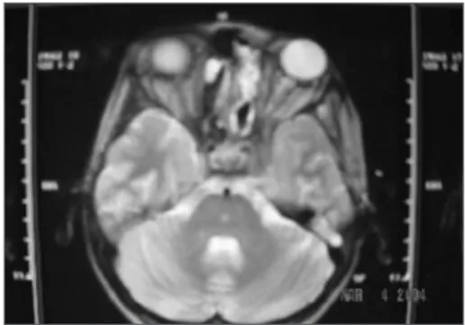
Skull and paranasal sinuses CT scan and MRI showed a soft tissue mass in the left nasal cavity, invading the ethmoidal, maxillary and cavernous sinuses and the ipsilateral orbit (Figure 1).
An endoscopic-guided tumor biop-sy showed mucormycosis. The patient was started in high doses of endovenous am-phothericin B liposome for 45 days. There was a mild improvement only. Anterior rhinoscopy showed a large brown-greenish mass occupying the entire left nasal cavity.
We decided to do a nasosinusal endoscopic exploration. During the proce-dure we noticed a large mass coming from the maxillary sinus and the left nasal cavity. We did an exhaustive surgical debridement and cleaning in the maxillary, ethmoidal and sphenoidal sinuses with hypertonic saline solution, and we also flushed the region with an amphothericin B solution. Histology revealed a zygomycosis by Mu-cor genus fungi.
The patient had a favorable outco-me and returned in one week for a review. Upon anterior rhinoscopy, we noticed a fungal mass. A new CT scan suggested
tumor recurrence. The patient was then submitted to 29 daily sessions of oxygen therapy in a hyperbaric chamber and the amphothericin B was exchanged for intra-venous itraconazole because of the long term use of the former, and she was also submitted to a rigorous diabetic control.
There was a mild improvement and stability on her condition, leading us to choose to do a second look endoscopic exploration. The left nasal cavity was broad and clear, without any evidence of infec-tion. The maxillary sinus ostium was open and there were no signs of mucormycosis.
The patient was discharged from the hospital in a stable condition and using oral itraconazole. The endoscopic view confirmed disease clearance.
She is currently asymptomatic. Her capillary glucose is strictly controlled. She has nerve palsy of the III and IV cranial nerves on the left, however with good visual acuity and no diplopia.
DISCUSSION
Mucormycosis or zygomycosis is a rare opportunistic disease caused by Mucorales fungi, which belong to the geni Mucor, Rhizopus, Rhizomucor, Absidia and Apophysomyces. The most virulent and frequent species is Rhizopus oryzae, which invades and clogs blood vessels, causing tissue schemia.1,2 This disease can
manifest itself in different clinical signs and symptoms, and the rhino-orbit-cerebral
zygomycosis is the most severe form, ma-nifesting itself usually in cases of diabetes mellitus of difficult control.3
Transmission is presumably throu-gh the inhalation pathway.
Many clinical conditions are asso-ciated with mucormycosis, but there are only two factors clearly associated with predisposing patients to the disease. The first is metabolic acidosis, and the second is disorders of neutrophils and monocytes.4
As a general rule, mucormycosis treatment is multimodal and includes the use of systemic antifungal drugs, surgical debridement and clinical control of the condition which is predisposing the patient to the disease.3
FINAL REMARKS
Proper Rhino-Orbit-Cerebral Mu-cormycosis control includes early diag-nosis, baseline disease control, treatment with antifungal systemic agents, proper sinus and orbital drainage and excision of the necrotic tissue, besides hyperbaric oxygen therapy.5
REFERENCES
1. Petrikkos G, Sklada A, Sambatakou H, Toskas A, Valopoulos G, Glannopoulou M, Katsllambros N. Mucormycosis: Ten-Year Experiance at a Terciary-Care Center in Greece. Eur J Clin Mi-crobiol Infect Dis. 2003;22:753-6.
2. Lueg EA, Ballagh RH, Forte, V. Analysis of the recent cluster of invasive fungal sinusitis at the Toronto Hospital for Sick Children. J Otolaryn-gol. 1996;25:366-70.
3. Eucker J, Sezer O, Graf B, et al. Mucormycoses. Mycoses. 2001;44:253-60.
4. Chimn RY, Diamond RD. Generations of chemo-tatic-factors by Rhizopus oryzae in the presence and absence of serum: relationship to hyphal damage mediated by human neutrophils and effects of hyperglycemia and ketoacidosis. Infect Immun. 1982;38:1123-9.
5. Hejny C, Kerrison J.B, Newman NJ, Cameron SM. Rhino-Orbital Mucormycosis in a Patient With Acquired Immunodeficiency Syndrome (AIDS) and Neutropenia. Am J Ophtalmol. 2001;132:111-2.
Keywords: amphotericin b, child, diabetes mellitus, endos-copy, mucormycosis.
1 MD. ENT resident physician - Brasilia University Hospital. 2 MD. Otorhinolaryngologist - Brasilia University Hospital. 3 MD. Otorhinolaryngologist - Brasilia University Hospital. 4 MD. ENT resident physician - Brasilia University Hospital.
5 MD. Radiologist - Brasilia University Hospital. 6 PhD. Professor - Head and Neck Surgery - Brasilia University Hospital.
Hospital Universitário de Brasília - Universidade Federal de Brasília
Send Correspondence to: Teixeira Raymundo Shin, QI 10, CONJ 10, CS 08 - Lago Norte Brasília DF 71525-100. Paper submitted to the BJORL-SGP (Publishing Management System – Brazilian Journal of Otorhinolaryngology) on June 03, 2007;
and accepted on November 11, 2007. cod. 0000
Braz J Otorhinolaryngol. 2009;75(4):619.