171 171 171 171 171 Mem Inst Oswaldo Cruz, Rio de Janeiro, Vol. 92(2): 171-179, Mar./Apr. 1997
Redescription of
Prosthenhystera obesa
(Diesing, 1850)
(Callodistomidae, Digenea) with New Host Records and
Data on Morphological Variability
Anna Kohn/*/
+, Berenice MM Fernandes/*,
Maria de Fatima D Baptista-Farias
Laboratório de Helmintos Parasitos de Peixes, Departamento de Helmintologia, Instituto Oswaldo Cruz, Av. Brasil 4365, 21045-900 Rio de Janeiro, RJ, Brasil
Prosthenhystera obesa (Diesing,1850) Travassos, 1922 from the gall bladder of Astyanax bimaculatus,
Caranx gibbosus, Galeocharax humeralis, Leporinus copelandii, Pimelodus fur, Pseudopimelodus
roosevelti, Salminus brevidens, Salminus maxillosus and from the new hosts, Cynopotamus amazonum
and Triurobrycon lundii is redescribed, demonstrating a large morphological variation, mainly in body
and testes size and shape. New hosts harbouring immature specimens of P. obesa are presented:
Brycon
sp., Leporellus vittatus, Pachyurus squamipinnis, Pimelodus clarias, Pseudoplatystoma corruscans
and Salminus hilarii. Scanning electron microscopy micrographies, original figures and measurements
of adult and immature specimens from different Brazilian hosts and localities are presented.
Key words: Prosthenhystera obesa - scanning electron microscopy - freshwater fish parasites - Brazil
Prosthenhystera obesa (Diesing, 1850) was
well described by Travassos (1922b) and Travassos
et al. (1928) and also recorded by other authors
from different hosts and localities in Brazil
(Travassos & Freitas 1941, Travassos & Kohn
1965, Kohn & Fernandes 1981, 1987, Pavanelli
et al. 1992), Mexico (Caballero & Jimmenez 1969)
and Colombia (Thatcher 1991).
The study of a large number of specimens,
collected mainly by Travassos since 1918, from
different hosts and localities in Brazil and from
specimens of Salminus maxillosus collected by
the authors from the Paraná River, in the localities
of Guaira in 1985 and Foz do Iguaçu in 1991,
allowed the redescription of the species and the
demonstration of the large morphological
variabil-ity of this parasite.
MATERIALS AND METHODS
Fish were collected with nets and kept alive
until they were examined. Prior to light
micros-copy, the specimens were fixed in AFA (alcohol,
formalin, acetic acid) under slight coverslip
pres-sure, stained in alcoholic-acid carmine, dehydrated
*Research fellow from Conselho Nacional de Desenvolvimento Científico e Tecnológico - CNPq
+Corresponding author. Fax: +55-21-260.4866/590.3545
E-mail: [email protected] Received 5 February 1996 Accepted 27 November 1996
in an alcohol series and mounted in Canada
bal-sam. Measurements were made using a calibrated
filar micrometer and are given in micrometres. For
scanning electron microscopy (SEM), specimens
were previously fixed in glutaraldeyde 2.5%,
post-fixed for 1 hr with 1% osmium tetroxide in 0.1M
phosphate buffer, dehydrated in graded ethanol,
critical point dried using CO
2, and coated with
gold. The observations were made using a Zeiss
DSM 940 scanning electron microscope. Eighty
six specimens from different hosts and localities,
were studied and deposited in the Helminthological
Collection of the Oswaldo Cruz Institute (CHIOC).
Part of the material deposited by Travassos in the
CHIOC, was preserved in Railliet and Henry’s
fluid, and part on whole mounts.
RESULTS
Prosthenhystera obesa (Diesing,1850)
Travassos, 1922
Figs 1-7
Synonym:
Pseudoprosthenhystera
micro-testiculata Kloss, 1966
Hosts: Astyanax bimaculatus (Linnaeus) (=
Cichlasoma bimaculatum (L); Brycon sp. (new
host record); Caranx gibbosus (Linnaeus);
Cynopotamus amazonum (Guenther) (new host
record);
Galeocharax humeralis (Valenciennes) (=
172 172 172 172
172 Redescription of Prosthenhystera obesa A Kohn et al.
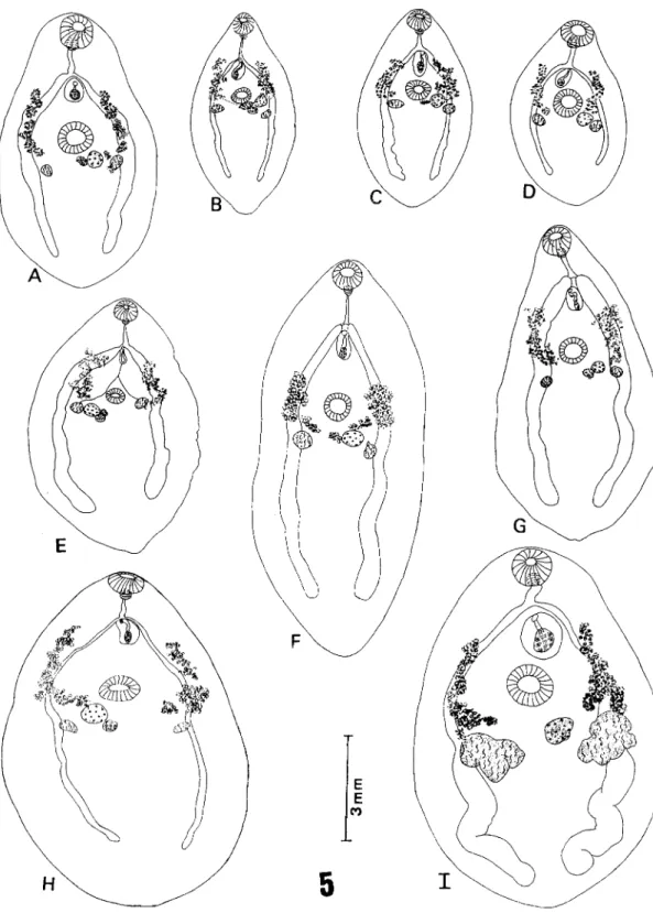
Prosthenhystera obesa from Salminus maxillosus. Fig. 1:total view, no.33.243b. Fig. 2: terminal genitalia no.33.243b. Fig.
3: photomicrography of eggs, no.33.244, bar= 0.04mm.Figs 4 A-C(figs in same scale): total view of mature specimens (uterus
173 173 173 173 173 Mem Inst Oswaldo Cruz, Rio de Janeiro, Vol. 92(2), Mar./Apr. 1997
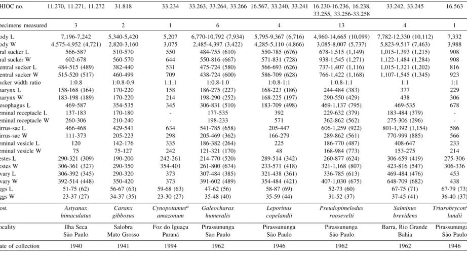
Fig. 5 (figs in same scale):Prosthenhystera obesa. Total view of mature specimens (uterus not represented) demonstrating
variation in body and testes size and shape of specimens from different hosts. A: no.16.563 from Triurobrycon lundii. B: no.
31.818a from Caranx gibbosus. C: no.33.234 from Cynopotamus amazonum. D: no. 33.256 from Pseudopimelodus roosevelti.
E: no. 11.270 from Astyanax bimaculatus. F: no. 33.266 from Galeocharax humeralis. G: no. 33.240 from Leporinus copelandii.
174 174 174 174
174 Redescription of Prosthenhystera obesa A Kohn et al.
Pimelodus fur (Lutk); Pseudopimelodus roosevelti
Borodin; Pseudoplatystoma corruscans (Agassiz)
(new
host record); Salminus brevidens (Cuvier);
Salminus
hilarii Cuvier & Valenciennes (new host
record);
Salminus maxillosus (Cuvier &
Valenciennes); Triurobrycon lundii Reinhardt (new
host record).
Site in hosts: gall bladder.
Data on the morphometric variation of 49 adult
specimens are summarized in Tables I and II and
of 21 immature specimens are presented in Table
III.
Redescription: body flattened, usually oval,
may be elliptical to round with rounded
pos-terior end, narrowing in forebody, with large
variation in size. Oral sucker rounded,
subter-minal. Ventral sucker muscular, pre-equatorial.
Suckers nearly of the same size, width ratio
within the range of 1:0.8-1:1. Pharynx small,
rounded. Oesophagus long, slender,
sur-rounded by glandular cells, extending back to
about the level of genital pore; at this point the
gut bifurcates, originating two narrow caeca,
slightly sinuous, ending blindly near the
poste-rior extremity of the body. Testes two,
irregu-lar in shape, usually smooth and smaller than
ovary, may also be lobed and larger than ovary,
lateral, symmetrically situated in ovarian zone
or below, intracaecal, caecal or extracaecal. Vas
efferentia extending from anteromedial margin
of testes to form short vas deferens in front of
acetabular level; vas deferens entering seminal
vesicle at the posterior region of cirrus-sac.
Cir-rus-sac median, between two suckers,
extend-ing back from genital pore, may reach
pre-ac-etabular zone, contains saccular or elongated
seminal vesicle, pars prostatica tubular
sur-rounded by gland cells and well developed
ejaculatory duct. Genital atrium small; common
genital pore mid-ventral, immediately below
oesophageal bifurcation. Ovary rounded to
oval, posterolateral to acetabulum, equatorial.
A large Mehlis’ gland and a well developed
seminal receptacle lie next to ovary; the
ovi-duct receives the common vitelline ovi-duct and the
Laurer’s canal which opens dorsally. Vitellaria
follicular, in two lateral fields, caecal,
intracaecal and extracaecal, may extend from
ovary level to mid-forebody. Uterus coiled,
intracaecal, caecal and extracaecal, filling most
of hindbody in mature specimens, reaching the
posterior extremity, extending to forebody, to
level of oral sucker, opening into genital atrium
through the muscular metraterm, which is
sur-rounded by glands. Eggs very small, oval,
oper-culated, present miracidium with an irregular
“v” or “8” shaped black spot (Fig. 3).
Excre-tory vesicle long and wide, Y-shaped. The
ex-cretory pore opens at the posterior end of the
body.
One specimen from S. maxillosus from the
Paraná River observed by SEM, showed round
shaped body (Fig. 6) and tegument with aciliated
papillae (Fig. 7) irregularly distributed on the
sur-face, more evident around the oral sucker and not
observed around the acetabulum, genital and
ex-cretory pores.
Scanning electron micrographs of Prosthenhystera obesa. Fig.
6: whole mount (ventral view), X 22. Fig. 7: anterior end of body with oral sucker showing ridge tegument and button-like papillae (arrow), X 150.
6
1
7
5
1
7
5
1
7
5
1
7
5
1
7
5
M
e
m
In
st
O
sw
al
d
o
C
ru
z,
R
io
d
e
Ja
n
e
ir
o
, V
o
l.
9
2
(2
),
M
ar
./A
p
r.
1
9
9
7
TABLE I
Original measurements (in mm) of Prosthenhystera obesa from Salminus maxillosus from different localities
CHIOC no. 2.147 3.279, 33.249 33.246, 33.253 33.247, 33.248 16.220, 16.224 33.252, 33.254 33.243 33.244
Specimens measured 1 2 2 2 2 3 2 1
Body L 14,027 11,092-15,040 16,315-23,160 8,757-17,412 13,615-17,712 9,517-17,900 (12,349) 7,740-7,860 15,977
Body W 8,157 4,960-7,632 6,545-8,570 6,507-7,632 9,367-10,530 6,170-9,930 (7,561) 3,497-3,572 10,155
Oral sucker L 1,305 1,045-1,091 1,387-1,530 1,076-1,137 1,137-1,290 566-1,499 (970) 733-769 1,259
Oral sucker W 1,244 1,045-1,183 1,484-1,660 1,091-1,395 1,275-1,305 877-1,407 (1,079) 806-879 1,499
Ventral sucker L 1,290 908-1,290 1,500-1,537 1,015-1,302 1,091-1,244 709-1,484 (1,023) 659-711 1,183
Ventral sucker W 1,259 1,030-1,350 1,350-1,660 1,122-1,422 1,198-1,275 908-1,575 (1,181) 689-696 1,575
Sucker width ratio 1:1 1:1 1:0.9-1:1 1:1 1:0.9-1:1 1:1 1:0.8 1:1
Pharynx L 361 260-407 271-453 466 336-438 229-535 (336) 251-306 392
Pharynx W 321 392-469 271-484 354 453-469 260-469 (335) 242-275 242
Oesophagus L 1,290 724-647 1,591-2,785 535-1,244 678-1,499 392-1,214 (692) 738-939 1,468
Seminal receptacle L 336 275 - 275 290-306 321-825 (538) 184
-Seminal receptacle W 484 275 - 392 469-535 321-525 (423) 122
-Cirrus-sac L 1,805 1,259 2,822 1,321-1,545 1,637 632-739 602 1,606
Cirrus-sac W 1,076 571 750 938-1,244 1,336 550-678 587 969
Seminal vesicle L 617 602 - - - 260-392 199-251 785
Seminal vesicle W 137 306 - - - 183-229 139-199 306
Testes L 586-602 535-602 709-900 846-1,061 423-816 244-787 (460) 326-480 602-617
Testes W 602-663 514-755 750-1,000 602-1,407 724-1,106 469-1,050 (695) 298-420 862-877
Ovary L 908 678 525-663 520-816 785-1,015 602-787 (664) 457-480 693
Ovary W 816 816 562-755 647-785 908-1,061 709-900 (788) 480-494 938
Eggs L 73-76 49-75 (58) 60-64 (62) 60-75 (68) 56-66 (62) 60-75 (65) 58-81 (70) 52-64 (57)
Eggs W 47-48 30-45 (34) 37-41 (39) 34-41 (37) 30-35 (34) 34-45 (38) 36-40 (38) 37-41 (38)
Locality Tibiriçá Lassance Porto Esperança Pirassununga Pirassununga Pirapora Guaira Foz do Iguaçu
São Paulo Minas Gerais Mato Grosso São Paulo São Paulo Minas Gerais Paraná Paraná
Date of collection 1918 1921 1922 1927 1946 1958 1985 1991
1
7
6
1
7
6
1
7
6
1
7
6
1
7
6
R
e
d
e
sc
rip
tio
n
o
f
P
ro
st
h
e
n
h
y
st
e
ra
o
b
e
sa
A
K
o
h
n
e
t a
l.
TABLE II
Original measurements (in mm) of Prosthenhystera obesa from different hosts and localities
CHIOC no. 11.270, 11.271, 11.272 31.818 33.234 33.263, 33.264, 33.266 16.567, 33.240, 33.241 16.230-16.236, 16.238, 33.242, 33.245 16.563
33.255, 33.256-33.258
Specimens measured 3 2 1 6 4 13 4 1
Body L 7,196-7,242 5,340-5,420 5,207 6,770-10,792 (7,934) 5,795-9,367 (6,716) 4,960-14,665 (10,099) 7,782-12,330 (10,112) 7,332
Body W 4,575-4,952 (4,721) 2,820-3,160 3,075 2,485-4,397 (3,422) 4,285-5,110 (4,866) 3,085-8,007 (5,737) 5,823-9,517 (7,463) 3,988
Oral sucker L 566-587 510-570 550 484-755 (610) 550-785 (676) 678-1,515 (1,149) 1,015-1,393 (1,215) 908
Oral sucker W 602-678 560-570 644 550-816 (667) 571-831 (728) 938-1,545 (1,271) 1,122-1,484 (1,284) 908
Ventral sucker L 484-515 (489) 382-440 531 475-724 (580) 566-693 (626) 737-1,407 (1,116) 1,015-1,321 (1,202) 816
Ventral sucker W 515-520 (517) 460-499 709 438-724 (600) 586-709 (628) 766-1,422 (1,168) 1,107-1,545 (1,345) 923
Sucker width ratio 1:0.8 1:0.8-0.9 1:1.1 1:0.8-1.0 1:0.8-1:1 1:0.8-1:1 1:1 1:1
Pharynx L 158-168 (164) 170-220 158 186-275 (227) 168-223 (186) 244-484 (383) 377 229
Pharynx W 183-198 (189) 170-220 214 198-290 (252) 168-225 (197) 290-550 (429) 438 306
Oesophagus L 469-587 354-535 345 306-831 (510) 183-709 (498) 469-1,137 (795) 469-535 678
Seminal receptacle L 137-183 170-180 - 177-535 392 229-632 (379) 183-484 (379)
-Seminal receptacle W 260-306 210-240 - 198-233 571 362-862 (562) 275-306 (296)
-Cirrus-sac L 466-468 429-541 634 541-785 (658) 205-447 606-1,259 (922) 801-1,392 (1,154) 586
Cirrus-sac W 111-373 205-223 298 205-469 (362) 166-279 289-862 (561) 770-999 (885) 566
Seminal vesicle L 120 142-176 335 186-382 (264) 225 186-770 (487) 408-647 233
Seminal vesicle W 75 75-127 242 121-321 (170) 48 168-984 (773) 153-275 214
Testes L 290-321 (309) 190-200 242-261 214-770 (520) 289-514 (342) 260-877 (624) 306-659 (419) 275-306
Testes W 306-361 (327) 290-350 354-401 261-800 (674) 233-571 (418) 321-1,168 (807) 423-816 (547) 306-336
Ovary L 306-392 (345) 290-320 373 307-484 (385) 321-438 (361) 336-785 (613) 469-484 (476) 453
Ovary W 392-514 (448) 350-420 373 391-602 (489) 354-484 (421) 407-1,030 (675) 648-709 (682) 438
Eggs L 51-75 (62) 56-67 (63) 59-68 (63) 47-62 (56) 58-87 (69) 52-73 (60) 67-75 (71) 67-79 (73)
Eggs W 23-37 (27) 34-37 (35) 23-30 (27) 35-48 (40) 35-59 (44) 31-52 (37) 37-45 (41) 36-40 (37)
Host Astyanax Caranx Cynopotamusa Galeocharax Leporinus Pseudopimelodus Salminus Triurobrycona
bimaculatus gibbosus amazonum humeralis copelandii roosevelti brevidens lundii
Locality Ilha Seca Salobra Foz do Iguaçu Pirassununga Pirassununga Pirassununga Barra, Rio Grande Pirassununga
São Paulo Mato Grosso Paraná São Paulo São Paulo São Paulo Bahia São Paulo
Date of collection 1940 1941 1994 1962 1946 1962 1962 1946
1
7
7
1
7
7
1
7
7
1
7
7
1
7
7
M
e
m
In
st
O
sw
al
d
o
C
ru
z,
R
io
d
e
Ja
n
e
ir
o
, V
o
l.
9
2
(2
),
M
ar
./A
p
r.
1
9
9
7
TABLE III
Original measurements (in mm) of immature specimens of Prosthenhystera obesa from different hosts and localities
CHIOC no. 16.988, 16.997 33.260 16.989, 16.990, 5.596, 12.028, 11.269 33.261 33.239, 33.250. 33.251 33.262 16.992,16.998 33.231-33.233
Specimens measured 4 1 4 4 1 1 5 1
Body L 1,560-3,381 (2,578) 2,642 1,468-2,376 (1,923) 3,102-6,573 (4,912) 3,010 7,120 2,972-6,441 (5,155) 3,880 Body W 1,015-2,285 (1,436) 1,501 923-2,030 (1,374) 1,852-3,228 (2,586) 2,500 3,180 1,612-4,247 (2,985) 1,390
Oral sucker L 261-484 (351) 373 251-345 (286) 447-550 469 831 499-831 (731) 466
Oral sucker W 298-499 (389) 373 279-345 (307) 503-678 514 831 499-862 (731) 466
Ventral sucker L 242-336 (283) - 242-279 (258) 522-632 520 816 469-800 (697) 513
Ventral sucker W 289-306 (295) - 242-317 (272) 522-693 535 755 469-800 (697) 513
Sucker width ratio 1:0.7-1 - 1:0.8-0.9 1:1 1:1 1:0.9 1:0.9-1 1:1
Pharynx L 102-153 139 84-111 (96) 168-214 (188) 153 275 137-244 (180)
-Pharynx W 112-137 121 102-135 (110) 168-205 (185) 168 290 153-260 (205)
-Oesophagus L 158-514 298 84-382 (202) 298-1,137 (654) 392 678 307-571 (470) 447
Cirrus-sac L - - 186 183-198 - 739 102-550 (416)
-Cirrus-sac W - - 130 107-183 - 306 111-423 (298)
-Testes L 56-229 (135) - 102-233 (155) 60-97 - 229-260 56-407 (235)
-Testes W 75-244 (143) - 93-195 (144) 75-78 - 198-244 45-453 (307)
-Ovary L 92-275 - 102-214 (142) - - 260 48-275 (159)
-Ovary W 76-244 - 111-214 (149) - - 198 86-321 (182)
-Host Brycon sp.a Leporellusa Pachyurusa Pimelodusa Pimelodus Pseudoplatystomaa Salminus Salminusa vittatus squamipinnis clarias fur corruscans brevidens hilarii
Locality Lagoa Juparanã Pirassununga Lagoa de Juparanã Porto Esperança Ilha Seca Pirapora Pirapora Pirassununga Espírito Santo São Paulo Espírito Santo Mato Grosso São Paulo Minas Gerais Minas Gerais São Paulo
Date of collection 1948 1947 1948 1925 1940 1957 1957 1962
178 178 178 178
178 Redescription of Prosthenhystera obesa A Kohn et al.
DISCUSSION
Diesing (1850, 1855) reported Distomum obesa
from specimens collected by Natterer from
Salminus brevidens
and Leporinus friderici
from the State of Mato Grosso and from
Xiphostoma cuvieri found in the State of Acre,
Brazil, with the following description.
“Corpus ellipticum erassum, supra planum,
subtus ventricosum. Os subterminale anticum
circulare. Acetabulum magnitudine oris,
subcentrale superum, apertura circulari. Penis
retractus, apertura genitali ampla, in medio inter
os et acetabulum. Longit. 3-7''' ; latit. 2-5''' ; erassit.
1 ½''' ”.
(Paiva in 1983 referred that the commonly named fish “dourado” from the São Francisco River belongs to the species Salminus brevidens and the ones from Southeast, Center-West and South of Brazil (Paraná and Paraguai River basins), belong to Salminus maxillosus. Considering this, S. brevidens men-tioned by Diesing (1850, 1855) from the State of Mato Grosso and by Travassos (1922a, 1922b) from the States of Mato Grosso and São Paulo, belongs to S. maxillosus.)
In 1920, during a meeting of the Brazilian
So-ciety of Sciences, Travassos proposed the new
ge-nus Prosthenhystera for D. obesum, with a
descrip-tion, without figures, of specimens from Salminus
maxillosus (= S. brevidens) and Leporinus sp. from
“Tibiriçá”, State of São Paulo (Travassos 1922a).
In another paper, Travassos (1922b) presented
original figures of P. obesa with the description
also based on histological sections.
In these papers, Travassos refers to the
mea-surements of P. obesa according to Diesing (1855)
as 3-7 mm long and 2-5 mm wide. Kloss (1966),
mentioned that these measurements must be
cor-rected to 3-7 and 2-5 austriac lines, which
corre-spond to 6.6-15.4 mm long and 4.4-11 mm wide.
In 1928, Travassos et al. published a large
pa-per about the helminthological fauna of the
fresh-water fishes of Brazil. In this paper, the authors
refer to new hosts for P. obesa, with the same data
as Travassos (1922a).
In 1941, Travassos and Freitas reported the
presence of adult specimens of P. obesa in
Astyanax bimaculatus (= C. bimaculatum). In the
present paper, original measurements of these
specimens are presented (Table II).
Pseudoprosthenhystera microtesticulata
Kloss, 1966 from
A. bimaculatus
and
A.
fasciatus,
was considered by Travassos et al. (1969) as a
syn-onym of P. obesa
.
The type specimen of P.
microtesticulata (no. 2.515) from “Museu de
Zoologia da Universidade de São Paulo” has been
examined by the authors. It represents, a very young
trematode, not well diaphanized. Parenchyma cells,
present over the entire body, and more condensed
in the borders, were erroneously described by Kloss
(1966) as vitellaria, the characteristic used by this
author to erect the new genus and species.
P. obesa presents a large variation in body
shape and size. The large range in body size of
worms from the same host and from different hosts,
from different localities, collected in different
pe-riods is demonstrated in Tables I and II.
The variation in body size of P. obesa was
also observed by Pavanelli et al. (1992) in three
specimens from S. maxillosus measuring 8.46 to
18.64 mm long by 5.65 to 9.88 mm wide.
Large morphological variation in size and shape
of body and in position and shape of testes was
already demonstrated by Travassos (1944) in other
gall bladder parasites of the family Dicrocoeliidae
as in Dicrocoelium dendriticum
, Eurytrema
coelomaticum, Lubens lubens (=E. (Lubens)
lubens), Platynosomum illiciens (= P. fastosum),
Zonorchis microrchis (Travassos 1944, pls: 3-4,
14, 17, 19-26, 27-30, 53-55).
This high variability was also confirmed in
other species of Digenea as in Mesocoelium
mo-nas by Freitas (1963), Plagiorchis koreanus and
P. verpertilionis by Groschaft and Tenora (1974),
Fasciolopsis buski by Roy and Tandon (1993),
and others.
The tegumental papillae now observed in P.
obesa by SEM, were also described in other
spe-cies as in Gorgoderina vitelliloba (see Hoole &
Michel 1981), Echinostoma revolutum (see Smales
& Blankespoor 1984), Gigantocotyle explanatum
(see Ahmad et al. 1988), Zygocotyle lunata (see
Irwin et al. 1991), Transversotrema licinum (see
Abdul-Salam & Sreelatha 1992), Fasciolopsis
buski (see Roy & Tandon 1993). At higher
mag-nification we observed in body surface, long and
slender structures resembling spines, not referred
to previously. As these structures were not
vis-ible using light microscopy or by SEM in low
magnification, we consider that further
observa-tion is required in order to confirm it.
ACKNOWLEDGEMENTS
179 179 179 179 179 Mem Inst Oswaldo Cruz, Rio de Janeiro, Vol. 92(2), Mar./Apr. 1997
REFERENCES
Abdul-Salam J, Sreelatha BNS 1992. The surface to-pography and ultrastructure of the tegument of the ectoparasitic digenean Transversotrema licinum.
Zool Anz228:48-261.
Ahmad M, Nizami WA, Hanna REB 1988. Topographi-cal studies of two digenetic trematodes of buffalo by scanning electron microscopy.ZoolAnz220: 59-64.
Caballero CE, Jimenez FG 1969. Presencia de
Prosthenhystera obesa (Diesing,1856) Travas-sos, 1920 (Trematoda, Digenea) en peces comestibles de agua dulce do Mexico. Rev Biol Trop15:283-287. Diesing KM 1850. Systema Helminthum1. 679 pp. Diesing KM 1855. Neunzehn arten von trematoden.
Denks Akad Wissen, Wien Math Naturw KI 10: 59-70.
Freitas JFT 1963. Revisão da família Mesocoeliidae Dollfus, 1933 (Trematoda). Mem Inst Oswaldo Cruz 61: 173-311.
Groschaft J, Tenora F 1974. Some remarks on the mor-phological variability of the species Plagiorchis vespertilionis (Müller,1780) and Plagiorchis koreanus Ogata,1938 (Trematoda, Plagiorchiidae) parazitizing bats. Acta Universit Agric 22: 115-130. Hoole D, Mitchell JB 1981. Ultraestructural observa-tions on the sensory papillae of juvenile and adult
Gorgoderina vitelliloba (Trematoda: Gorgoderidae).
Int J Parasitol11: 411-417.
Irwin SWB, McCloughlin TJJ, Fried B 1991. Scanning and transmission electron microscopical observations on the tegument of excysted metacercariae and adults of Zygocotyle lunata. J Helminthol65: 270-274. Kloss GR 1966. Helmintos parasitos de espécies
simpátricas de Astyanax (Pisces, Characidae). 1. Pap Avuls Dep Zool18: 189-219.
Kohn A, Fernandes BMM 1981. The adult form of
Himasthla piscicola Stunkard,1960 and other trema-todes from Brazilian freshwater fishes. J Helminthol 55: 85-87.
Kohn A, Fernandes BMM 1987. Estudo comparativo dos helmintos parasitos de peixes do rio Mogi Guassu, coletados nas excursões realizadas entre 1927 e 1985. Mem Inst Oswaldo Cruz82: 483-500. Paiva MP 1983. Peixes e pescas de águas interiores do
Brasil. Ed. Editerra, 158 pp.
Pavanelli GC, Arana S, Alexandrino de Pérez AC, Machado MH, Matushima ER, Tanaka LK, Dias PG, Sato SK 1992. Parasitose por Prosthenhystera obesa
(Diesing,1850) (Trematoda-Callodistomidae) em vesícula biliar de “dourado”, Salminus maxillosus
(Pisces-Salmininae). SIMBRAq. 7 EMBRAPOA2.
Peruibe Anais: 167-172.
Roy B, Tandon V 1993. Morphological and microtopographical strain variations among Fasci-olopsis buski originating from different geographi-cal areas. Acta Parasitol 38: 72-77.
Smales LR, Blankspoor HD 1984. Echinostoma revolutum (Froelich, 1892) Looss, 1899 and
Istimiophora melis (Schrank,1788) Luhe, 1909 (Echinostomatinae, Digenea): Scanning electron microscopy of the tegumental surfaces. J Helminthol 58: 187-195.
Thatcher VE 1991. Amazon Fish Parasites. Amazoniana 11: 263-571.
Travassos L 1922a. Contribuições para o conhecimento da fauna helmintológica brasileira - XIV. Espécies brasileiras da família Gorgoderidae Looss, 1901.
Brasil Médico36: 17-20.
Travassos L 1922b. Contribuições para o conhecimento da fauna helmintológica brasileira - XVII. Gorgoderidae brasileiras. Mem InstOswaldo Cruz 15: 220-234.
Travassos L 1944. Relatório da excursão do Instituto Oswaldo Cruz ao Município de Santa Teresa, no Estado do Espírito Santo, em agôsto e setembro de 1943. Mem Inst Oswaldo Cruz40: 121-128. Travassos L, Freitas JFT 1941. Relatório da terceira
excursão à zona da Estrada de Ferro Noroeste do Brasil, realizada em fevereiro e março de 1940. II. -Pesquisas helmintológicas. Mem Inst Oswaldo Cruz 35: 610-634.
Travassos L, Kohn A 1965. Lista dos helmintos parasitos de peixes encontrados na Estação Experimental de Biologia e Piscicultura de Emas, Pirassununga, Estado de São Paulo. Pap Avuls Dep Zool 17: 35-52. Travassos L, Artigas P, Pereira C 1928. Fauna
helmintológica dos peixes de água doce do Brasil.
Arch Inst Biol S. Paulo 1: 5-68.
180 180 180 180