www.cbpv.com.br/rbpv
Metazoan endoparasites of
Pygocentrus nattereri
(Characiformes: Serrasalminae) in the Negro River,
Pantanal, Brazil
Endoparasitas metazoários de
Pygocentrus nattereri
(Caraciformes: Serrasalminae)
no rio Negro, Pantanal, Brasil
Wagner Vicentin1*; Kelly Regina Ibarrola Vieira1; Luiz Eduardo Roland Tavares2; Fábio Edir dos Santos Costa3; Ricardo Massato Takemoto4; Fernando Paiva2
1Programa de Pós-graduação em Ecologia e Conservação, Universidade Federal de Mato Grosso do Sul – UFMS, Campo Grande, MS, Brasil
2Centro de Ciências Biológicas e da Saúde, Universidade Federal de Mato Grosso do Sul – UFMS, Campo Grande, MS, Brasil
3Laboratório de Ictiologia, Centro de Pesquisas em Biodiversidade, Universidade Estadual de Mato Grosso do Sul – UEMS, Dourados, MS, Brasil
4Núcleo de Pesquisas em Limnologia, Ictiologia e Aquicultura – NUPÉLIA, Laboratório de Ictioparasitologia, Universidade Estadual de Maringá – UEM, Maringá, PR, Brasil
Received January 7, 2013 Accepted May 6, 2013
Abstract
In the period of October 2007 to August 2008, 152 specimens of Pygocentrus nattereri were caught in the Negro
River in the Nhecolândia region, central Pantanal wetland, State of Mato Grosso do Sul, Brazil. The specimens were necropsied and a total of 4,212 metazoan endoparasites were recovered, belonging to 10 taxons: Procamallanus
(Spirocamallanus) inopinatus, Philometridae gen. sp., Eustrongylides sp., Brevimulticaecum sp., Contracaecum sp.
(Nematoda), Echinorhynchus paranensis (Acanthocephala), Leiperia gracile, Sebekia oxycephala, Subtriquetra sp. 1 and Subtriquetra sp. 2 (Pentastomida). This is the first record of two parasite species from P. nattereri: E. paranensis and L. gracile.
Keywords:Pygocentrus nattereri, Echinorhynchus paranensis, Leiperia gracile, helminths, freshwater fishes, piranhas.
Resumo
No período de outubro de 2007 a agosto de 2008, 152 espécimes de Pygocentrus nattereri foram capturados no rio Negro na região da Nhecolândia, parte central do Pantanal, Mato Grosso do Sul, Brasil. Os espécimes foram necropsiados e um total de 4.212 endoparasitas metazoários foram colhidos, pertencentes a 10 táxons: Procamallanus
(Spirocamallanus) inopinatus, Philometridae gen. sp., Eustrongylides sp., Brevimulticaecum sp., Contracaecum sp. (Nematoda), Echinorhynchus paranensis (Acanthocephala), Leiperia gracile, Sebekia oxycephala, Subtriquetra sp. 1 e
Subtriquetra sp. 2 (Pentastomida). Este é o primeiro registro de duas espécies de parasitas em P. nattereri: E. paranensis
e L. gracile.
Palavras-chave: Pygocentrus nattereri, Echinorhynchus paranensis, Leiperia gracile, helmintos, peixes de água doce, piranhas.
*Corresponding author: Wagner Vicentin
Programa de Pós-graduação em Ecologia e Conservação, Universidade Federal de Mato Grosso do Sul - UFMS, CEP 79070-900, Campo Grande, MS, Brasil
e-mail: wagnervicentin.bio@gmail.com
Introduction
Pygocentrus nattereri Kner, 1858 is a gregarious and piscivorous species (SAZIMA; MACHADO, 1990). Its wide geographical distribution includes the tropical and neotropical regions of South America (Argentina, Bolivia, Brazil, Colombia, Ecuador, Guyana, Paraguay, Peru and Uruguay), mainly in the Amazon
and Paraguay-Paraná river basins, coastal rivers in northeastern Brazil, and the Essequibo River basin (JÉGU, 2003).
Most information on its parasite fauna consists of records of 34 ectoparasite species (THATCHER, 2006; EIRAS et al., 2010). Only five described and five undescribed endoparasite species have been reported in P. nattereri: the nematodes Eustrongylides ignotus
Jäegerskiold, 1909 larvae (MORAVEC, 1998), Eustrongylides sp. larvae (BARROS et al., 2010, EIRAS et al., 2010), Contracaecum
sp. larvae (PAVANELLI et al., 2004), Procamallanus (S.)
inopinatus Travassos, Artigas & Pereira, 1928 (THATCHER, 2006), Brevimulticaecum sp. larvae (VIEIRA et al., 2010),
Procamallanus sp. (BARROS et al., 2010), Philometra nattereri n. sp. (CÁRDENAS et al., 2012); the acanthocephalan Acanthocephala fam. gen. sp. (EIRAS et al., 2010); the cestode Proteocephalus serrasalmus Rego & Pavanelli, 1990 (REGO; PAVANELLI, 1990); and larvae of the pentastomids Leiperia sp. (EIRAS et al., 2010) and Sebekia oxycephala Sambon, 1922 (REGO; EIRAS, 1989).
Because of the lack of knowledge regarding the endoparasite fauna in this host, the aim of this study was to report and describe the species of metazoan endoparasites found in P. nattereri in the Negro River, Pantanal wetland, State of Mato Grosso do Sul, Brazil.
Materials and Methods
From October 2007 to August 2008, 152 specimens of P. nattereri were collected, pithed immediately after capture and preserved under refrigeration until the moment of necropsy. The necropsy, specimen collection, preparation and conservation of the endoparasites were performed according to Eiras et al. (2006).
Hosts measured 19.89 ± 3.11 (11.4-24.8) cm in standard length and weighed 376.36 ± 179.35 (49-853) g. The fish were caught with hooks and cast nets of different mesh sizes, in the main channel of the middle Negro River (19° 34’ 29.2” S and 56° 14’ 37.1” W), a tributary of the Paraguay River in the Nhecolândia subregion of the central Pantanal wetland, state of Mato Grosso do Sul, Brazil.
Some parasite specimens were prepared for observation by means of scanning electron microscopy (SEM) according to Chiarini-Garcia (1997). Measurements of specimens in light microscopy (LM) were made with Leica (LAS LeicaTM) software. All measurements are in millimeters; means are followed by the range in parentheses.
These parasites were compared with specimens deposited in the Helminthological Collection of the Oswaldo Cruz Institute
(CHIOC). Representative specimens were deposited in the Zoological Reference Collection at the Universidade Federal do Mato Grosso do Sul, together with a host voucher specimen (ZUFMS-PIS No. 3087). Parasitological descriptors were calculated according to Bush et al. (1997). The mean values of descriptors are followed by the standard deviation (±).
Results and Discussion
Of 152 P. nattereri examined, 84% had at least one species of metazoan endoparasite: 35% were parasitized by one species, 24% by two, 16% by three, 7% by four, and 1% by five species. Ten species of endoparasites were represented by the total of 4,212 specimens.
Nematoda
Camallanidae Railliet & Henry, 1915
Procamallanus Baylis, 1923
Procamallanus(Spirocamallanus) inopinatus Travassos, Artigas & Pereira, 1928 (ZUFMS-INV = 008)
Site of infection: cecum and intestine; prevalence 22.37%; mean abundance 0.36 ± 0.82; mean intensity 1.64 ± 0.98; range of variation: 1-5.
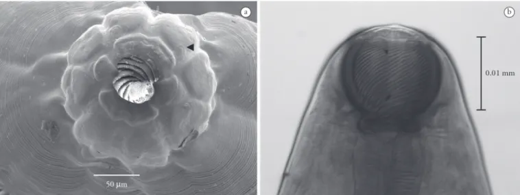
Based on 10 adult female specimens: Body 19.72 (12.35-24.28) long, 0.678 (0.546-0.806) largest width; oral opening surrounded by four cephalic papillae (Figure 1a) and two amphids; two median teeth (dorsal and ventral); buccal capsule including basal ring 0.141 (0.131-0.145) length and 0.16 (0.146-0.17) largest width, with 20-23 spiral thickenings in buccal capsule (Figure 1b); muscular-glandular oesophagus, short muscular portion 0.517 (0.49-0.59) length, 0.192 (0.17-0.21) largest width, oesophagus base 0.091 (0.08-0.1) width; glandular portion 0.869 (0.784-0.963) length, 0.241 (0.181-0.296) largest width, base 0.112 (0.095-0.143) width; nerve ring 0.276 (0.27-0.286) and excretory pore 0.365 (0.325-0.387) to anterior end; vulva 9.35
(7.562-10.48) to posterior end, equal to 45.54% (43.16-47.8%) of total body length; tail conical 0.243 (0.204-0.305) length.
Remarks: The number of spiral thickenings in the buccal capsule of the specimens examined is close to that reported by Moravec (1998). However, the number of spiral thickenings differs among several reports, and these differences are considered as intraspecific variability (MORAVEC et al., 1997). The specimens described by Petter and Dlouhy (1985) showed 14-25 spiral thickenings. Rodrigues et al. (1991) and Moravec et al. (1993, 1997) reported 15-19, 8-17 and 13-22 spiral thickenings, respectively.
Moravec et al. (1997) reported the presence of eight cephalic papillae arranged in two circles around the oral opening, differing from the present specimens which have only four cephalic papillae. Some specimens were compared to voucher specimen CHIOC 31.324, parasitic in Leporinus sp., from the Machado River, state of Rondônia, Brazil (GIESE et al., 2009). Similarities in the number of the spiral thickenings were observed, and although the voucher specimen was not measured, it was visibly longer and wider than our specimens. This voucher specimen is permanently mounted, and due to the impossibility of en face observation and the poor
conservation state of the specimen, it was not possible to observe the number of cephalic papillae.
Several fish species were reported as being parasitized by
P. (S.) inopinatus (MORAVEC, 1998; PAVANELLI et al., 2004), including P. nattereri (EIRAS et al., 2010).
Philometridae Baylis & Daubney, 1926 Philometridae gen. sp. (ZUFMS-INV = 013)
Site of infection: body cavity; prevalence 0.66%; mean abundance 0.006 ± 0.08; intensity 1.
Based on one adult female specimen: Long and slenderbody, 15.39 length, 0.71 width; cuticle smooth, thin and fragile; mouth simple, buccal capsule absent; six cephalic papillae around oral opening; female gravid, viviparous, bulky uterus. Larvae 0.337 (0.189-0.452) length and 0.017 (0.014-0.230) width.
Remarks: Genus and species determination was not possible owing to the poor condition of this specimen. The family identification is according to the features of an adult female philometrid as described by Moravec (1998): anterior end rounded, peribuccal ring absent; mouth simple, without buccal capsule; oral opening surrounded by 6-8 cephalic papillae. The vulva and vagina were not observed in this specimen; Moravec (1998) mentioned that these structures are atrophied in gravid females. Recently, Philometra nattereri was described parasitizing
P. nattereri in Amazonia (CÁRDENAS et al., 2012). Although the specimen recorded in the present study has not been identified, it differs in some aspects from the one described by Cárdenas et al. (2012), who reported a narrower range of widths (0.47-0.65) and 14 cephalic papillae. The infection site, also different, was reported as in the oculo-orbits and nasal cavity.
Dioctophymatidae Railliet, 1915
Eustrongylides Jägerskiöld, 1909
Eustrongylides sp. (ZUFMS-INV 015)
Site of infection: mesentery; prevalence 1.32%; mean abundance 0.013 ± 0.11; mean intensity 1.
Based on two larval specimens: Body brownish, 58.27 (49.5-66.69) length, 0.70 (0.692-0.72) width; anterior end rounded; buccal cavity 0.057 (0.048-0.067) long, surrounded
by cephalic papillae arranged in circle; cylindrical oesophagus 3.4 (2.2-4.7) long; nerve ring 0.50 (0.09-0.11) from anterior end; anus posterior and terminal.
Remarks: The body length and width measurements are close to those reported for larval Eustrongylides ignotus Jäegerskiold, 1909, while the oesophagus length is close to those of larvae of
E. tubifex (Nitzsch, 1819) (MORAVEC, 1998) (we found no report of the oesophagus length in E. ignotus). The buccal cavity is longer than that of E. tubifex (0.012) and closer to that reported for E. ignotus (0.060-0.097). Moravec et al. (1997), studying nematodes of freshwater fishes in Venezuela, described one larva of Eustrongylides sp., and reported a buccal cavity length (0.285) five times longer than in the present specimens.
It was not possible to count the cephalic papillae or to determine if the papillae of the outer circle were larger than the papillae of the inner circle, because of the condition of the specimens; these features enable the species determination of Eustrongylides according
to Moravec (1998) and Thatcher (2006). Species determination of larval specimens belonging to the genus EustrongylidesJägerskiöld,
1909 is problematic, and the surest way to identify the species is to obtain adult forms from experimental infection of birds, which are the definitive hosts (MORAVEC, 1998). Larval stages of this genus are recorded in a wide variety of fish species (MORAVEC, 1998; PAVANELLI et al., 2004), and larvae of E. ignotus were previously recorded in high prevalence in P. nattereri (= Serrasalmus nattereri) (MORAVEC, 1998).
Acanthocheilidae Wülker, 1929
Brevimulticaecum Mozgovoy, 1951
Brevimulticaecumsp. larvae (ZUFMS-INV = 009) Site of infection: stomach tissue and mesentery; prevalence 19.08%; mean abundance 22.36±71.24; mean intensity 117.24 ± 125.91; range of variation: 1-485.
Based on 10 larval specimens: Body 3.75 (3.07-4.1) length, 0.1 (0.072-0.137) width; ratio body length/width 2.6% (1.9-3.5); anterior end with four papillae, two subdorsal and two subventral and two tooth-like prominences; amphids between the lateral subdorsal and subventral papillae; oesophagus 0.401 (0.156-0.59) length, 0.018 (0.011-0.024) width at the base; comprising 10.48% (5.15-15.66) of body length; right ventricular lobes 0.06 (0.044-0.072) length, 0.041 (0.038-0.047) width; ratio right ventricular lobes length/oesophagus length 0.163 (0.103-0.322); left ventricular lobes 0.061 (0.043-0.075) length, 0.04 (0.036-0.044) width; ratio left ventricular lobes length/oesophagus length 0.165 (0.098-0.308); intestinal caecum 0.4 (0.329-0.486) length, 0.036 (0.032-0.04) width; nerve ring 0.183 (0.15-0.25) and excretory pore 0.198 (0.17-0.21) from anterior extremity; excretory nucleus situated about halfway along the intestinal caecum, 0.391 (0.28-0.48) from anterior extremity; conical tail 0.075 (0.041-0.08) length.
Remarks: The measurements indicate a wider range of values than reported by Moravec (1998). The first report of a fish parasitized by Brevimulticaecum sp. in the adult stage was in a stingray Paratrygon motoro (Müller & Henle, 1841) in the
Salobra River, Mato Grosso by Rego (1979). This species was described as Brevimulticaecum regoi by Sprent (1990). In the
Neotropical region, larvae of Brevimulticaecum sp. have been
1997); in Leporinus friderici (Bloch, 1794), L. lacustris Amaral Campos, 1945, L. obtusidens (Valenciennes, 1837), L. elongatus
Valenciennes, 1850 (GUIDELLI et al., 2006) and in Potamotrygon falkneri Castex & Maciel, 1963 (LACERDA et al., 2008) in the Upper Paraná River floodplain; and recently in the Upper Paraguay River basin, in Gymnotus inaequilabiatus (Valenciennes, 1839),
Hoplias aff. malabaricus (Bloch, 1794), Hemisorubim platyrhynchos
(Valenciennes, 1840), Myleus levis (Eigenmann & McAtee, 1844),
Pseudoplatystoma corruscans (Spix and Agassiz, 1829), P. nattereri
Kner, 1858 (VIEIRA et al., 2010) and Serrasalmus marginatus
Valenciennes, 1837(VIEIRA et al., 2010; VICENTIN et al., 2011). Some of the present specimens were compared with the voucher specimen CHIOC 36.977, from Potamotrygon falkneri
Castex & Maciel, 1963, collected in the Paraná River, State of Paraná, Brazil (LACERDA et al., 2008). In this specimen, the ventriculus could not be observed due to the overlapping of the body parts in the permanent mount.
Anisakidae Raillet & Henry, 1912
ContracaecumRailliet & Henry, 1912
Contracaecumsp. (ZUFMS-INV = 010)
Site of infection: stomach tissue, cecum and mesentery; prevalence 61.84%; mean abundance 3.55 ± 7.94; mean intensity 5.75 ± 9.46; range of variation: 1-74.
Based on 10 larval specimens: Body 20.19 (15.11-27.25) length, 0.695 (0.617-0.798) width; ratio body length/width 3.5% (2.5-4.08); transversely striated cuticle; larval tooth in anterior end below subventral lips; excretory pore in anterior end, between subventral lips; nerve ring 0.22 (0.16-0.28) from anterior extremity; narrow oesophagus 2.103 (1.7-2.93) long, comprising 10% (7-15) of body length; ventriculus 0.156 (0.114-0.243) length, 0.125 (0.076-0.169) width; ratio ventriculus/oesophagus length 0.07 (0.05-0.11); posterior ventricular appendix 0.51 (0.40-0.77) length; intestinal cecum 1.1 (1.42-2.74) length; ratio of ventriculus length and ventricular appendix length to intestinal cecum length 0.09 (0.05-0.14) and 0.29 (0.21-0.49), respectively; conical tail 0.20 (0.14-0.26).
Remarks: The measurements of Contracaecum sp. larvae in this study are close to those of Contracaecum sp. type 2 of Moravec (1998), with differences in the length and width of the ventriculus and in the length of the ventricular appendix (greater in the present specimens). Some specimens were compared with the voucher specimen CHIOC 35.521, from Geophagus brasiliensis (Quoy & Gaimard, 1824), collected in the Guandu River, State of Rio de Janeiro, Brazil (AZEVEDO et al., 2006), which is larger than the specimens found in the present study. Contracaecum sp. larvae have been reported in several fish species (MORAVEC et al., 1997; MORAVEC, 1998; MARTINS et al., 2005), including
P. nattereri (EIRAS et al., 2010).
Acanthocephala
Echinorhynchidae Cobbold, 1879
Echinorhynchus Zoega in Müller, 1776
Echinorhynchus paranensis Machado Filho, 1959
(ZUFMS-INV = 014)
Site of infection: intestine; prevalence 0.66%; mean abundance 0.006 ± 0.08; intensity 1.
Based on one immature adult female specimen: Trunk elongate, slender, 12.19 long, 2.31 largest width; anterior end wider than
posterior end; proboscis 1.38 long, 0.36 wide, armed with 14 longitudinal rows of 11 hooks each; proboscis receptacle double-walled, 1.56 length, 0.47 width; lemnisci 1.001 length; ovijector 0.89 length, selector apparatus 0.544 length.
Remarks: The dimensions of the trunk, proboscis and proboscis receptacle, number of rows and hooks, as well as the dimensions of the lemnisci and ovijector, are very similar to those reported for
E. paranensis by Machado Filho (1959). According to Machado Filho
(1959), the main difference observed between E. paranensis and other congeneric species is the presence of 14 longitudinal rows with 11 hooks per row on the proboscis. This specimen was considered adult, because it had well-developed reproductive structures, but non-gravid because it did not contain eggs. This is the first report of this species in P. nattereri.
Pentastomida Diesing, 1836 Sebekidae Sambon, 1922
LeiperiaSambon, 1922
Leiperia gracileDiesing, 1835
Site of infection: body cavity; prevalence 3.29%; mean abundance 0.03 ± 0.17; intensity 1.
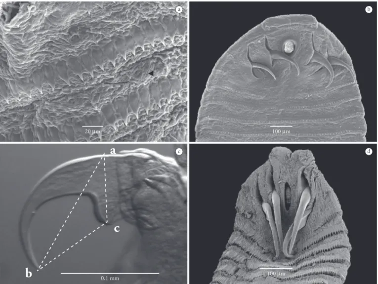
Based on 6 larval specimens: Body with anterior end rounded and tapering slightly at the posterior end, 21.53 (20.7-22.11) length, 0.89 (0.882-0.924) width, with 103 (100-107) distinct annuli with spines on their edges, spines 0.012 (0.009-0.014) length; anterior end with two pairs of double-hooks (Figure 2a), each with a principal hook and another smaller accessory; anterior pair of hooks with principal hook 0.24 (0.228-0.245) total length, and accessory hook 0.169 (0.144-0.178) total length; anterior pair of hooks, blade length (ab) = 0.201 (0.15-0.211), base length (ac) = 0.104 (0.091-0.13), base to extremity of hook blade (bc) = 0.84 (0.068-0.11) (Figure 2b); posterior pair of hooks, principal and accessory hooks measuring, respectively, 0.245 (0.24-0.26) and 0.141 (0.139-0.169) total length; blade length (ab) = 0.19 (0.18-0.234), base length (ac) = 0.098 (0.06-0.121), base to extremity of hook blade (bc) = 0.119 (0.103-0.134); oral cadre slightly chitinized, 0.253 (0.237-0.268) length, 0.131 (0.109-0.152) width, located between the pairs of hooks; anus posterior and terminal.
Remarks: The measurements of these specimens are in agreement with those reported for larval stages of L. gracile in
Salminus brevidens (syn. = S. brasiliensis, Cuvier, 1819)and are slightly larger than those for larvae found in H. malabaricus and
Brachyplatystoma sp. Bleeker, 1862 described by Rego and Eiras (1989). The principal hooks of these specimens are larger than the accessory hooks, differing from specimens found in S. brevidens
and H. malabaricus, where the accessory hooks were larger than
the principal hooks (REGO; EIRAS, 1989). Some individuals were compared to the voucher specimens CHIOC 32.446, parasitic in Brachyplatystoma sp., from the Salobra River, State of Mato Grosso, Brazil; CHIOC 30.353; parasitic in H. malabaricus, from the State of Espírito Santo, Brazil; and CHIOC 29.889a-b, parasitic in Salminus brevidens from the Salobra River (REGO; EIRAS, 1989).
them, including the body length, number of annuli (more than 100) and the characteristics of the oral cadre, although the accessory hooks visually appeared to be smaller than the principal hooks in the voucher specimens. The report of this parasite in P. nattereri
by Eiras et al. (2010) was based on the dissertation that reported the first record in this host and formed the basis of the present study, but Eiras et al. (2010) did not report any description of specimens, collection locality, or parasitological descriptors.
Sebekidae Sambon, 1922
SebekiaSambon, 1922
Sebekia oxycephalaDiesing, 1835 (ZUFMS-INV = 007) Site of infection: body cavity; prevalence 29.61%; mean abundance 0.69±1.41; mean intensity 2.33 ± 1.70; range of variation: 1-8.
Based on 10 larval specimens: Body with anterior end rounded and tapering slightly at the posterior end, 6.7 (5.136-9.463) length, 0.832 (0.68-1.03) width, with 63 (56-75) distinct annuli with spines on their edges, spines 0.022 (0.017-0.025) length (Figure 3a); anterior end with two pairs of double-hooks, each with a principal hook and another smaller accessory (Figure 3b); anterior pair of hooks with principal hook 0.084 (0.061-0.125) total length and accessory hook 0.071 (0.048-0.113) total length; blade length (ab) = 0.073 (0.057-0.102), base length (ac) = 0.046 (0.028-0.079), base to extremity of hook blade (bc) = 0.036 (0.026-0.047); posterior pair of hooks with principal and accessory hooks measuring, respectively, 0.086 (0.06-0.121) and 0.067 (0.034-0.102) total length; blade length (ab) = 0.08 (0.059-0.102), base length (ac) = 0.048 (0.038-0.06), base to
Figure 2. Micrographs of Leiperia gracile: A – SEM micrograph of anterior end, ventral view. B – LM micrograph showing the main and accessory hooks; (ab) = length of hook; (ac) = hook base; (bc) = length from base to extremity of hook blade.
extremity of hook blade (bc) = 0.052 (0.03-0.088); oral cadre slightly chitinized, 0.134 (0.115-0.16) length, 0.066 (0.047-0.078) width, located between pairs of hooks; anus posterior and terminal.
Remarks: The measurements and some morphological features of S. oxycephala are close to those reported by Rego and Eiras (1989), mainly in relation to body length, hooks and number of annuli. In this study, specimens of S. oxycephala had the principal hooks larger than the accessory hooks, differing from the description by Rego and Eiras (1989) for this same species in P. nattereri. Some of the specimens were compared to voucher specimens CHIOC 32.445, parasitic in P. nattereri, from the Cuiabá River,
State of Mato Grosso, Brazil and CHIOC 32.447, parasitic in
P. corruscans, also from the Cuiabá River (REGO; EIRAS, 1989). These specimens were similar in the shape of hooks, oral cadre, body dimensions and number of annuli.
Despite the observed similarity between the larvae of
S. oxycephala and L. gracile, these larvae can be distinguished mainly by the body size, approximately three times longer; by the number of body annuli, approximately twice as numerous in
L. gracile; and by the shape of the oral cadre, oval to elongate in
Sebekia spp. and U-shaped in Leiperia spp. (OVERSTREET et al., 1985; REGO; EIRAS, 1989; JUNKER et al., 2000).
Subtriquetridae Fain, 1961
SubtriquetraSambon, 1922
Subtriquetra sp. 1 (ZUFMS-INV = 011)
Site of infection: swim bladder; prevalence 6.58%; mean abundance 0.09 ± 0.48; mean intensity 1.5 ± 1.26; range of variation: 1-5.
Based on 10 larval specimens: Larvae with bright red body color while in the host; body elliptical, ventrally flattened and dorsally convex; anterior end wider than posterior end; body 2.10 (1.562-2.528) length, 0.651 (0.556-0.779) largest width, with 30 (28-33) distinct annuli with spines on their edges, spines 0.025 (0.022-0.029) length, chloride cells arranged in single row in each annulus (Figure 4a); anterior end with two symmetrical cephalic papillae, and two pairs of single hooks (Figure 4b); anterior pair of hooks 0.164 (0.129-0.206) total length, blade length (ab) = 0.151 (0.111-0.201), base length (ac) = 0.109 (0.073-0.138),
base to extremity of hook blade (bc) = 0.076 (0.054-0.092) (Figure 4c); posterior pair of hooks 0.174 (0.131-0.22) total length, blade length (ab) = 0.162 (0.122-0.213), base length (ac) = 0.107 (0.076-0.126), base to extremity of hook blade (bc) = 0.08 (0.057-0.107); oral cadre 0.128 (0.112-0.141) length, 0.068 (0.057-0.078) width, located between the first pairs of hooks.
Subtriquetra sp. 2 (ZUFMS-INV = 012)
Site of infection: swim bladder; prevalence 22.37%; mean abundance 0.55 ± 1.69; mean intensity 2.5±2.86; range of variation: 1-15.
Based on 10 larval specimens (Figure 4d): Larvae with whitish body while in the host; body elliptical, slightly flattened; anterior end wider than posterior; body 2.56 (2.193-3.21) length, 0.891 (0.781-1.08) largest width, with 30 (28-34) distinct annuli with spines on their edges, spines 0.029 (0.028-0.032) length; chloride cells arranged in single row in each annulus; anterior end with two symmetrical cephalic papillae, and two pairs of single hooks; anterior pair of hooks 0.211 (0.176-0.256) total length, blade length (ab) = 0.197 (0.164-0.231), base length (ac) = 0.14 (0.115-0.153), base to extremity of hook blade (bc) = 0.086 (0.077-0.101); posterior pair of hooks 0.199 (0.157-0.253) total length, blade length (ab) = 0.191 (0.154-0.237), base length (ac) = 0.133 (0.113-0.157), base to extremity of hook blade (bc) = 0.082 (0.066-0.098); oral cadre 0.153 (0.125-0.175) length, 0.097 (0.083-0.110) width, located between the first pairs of hooks.
Remarks: According to Junker and Boomker (2006), the
features observed in these two morphospecies are in agreement with those of the genus Subtriquetra Sambon, 1922 including the elliptical and dorsally flattened body, and the rounded oral cadre located between two pairs of simple, slender and sharply pointed hooks.
Records of larval stages of species of Subtriquetra parasitizing fish are scarce, and are mainly from South America, which prevented the specific identification of this morphospecies. These two morphotypes were considered distinct because of the differences in the shape of the oral cadre, hooks and spines, structures that were larger in Subtriquetra sp. 2.
Some collected specimens were compared to some of the voucher specimens of the genus Subtriquetra, CHIOC 17.797,
parasitic in H. malabaricus, from the Juparanã Lagoon, State of Espírito Santo, Brazil (TRAVASSOS; FREITAS, 1940) and CHIOC 11.424, parasitic in P. nattereri, from the Salobra River, State of Mato Grosso do Sul, Brazil (unknown publication). These parasites were similar in shape and dimensions of the body and oral cadre, and in the presence of ventral papillae on the anterior end of the body.
Acknowledgments
The authors thank Marcelo Knof, PhD, curator of the Helminthological Collection of the Oswaldo Cruz Institute. This study was partially supported by the Fundação de Apoio ao Desenvolvimento do Ensino, Ciência e Tecnologia do Estado de Mato Grosso do Sul (FUNDECT) and by the Centro de Pequisas do Pantanal (CPP). W. Vicentin and K.R.I. Vieira were supported
by student fellowships from FUNDECT and Conselho Nacional de Desenvolvimento Científico e Tecnológico (CNPq), respectively.
References
Azevedo RK, Abdallah VD, Luque JL. Ecologia da comunidade de metazoários parasitos do acará Geophagus brasiliensis (Quoy & Gaimard, 1824) (Perciformes: Cichlidae) do rio Guandu, Estado do Rio de Janeiro, Brasil. Acta Sci Biol Sci 2006; 28(4): 403-411.
Barros LA, Mateus LAF, Braum DT, Bonaldo J. Aspectos ecológicos de endoparasitos de piranha vermelha (Pygocentrus nattereri, Kner, 1860) proveniente do rio Cuiabá. Arq Bras Med Vet Zootec 2010; 62(1): 228-231. http://dx.doi.org/10.1590/S0102-09352010000100033
Bush AO, Lafferty KD, Lotz JM, Shostak AW. Parasitology meets ecology on its own terms: Margolis et al. revisited. J Parasitol 1997; 83(4): 575-583.
Cárdenas MQ, Moravec F, Fernandes BMM, Morais AM. A new species of Philometra Costa, 1845 (Nematoda: Philometridae) from the freshwater fish (red piranha) Pygocentrus nattereri Kner (Characidae) in Amazonia, Brazil. Syst Parasitol 2012; 83(2): 137-144. http://dx.doi. org/10.1007/s11230-012-9377-4
Chiarini-Garcia H. Microscopia Eletrônica de Varredura. Belo Horizonte: Universidade Federal de Minas Gerais; 1997.
Eiras JC, Takemoto RM, Pavanelli GC. Métodos de Estudo e Técnicas Laboratoriais em Parasitologia de Peixes. Maringá: Eduem; 2006. Eiras JC, Takemoto RM, Pavanelli GC. Diversidade dos parasitas de peixes de água doce do Brasil. Maringá: Clichetec; 2010.
Giese EG, Santos JN, Lanfredi RM. A new species of Camallanidae from Ageneiosus ucayalensis (Pisces: Siluriformes) from Pará State, Brazil.
J Parasitol 2009; 95(2): 407-412. http://dx.doi.org/10.1645/GE-1680.1 Guidelli G, Tavechio WLG, Takemoto RM, Pavanelli GC. Fauna parasitária de Leporinuslacustris e Leporinus friderici (Characiformes, Anostomidae) da planície de inundação do alto rio Paraná, Brasil. Acta Sci Biol Sci 2006; 28(3): 281-290. http://dx.doi.org/10.4025/actascibiolsci. v28i3.228
Jégu M. Subfamily Serrasalminae. In: Reis RE, Kullander SO, Ferraris-Junior CJ. Check List of the Freshwater Fishes of South and Central America. Porto Alegre: EdiPUCRS; 2003. p. 182-196.
Junker K, Boomker J. A check-list of the pentastomid parasites of crocodilians and freshwater chelonians. Onderstepoort J Vet Res 2006; 73(1): 27-36. http://dx.doi.org/10.4102/ojvr.v73i1.167 Junker K, Boomker J, Swanepoel D, Taraschewski H. Leiperia cincinnalis Sambon, 1922 (Pentastomida) from Nile crocodiles
Crocodylus niloticus in the Kruger National Park, South Africa, with a description of the male. Syst Parasitol 2000; 47(1): 29-41. http://dx.doi. org/10.1023/A:1006306507207
Martins ML, Onaka EM, Fenerick J Jr. Larval Contracaecum sp. (Nematoda: Anisakidae) in Hoplias malabaricus and Hoplerythrinus unitaeniatus (Osteichthyes: Erythrinidae) of economic importance in occidental marshlands of Maranhão, Brazil. Vet Parasitol 2005; 127(1): 51-59. http://dx.doi.org/10.1016/j.vetpar.2004.09.026
Moravec F. Nematodes of Freshwater Fishes of the Neotropical Region. Prague: Institute of Parasitology, Academy of Sciences of the Czech Republic; 1998.
Moravec F, Kohn A, Fernandes BMM. Nematode parasites of fishes of the Paraná River, Brazil. Part 3. Camallanoidea and Dracunculoidea.
Folia Parasitol 1993; 40(3): 211-229.
Moravec F, Prouza A, Rovero R. Some nematodes of freshwater fishes in Venezuela. Folia Parasitol 1997; 44(1): 33-47.
Overstreet RM, Self JT, Vliet KA. The pentastomid Sebekia mississipiensis
sp. n. in the American alligator and other hosts. Proc Helminthol Soc Wash 1985; 52(2): 266-277.
Pavanelli GC, Machado MH, Takemoto RM, Guidelli GM, Lizama MAP. Helminth fauna of fishes: diversity and ecological aspects. In: Thomaz SM, Agostinho AA, Hahn NS. The Upper Paraná River and Its Floodplain: Physical Aspects, Ecology and Conservation. Netherlands: Backhuys Publishers; 2004. p. 309-329.
Petter AJ, Dlouhy C. Nématodes de Poissons du Paraguay. III.
Camallanina. Description d’une espèce et d’une sous-espèce nouvelles de la famille des Guyanemidae. Rev Suisse Zool 1985; 92(1): 165-177. Rego AA. Contribuição ao conhecimento dos helmintos de raias fluviais Paratrygonidae. Rev Bras Biol 1979; 39(4): 879-890.
Rego AA, Eiras J. Identificação das larvas de Sebekia e Leiperia
(Pentastomida) histopatologia em peixes de rios. Rev Bras Biol 1989; 49(2): 591-595.
Rego AA, Pavanelli GC. Novas espécies de cestóides proteocefalídeos: parasitas de peixes não Siluriformes. Rev Bras Biol 1990; 50(1): 91-101. Rodrigues HO, Pinto RM, Noronha D. Key to the species of Brazilian
Procamallanus with general considerations (Nematoda, Camallanoidea).
Mem Inst Oswaldo Cruz 1991; 86(1): 107-113. http://dx.doi. org/10.1590/S0074-02761991000100017
Sazima I, Machado FA. Underwater observations of piranhas in western Brazil. Environ Biol Fishes 1990; 28(1-4): 17-31. http://dx.doi. org/10.1007/BF00751026
Sprent JFA. Some ascaridoid nematodes of fishes: Heterochelinae. Syst Parasitol 1990; 16(2): 149-161. http://dx.doi.org/10.1007/BF00009613 Thatcher VE. Aquatic Biodiversity in Latin America: Amazon Fish Parasites. Moscow: Pensoft; 2006.
Travassos L, Freitas JFT. Pesquisas Helmintológicas – II. Mem Inst Oswaldo Cruz 1940; 35: 610-633.
Vicentin W, Vieira KRV, Costa FES, Takemoto RM, Tavares LER, Paiva F. Metazoan endoparasites of Serrasalmus marginatus (Characiformes: Serrasalminae) in the Negro River, Pantanal, Brazil. Rev Bras Parasitol Vet 2011; 20(1): 61-63. http://dx.doi.org/10.1590/S1984-29612011000100012