Anti-inflammatory effect of a sulphated polysaccharide
fraction extracted from the red algae
Hypnea musciformis
via the suppression of neutrophil migration by the nitric
oxide signalling pathway
Tarcisio Vieira de Britoa, Rafael da Silva Prudêncioa, Adriano Bezerra Salesa,
Francisco das Chagas Vieira Júniora, Starley Jone Nogueira Candeiraa, Álvaro Xavier Francob, Karoline Sabóia Aragãob, Ronaldo de Albuquerque Ribeirob,
Marcellus Henrique Loiola Ponte de Souzab, Luciano de Sousa Chavesc, Ana Lúcia Ponte Freitasc, Jand-Venes Rolim Medeirosaand André Luiz dos Reis Barbosaa
aLAFFEX – Laboratory of Experimental Physiopharmacology, Biotechnology and Biodiversity Center Research (BIOTEC), Federal University of
Piauí-CMRV, Parnaíba,bLAFICA – Laboratory of Pharmacology of Inflammation and Cancer, Department of Physiology and Pharmacology and cLaboratory of Proteins and Carbohydrates of Marine Algae, Department of Biochemistry and Molecular Biology, Federal University of Ceará,
Fortaleza, Brazil
Keywords
inflammatory process; nitric oxide; polysaccharide
Correspondence
André Luiz dos Reis Barbosa,
BIOTEC/LAFFEX/UFPI, Av. São Sebastião, n°2819, CEP 64202-020, Parnaíba, PI, Brazil. E-mail: andreluiz@ufpi.edu.br
Received June 26, 2012 Accepted December 19, 2012
doi: 10.1111/jphp.12024
Abstract
Objectives The aim of this study was to evaluate the anti-inflammatory effect of a sulphated polysaccharide fraction (PLS) extracted from the alga Hypnea
musci-formisand investigate the possible involvement of the nitric oxide (NO) pathway
in this effect.
Methods The anti-inflammatory activity of PLS was evaluated using inflamma-tory agents (carrageenan and dextran) to induce paw oedema and peritonitis in Swiss mice. Samples of paw tissue and peritoneal fluid were removed to determine myeloperoxidase (MPO) activity, NO3/NO2 levels, and interleukin-1b (IL-1b)
level. The involvement of NO in the modulation of neutrophil migration in carrageenan-induced paw oedema or peritonitis was also investigated.
Key findings Compared with vehicle-treated mice, mice pretreated with PLS (10 mg/kg) inhibited carrageenan-induced and dextran-induced oedema; it also inhibited total and differential peritoneal leucocyte counts in a model of peritoni-tis. These PLS effects were reversed by l-arginine treatment and recovered with
the administration of a NO synthase blocker (aminoguanidine). Furthermore, PLS reduced the MPO activity, decreased IL-1b levels, and increased NO3/NO2
levels in the peritoneal cavity.
Conclusions PLS reduced the inflammatory response by modulating neutrophil migration, which appeared to be dependent on the NO pathway.
Introduction
The search for natural products with pharmacological properties has significantly contributed to the discovery of substances with important applications.[1,2] Thus, marine
algae are valuable sources of diverse structurally bioactive compounds such as carotenoids, pigments, polyphenols, enzymes, and diverse functional polysaccharides.[3,4]
Many species of seaweed (marine macroalgae) are used as food and they have also found use in traditional medicine because of their perceived health benefits. Seaweeds are rich
sources of sulphated polysaccharides, including some that have become valuable additives in the food industry because of their rheological properties as gelling and thick-ening agents (e.g. carrageenan).[5]
The red seaweed Hypnea musciformis synthesizes sul-phated polysaccharide fraction (PLS), which is composed of a backbone structure of alternatingb-d-galactosyl anda-d
-galactosyl residues. The carrageenan from H. musciformis
has been reported as being a k-carrageenan, a family of
And Pharmacology
carrageenans which has a sulphate group at the carbon 4 of theb-d-galactopyranosyl residue, and a 3,6-anhydro bridge
ona-d-galactose.[6,7]
Many types of sulphated polysaccharides are recognized as having a number of biological activities, including anticoagulant, gastroprotective, antinociceptive, and anti-inflammatory, which might give them relevance in pharmaceutical applications.[8–10] However, despite the
wealth of marine flora, the anti-inflammatory activity of sulphated polysaccharides from marine organisms have not been well explored.
The inflammatory process consists of diverse physiologi-cal and pathologiphysiologi-cal activities. A key characteristic of the inflammatory reaction is the migration of leucocytes from the blood into the tissues, which occurs in a sequence of steps. Locally produced inflammatory mediators, including tumour necrosis factor-a(TNF-a) and interleukin-1b (IL-1b), activate vascular endothelial cells and upregulate key adhesion molecules that mediate tethering, rolling, cell adhesion, and extravasation of leucocytes. The leucocytes then migrate towards the site of tissue inflammation.[11,12]
However, the nitric oxide (NO) released by either constitu-tive NO synthase (cNOS) or inducible NO synthase (iNOS) during the inflammatory process down-modulates the migration of neutrophils to inflammatory sites as a result of decreased rolling and adhesion of the neutrophils on the endothelium. In addition, the released NO plays an important major role in the apoptosis of migrated neutrophils.[13,14]
Given that marine sulphated polysaccharides are impor-tant sources of new chemical substances that may have anti-inflammatory activity and therapeutic effects, this study aimed to evaluate the anti-inflammatory effect of a sul-phated polysaccharide fraction (PLS) extracted from the red algaeH. musciformisand to investigate the possible involve-ment of the NO signalling pathway in this effect.
Materials and Methods
Extraction of the polysaccharide fraction of H. musciformis
The marine red algaeH. musciformiswas collected at Flech-eiras Beach, Trairí, Ceará, Brazil. The samples were cleaned of epiphytes, washed with distilled water and stored at -20°C. The extraction procedure of polysaccharides was performed according to Fariaset al.[15]The dried tissue (5 g)
was milled and suspended in 250 ml 0.1msodium acetate
buffer (pH 6.0) containing 510 mg papain (E. Merck, St Louis, MO, USA), 5 mmethylene diamine tetra acetic acid
(EDTA), 5 mmcysteine and incubated at 60°C for 12 h. The
residue was removed by filtration and centrifugation (2700g
for 25 min at 4°C) and the sulphated polysaccharides were precipitated by addition of 48 ml 10% cetylpyridinium
chloride (CPC, Sigma Chemical, St Louis, MO, USA). The mixture was centrifuged (2700gfor 25 min at 4°C) and the polysaccharides (k-carrageenan) in the pellet were washed with 200 ml 0.05% cetylpyridinium chloride solution, dis-solved in 174 ml 2mNaCl/ethanol (100 : 15, v/v) solution,
and precipitated with 200 ml 70% ethanol (v/v) for 12 h at 4°C. After further centrifugation (2700g; 4°C; 25 min) the precipitate was washed twice with 200 ml absolute ethanol and dried with acetone under hot air flow (60°C).
Chemical characterization of the polysaccharide fraction extracted from H. musciformis
The chemical characterization had been determined previ-ously.[6,7] Total sugar content of each fraction was
deter-mined according to the method of Duboiset al.[16]Protein
content was measured by the Bradford method.[17]Sulphate
content in polysaccharides was determined by the barium chloride-gelatin method and the monosaccharide composi-tion of red seaweed galactans was obtained by reductive hydrolysis.[18,19]
Animals
Male Swiss mice (25–35 g) were from the Central Animal Facility of the Federal University of Piauí. Experiments were approved under N°23111.011979/11-80 in 2011 by the Ethics Committee of the Federal University of Piauí, Brazil.
Drugs and reagents
The following drugs and reagents were used: carrageenan (Sigma Aldrich, St Louis, MO, USA), dextran sulphate (Sigma Aldrich), indometacin, aminoguanidine and
l-arginine (Sigma Aldrich). These drugs were dissolved in
sterile saline (0.9% NaCl).
Carrageenan-induced or dextran-induced paw oedema
Doses of PLS (2.5, 5 or 10 mg/kg, i.p.) were administered to the mice. One hour later, carrageenan (500mg per paw; 50ml) or dextran(500mg per paw; 50ml) were administered by subplantar injection into the right paw.
% inhibition of oedema
Vt Vo Control Vt Vo Treated Vt Vo Co
=( − ) −( − )
−
( ) nntrol ×100
where Vo is the basal volume and Vt is the final volume measured at the indicated times.
Myeloperoxidase activity
The extent of neutrophil accumulation in the mouse paw was measured by myeloperoxidase (MPO) activity evalua-tion. Briefly, 50–100 mg hind paw tissue was homogenized in 1 ml potassium buffer with 0.5% hexadecyltrimethylam-monium bromide for each 50 mg tissue. The homogenate was centrifuged at 40 000gfor 7 min at 4°C. MPO activity in the resuspended pellet was assayed by measuring the change in absorbance at 450 nm using o-dianisidinedihydrochloride and 1% hydrogen peroxide. The results were reported as the MPO units/mg tissue. A unit of MPO (UMPO) activity was defined as that converting 1mmol hydrogen peroxide to water in 1 min at 22°C.
Measurement of cytokine IL-1b
Samples of peritoneal fluid was collected and the level of IL-1bwas evaluated using sandwich enzyme-linked immu-noabsorbent assay (ELISA). ELISA kits for IL-1bwere from the National Institute for Biological Standards and Control (Potters Bar, UK).
These ELISA methods consistently detected a level of IL-1bover 4000 pg/ml and did not cross-react with other cytokines. The results were expressed as picograms (pg/ml) of each cytokine per peritoneal cavity washed.
Peritonitis model in mice
Mice were injected intraperitoneally with 250ml sterile saline or indometacin 10 mg/kg or PLS 10 mg/kg. One hour later, the animals were injected intraperitoneally with 250ml carrageenan (500mg per cavity) into the peritoneal cavity. Mice were killed by decapitation under anaesthesia 4 h later and the peritoneal cavity was washed with 1.5 ml heparinized phosphate buffered saline (PBS) to harvest peritoneal cells. Total cell counts were performed in a Neu-bauer chamber, and differential cell counts (100 cells total) were carried out on cytocentrifuge slides stained with hae-matoxylin and eosin. The results were presented as the number of neutrophils per ml of peritoneal exudate.
Actions of aminoguanidine andL-arginine on the inhibitory effect of PLS on
peritonitis induced by carrageenan
Mice were injected intraperitoneally with 250ml sterile saline or indometacin 10 mg/kg or PLS 10 mg/kg. Animals
were then co-treated subcutaneously with l-arginine
(500 mg/kg; 250ml) plus aminoguanidine (50 mg/kg; 250ml) or aminoguanidine (50 mg/kg; 250ml) only, and 30 min later carrageenan (500mg per cavity) was injected intraperitoneally and the neutrophil migration was deter-mined. The determination of neutrophil migration into the peritoneal cavity was done as described previously in the peritonitis model in mice.
Actions of aminoguanidine andL-arginine on the inhibitory effect of PLS on paw oedema induced by carrageenan
Mice were injected orally with 500ml sterile saline or PLS 10 mg/kg. Animals were then co-treated subcutaneously withl-arginine (500 mg/kg; 250ml) plus aminoguanidine
(50 mg/kg; 250ml) or treated subcutaneously with amino-guanidine (50 mg/kg; 250ml) only, and 30 min later carra-geenan (500mg per paw) was injected into the right plantar surface and the paw oedema was determined. The measure-ment of volume of the legs was done as described in carrageenan-induced or dextran-induced paw oedema.
Action of PLS on peritoneal fluid levels of nitric oxide (NO3/NO2) in peritonitis model
The animals received injections of 250ml sterile saline or PLS 10 mg/kg into the peritoneal cavity. One hour later, the animals were injected intraperitoneally with 250ml car-rageenan (500mg per cavity) into the same cavity. Four hours later mice were killed by decapitation under anaes-thesia and the peritoneal cavity was washed with 1.5 ml PBS to harvest samples of peritoneal fluid. Peritoneal fluid of animals was incubated in a microplate with nitrate reductase (0.016 U per well) for 12 h to convert NO3 to
NO2. Nitric oxide production was determined by
measur-ing nitrite concentrations in an ELISA plate reader at 540 nm using the Griess method.[21]Results were expressed
as micromoles of nitrite using the internal standard curve.
Statistical analysis
Results are expressed as mean⫾SEM from at least five animals per group. Statistical analysis was performed using analysis of variance followed by Bonferroni post hoc test, when appropriate. Statistical significance was set atP<0.05.
Results
Effect of PLS on carrageenan-induced paw oedema in mice
surface) induced severe paw oedema within 1 h of injection and was maintained until 4 h after injection. Indometacin (10 mg/kg) administration significantly decreased paw oedema throughout the experimental period (*P<0.05), with maximal inhibition of 95.74%. Similarly, PLS (5 and 10 mg/kg, i.p.) induced long-lasting inhibition of paw oedema at all intervals. At 3 h, compared with the carra-geenan group, the animals pretreated with 5 and 10 mg/kg PLS showed 74.46% and 93.62% reduction in oedema, respectively. Table 1 shows that PLS prevented carrageenan-induced paw oedema (500mg per paw/50ml) in a dose-dependent manner, with maximal inhibitory effect exerted at a dose of 10 mg/kg (3 h: 0.006⫾0.006 ml; 4 h: 0.012⫾0.005 ml). Therefore, this dose was selected for studying the possible mechanisms of action involved in PLS-mediated decrease in inflammatory response.
Effect of sulphated polysaccharide on paw oedema induced by dextran
Figure 1 shows that injection of dextran (0.0960⫾ 0.010 ml) resulted in an increase in oedema over time, peaking approximately 30 min after injection. By contrast, saline injected in the paw did not induce paw oedema. Pre-treatment of the animals with PLS (10 mg/kg) 30 min before dextran injection effectively inhibited oedema (inhi-bition at 30 min: 97.91%; Figure 1).
Effect of sulphated polysaccharide fraction on carrageenan-induced myeloperoxidase activity in paw tissue
Figure 2 shows that PLS (10 mg/kg) inhibited neutrophil infiltration, which was evident from the MPO activity measured in the mouse paws. The carrageenan subplantar determined MPO activity in the concentration of 37.39⫾8.23 UMPO/mg plantar tissue, while the group treated with PLS presented an activity of 4.73⫾ 0.68 UMPO/mg plantar tissue, which was equivalent to a reduction of 87.34%.
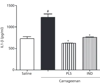
Effect of sulphated polysaccharide fraction on carrageenan-induced cytokine
production in peritonitis
The intraperitoneal administration of carrageenan was found to induce a marked increase in IL-1bconcentrations in the peritoneal exudate (Figure 3). The level of IL-1bin the peritoneal cavity of control animals (saline group) was 713⫾35.3 pg/ml and increased to 1230.0⫾76.94 pg/ml after carrageenan injection. Compared with the carra-geenan group, the animals pretreated with PLS (10 mg/kg, i.p.) showed significantly decreased IL-1b peritoneal con-centration (621.1⫾9.2 pg/ml).
Table 1 Effect of sulphated polysaccharide fraction on carrageenan-induced paw oedema in mice
Treatment
Dose (mg/kg)
Paw oedema (ml)
1 h 2 h 3 h 4 h
Control (carrageenan) 0.086⫾0.004 0.088⫾0.008 0.094⫾0.002 0.104⫾0.004
Saline 0.008⫾0.003* 0.012⫾0.005* 0.008⫾0.003* 0.004⫾0.002*
Indometacin 10 0.022⫾0.005* (74.42%) 0.004⫾0.400* (95.44%) 0.004⫾0.002* (95.74%) 0.016⫾0.005* (84.61%) PLS 2.5 0.036⫾0.010* (58.14%) 0.066⫾0.019 (25.00%) 0.060⫾0.011* (36.17%) 0.080⫾0.018 (23.076%) 5 0.036⫾0.008* (58.13%) 0.018⫾0.008* (79.54%) 0.024⫾0.009* (74.46%) 0.018⫾0.009* (82.69%) 10 0.030⫾0.008* (65.12%) 0.008⫾0.003* (90.91%) 0.006⫾0.006* (93.62%) 0.012⫾0.005* (88.46%)
PLS, sulphated polysaccharide fraction. Values of paw oedema are expressed as mean⫾SEM (n=5). The % inhibition of paw oedema is indicated in parentheses. *P<0.05 compared with control (one-way analysis of variance followed by the Bonferroni post hoc test).
0.15
0.10
0.05
Change in volume (
µ
l)
0.00
Time
Saline Dextran PLS 10 mg/kg IND
0 1 2
*
* *
*
* ** * *
3 4 5
Anti-inflammatory effect of sulphated polysaccharide fraction on
carrageenan-induced peritonitis in mice
As shown in Figure 4a, compared with the carrageenan group, the PLS-administered group showed significantly reduced peritoneal leucocyte count (7970.0⫾1078.0 vs 550.0¥103
⫾97.47¥103cells/ml). Furthermore, the same
50
#
*
Carrageenan (500 µg per paw)
Saline PLS IND
* 40
30
UMPO/mg of tissue
20
10
0
Figure 2 Effect of sulphated polysaccharide fraction on carrageenan-induced myeloperoxidase activity in paw tissue. Saline (s.c.) or carra-geenan (500mg per paw) was injected into the plantar surface of mice. One hour before this injection, animals had been treated with indometacin (IND; 10 mg/kg, i.p.) or sulphated polysaccharide fraction (PLS; 10 mg/kg, i.p.). Myeloperoxidase (MPO) activity was detected in the paw tissue after 4 h. The results are expressed as the mean⫾SEM MPO units (UMPO)/mg of tissue. *P<0.05 compared with carra-geenan group; #P<0.05 compared with saline plus carrageenan group with saline group. Statistical analysis was performed using analysis of variance followed by Bonferroni post hoc test.
#
*
*
Carrageenan
Saline PLS IND
1500
1000
500
0
IL1-β
(pg/ml)
Figure 3 Effect of sulphated polysaccharide fraction on carrageenan-induced cytokine production in peritonitis. The level of interleukin (IL)-1b in the peritoneal cavity was measured 4 h after carrageenan injection. Mice were orally administered sulphated polysaccharide frac-tion (PLS; 10 mg/kg) or indometacin (IND; 10 mg/kg), followed by injection of 250ml carrageenan (500mg per cavity, i.p.) after 1 h. Each point represents the mean⫾SEM values obtained from five animals. *P<0.05 compared with carrageenan group; #P<0.05 compared with saline group. Statistical analysis was carried out using one-way analysis of variance followed by Bonferroni post hoc test.
#
* *
Carrageenan (500 µg per cavity)
Saline PLS 10 IND
10000 (a)
8000
6000
4000
2000
0
Total leucocytes (
×
10
3 ml)
#
*
*
Carrageenan (500 µg per cavity)
Saline PLS 10 IND
(b) 6000
4000
2000
0
Neutrophils
×
10
3 ml per cavity
dose of PLS significantly reduced neutrophil migration into the peritoneal cavity (208.6¥103
⫾46.84¥103cells/
ml; Figure 4b) compared with that in the carrageenan group (4679.0¥103
⫾317.3¥103cells/ml). This result was
consistent with the fact that neutrophils are the most abun-dant cells in primary inflammatory exudates.
Effect of aminoguanidine andL-arginine on the inhibitory effect of sulphated
polysaccharide fraction on neutrophil migration
Figure 5a shows that aminoguanidine when co-admini-stered with PLS plus carrageenan increased the leucocyte count (4800⫾738.7¥103cells/ml) relative to that in the
PLS plus carrageenan group (1325.0⫾78.26¥103cells/
ml). l-Arginine when co-administered with PLS and
aminoguanidine decreased the leucocyte count (425.0⫾156.1¥103cells/ml) relative to that in the
amino-guanidine plus PLS group (4800⫾738.7¥103cells/ml).
The same dose of aminoguanidine also significantly increased neutrophil migration into the peritoneal cavity (Figure 5b; 4788.0⫾737¥103cells/ml) relative to that in
the PLS plus carrageenan group (1290.0¥103
⫾85.73¥ 103cells/ml). When co-administered with PLS and
amino-guanidine, l-arginine resulted in a decrease in the
neu-trophil count (424.0⫾154.1¥103cells/ml) relative to that
in the aminoguanidine plus PLS group (4788.0⫾737¥ 103cells/ml).
Effect of aminoguanidine andL-arginine on the inhibitory effect of sulphated
polysaccharide fraction on
carrageenan-induced paw oedema
As shown in Figure 6, when co-administered with PLS, aminoguanidine increased the volume of paw oedema (first hour: 0.0575⫾0.0014 ml; second hour: 0.0500⫾0.009 ml; third hour: 0.0460⫾0.006 ml) relative to the volumes in the PLS plus carrageenan group (first hour: 0.002⫾0.001 ml; second hour: 0.000⫾0.000 ml; third hour: 0.000⫾0.000 ml).l-Arginine when co-administered
with PLS and aminoguanidine decreased the volume of paw oedema (first hour: 0.005⫾0.004 ml; second hour: 0.004⫾0.002 ml; third hour: 0.0025⫾0.002 ml) relative to the volumes in the aminoguanidine plus PLS group.
Effect of sulphated polysaccharide fraction on peritoneal fluid levels of nitric oxide (NO3/NO2) in a peritonitis model
As shown in Figure 7, the PLS plus carrageenan group showed an increased level of NO3/NO2in peritoneal fluid
(0.1022⫾0.006mm) relative to that in the carrageenan
group (0.756⫾0.003mm) or the saline group (0.0778⫾ 0.005mm).
Discussion
Natural products of algae origin are used in folk medicine all over the world and exhibit a wide range of
pharmaco-(a) 8000
6000
4000
2000
0
Total leucocytes (
×
10
3 ml)
(b) 6000
4000
2000
0
Neutrophils
×
10
3 ml per cavity
*
* #
#
a
a
b* c*
b* c*
AG PLS Carrageenan
Saline L-arg IND
AG PLS Carrageenan
Saline L-arg IND
logical activities. Over the years, natural products have con-tributed enormously to the development of important therapeutic drugs used currently in modern medicine.[22,23]
Polysaccharides extracted from algae can play a relevant role in biomedical and pharmaceutical applications, particularly in the field of drug delivery. In this study, we sought to investigate the possible anti-inflammatory effect of PLS extracted from the marine red algaeH. musciformisby using pharmacological tools and molecular procedures.
Our results clearly demonstrated that PLS has an anti-inflammatory effect in mice models of inflammation (paw oedema, peritonitis, MPO, cytokine levels). The administra-tion of PLS (5 and 10 mg/kg, i.p.) induced long-lasting inhibition of paw oedema at all intervals. PLS prevented carrageenan-induced and dextran-induced paw oedema. Carrageenan-induced paw oedema involves several chemi-cal mediators, including histamine, serotonin, bradykinin, and prostaglandins.[24,25] In this model, the oedema is
believed to be biphasic, with the first phase being mediated by the release of histamine and serotonin, followed by the subsequent release of bradykinin, NO, and prostaglandins. The late oedema phase is dependent on cytokine produc-tion by resident cells and neutrophil infiltraproduc-tion.[26–28] In
contrast, dextran-induced paw oedema is mediated by increased vascular permeability induced by mast cell degranulation of histamine and serotonin.[29]The
oedema-tous fluid induced by dextran injection contains little protein and few neutrophils.[30]Therefore, we can infer that
the anti-oedematogenic action of PLS might be because of the differential inhibition of the mediators involved in inflammatory events and modulation of neutrophil infiltra-tion into the inflamed plantar tissue.
The paw inflammatory response induced by carrageenan was accompanied by an intense neutrophil infiltrate.[31,32]
MPO activity is commonly considered an indicator of neu-trophil infiltration. Our results demonstrated that PLS (10 mg/kg) inhibited neutrophil infiltration, which was evident from the reduced MPO activity measured in the mouse paws. These results suggested that the anti-oedematogenic effect of PLS was related to inflammatory events involving neutrophil migration.
The late oedema phase induced by carrageenan is known to be dependent on cytokine production by resident cells and neutrophils.[26–28] Our results demonstrated that PLS
decreased the level of IL-1bin the peritoneal fluid.
IL-1b is a potent pro-inflammatory cytokine that has multiple effects, including activation of inflammatory cells, induction of several inflammatory proteins, cytotoxicity,
Change in volume (ml)
0.15
0.12
0.09
0.06
0.03
0.00
Saline Carrageenan PLS
AG
AG + L-arginine
* * *
* *
* **
# # #
Time
0 1 2 3 4 5
Figure 6 Effect of aminoguanidine and L-arginine on the inhibitory effect of sulphated polysaccharide fraction on carrageenan-induced paw oedema. Paw oedema was induced by carrageenan injection (500mg per paw/50ml) into the plantar right paw: paw volume was measured 0, 1, 2, 3, and 4 h after carrageenan injection. Sulphated polysaccharide fraction (PLS; 10 mg/kg, i.p.) was administered 1 h before the inflammatory stimulus. The mice were then cotreated with L-arginine (500 mg/kg, s.c.) plus aminoguanidine (AG; 50 mg/kg, s.c.) or treated with aminoguanidine (AG; 50 mg/kg, s.c.) alone. After 30 min, carrageenan (500mg per paw) was injected into the plantar surface. Values are given as mean⫾SEM (n=5). #P<0.05 compared with PLS plus carrageenan plus L-arginine group; *P<0.05 compared with carrageenan group or the AG+PLS+carrageenan group. Statisti-cal analysis was performed using analysis of variance followed by Bon-ferroni post hoc test.
0.15
0.10
0.05
0.00
NO
3
/NO
2
(
µ
M
)
*
Saline PLS 10
Carrageenan
Figure 7 Effect of sulphated polysaccharide fraction on serum levels of nitric oxide (NO3/NO2) in a peritonitis model. Carrageenan was
oedema formation, and neutrophil migration.[33]On these
effects, we could infer that the anti-inflammatory effect of PLS might occur through the inhibition of cytokines involved in carrageenan-induced peritonitis.
In the neutrophil migration, our results demonstrated that PLS significantly reduced peritoneal leucocyte and neu-trophil counts. Carrageenan induces neuneu-trophil migration into the mouse peritoneal cavity through an indirect mechanism that involves the activation of macrophages and the release of cytokines into the peritoneal cavity.[34]In this
study, we have shown for the first time that PLS administra-tion prevented the carrageenan-induced increase in perito-neal levels of IL-1b. These results suggested that the anti-inflammatory effect of PLS may occur through the inhibition of cytokines involved in carrageenan-induced peritonitis.
During the inflammatory process, concomitant with the release of neutrophil chemotactic factors such as cytokines and chemokines, NO is produced by either cNOS or iNOS and results in the down-modulation of neutrophil recruit-ment to the inflammatory site.[35,36]Based on this
informa-tion we verified the influence ofl-arginine/NOS pathway in
the neutrophil migration-induced by carrageenan. Our results demonstrated that aminoguanidine when co-administered with PLS increased the leucocyte and neu-trophil counts, and l-arginine when co-administered with
PLS and aminoguanidine decreased leucocyte and neu-trophil counts.
The effects of the NOS inhibitors on neutrophil migra-tion appear to be a consequence of the inhibimigra-tion of the
l-arginine/NOS pathway because the enhancement of
neu-trophil migration was reversed by co-treatment with
l-arginine, the NOS substrate. The fact that the inhibition
of NO production enhances neutrophil migration suggests that during the inflammatory process, simultaneous with the release of neutrophil chemotactic factors, NO is pro-duced and that it down-modulates the recruitment of neu-trophils to the inflammatory site.[37,38]On the basis of these
results, we can infer that thel-arginine/NOS pathway
par-ticipated in the anti-inflammatory effect of PLS by modu-lating the migration of neutrophils into the inflammatory site.
To strengthen our hypothesis, we performed the carrageenan-induced paw oedema experiment with
l-arginine/NOS pathway modulation. Our results clearly
demonstrated that aminoguanidine when co-administered with PLS increased the volume of paw oedema and
l-arginine when co-administered with PLS and
aminogua-nidine decreased the carrageenan-induced paw oedema.
The late oedema phase induced by carrageenan is known to be dependent on cytokine production by resident cells, neutrophil infiltration, the release of neutrophil-derived mediators, as well as the production of neutrophil-derived free radicals, such as hydrogen peroxide, superoxide, and hydroxyl radicals, and the production of malondialdehyde is due to the attack of plasma membranes by free radicals.[26–28,39,40]Our results suggested that the
administra-tion of PLS may lead to increased producadministra-tion of endog-enous NO, which results in a reduced inflammatory response by decreasing the migration of neutrophils to the focus of the inflammatory site.
In the evaluation of peritoneal fluid levels of NO (NO3/
NO2) in a mouse peritonitis model, our results
demon-strated that PLS increased serum level of NO3/NO2 in
peritoneal fluid. The measurement of NO3/NO2 levels is a
method that is used to quantify endogenous NO produc-tion.[41]The results obtained from this study suggested that
during the inflammatory process, NO released by either cNOS or iNOS, during the PLS administration, down-modulated the migration of neutrophils to inflammatory sites.
Conclusions
PLS effectively reduced carrageenan-induced and dextran-induced paw oedema in mice, as well as carrageenan-induced peritonitis. Moreover, PLS modulated the production and release of pro-inflammatory cytokines and caused an increase in the levels of NO. This anti-inflammatory effect of PLS appeared to be mediated by the activation of the l-arginine/NOS pathway. These
observa-tions raise the possibility that the administration of polysac-charides may represent new strategies for the treatment of inflammatory disease.
Declarations
Conflict of interest
The Author(s) declare(s) that they have no conflicts of interest to disclose.
Funding
We thank UFPI/CNPq for fellowship support.
Acknowledgement
References
1. Sousa FCFet al. Medicinal plants and their bioactive constituents: a scien-tific review of bioactivity and poten-tial benefits in the anxiety disorders in animal models. Rev Bras Farmacogn 2008; 18: 642–654.
2. Corrêa MFP et al. Natural products from plant origin potentially useful in the asthma therapy.Rev Bras Farma-cogn2008; 18: 785–797.
3. Karnjanapratum S, You S. Molecular characteristics of sulfated polysaccha-rides from Monostroma nitidumand their in vitro anticancer and immu-nomodulatory activities. Int J Biol Macromol2010; 48: 311–318. 4. Wijesekara Iet al. Biological activities
and potential health benefits of sul-fated polysaccharides derived from marine algae.Carbohydr Polym2011; 84: 14–21.
5. Kusaykin Met al. Structure, biological activity, and enzymatic transforma-tion of fucoidans from the brown sea-weeds.Biotechnol J2008; 3: 904–915. 6. Creer CW et al. Analysis of
carra-geenan from Hypnea musciformis by using k and l – carrageenanases and 13c-n.m.r. spectroscopy. Carbohydr Res1984; 129: 189–196.
7. Aziza Met al. Seasonal variation of the growth, chemical composition and carrageenan extracted from Hypnea musciformis (Wulfen) Lamouroux harvested along the Atlantic coast of Morocco.Sci Res Essays2008; 2: 509– 514.
8. Cumashi Aet al. A comparative study of the anti-inflammatory, anticoagu-lant, antiangiogenic, and antiadhesive activities of nine different fucoidans from brown seaweeds. Glycobiology 2007; 17: 541–552.
9. Silva ROet al. Sulfated-polysaccharide fraction from red algae Gracilaria caudate protects mice gut against ethanol-induced damage. Mar Drugs 2011; 9: 2188–2200.
10. Rocha ACCet al. Relevance of tumor necrosis factor-afor the inflammatory and nociceptive responses evoked by carrageenan in the mouse paw. Br J Pharmacol2006; 148: 688–695.
11. Castellheim A et al. Innate immune responses to danger signals in systemic inflammatory response syndrome and sepsis.Scand J Immunol2009; 69: 479– 491.
12. Shin DQ, Targan SR. Immunopatho-geneses of inflammatory bowel disease.World J Gastroenterol2008; 21: 390–400.
13. Albina JEet al. Nitric oxide mediated apoptosis in murine peritoneal mac-rophages.J Immunol1993; 150: 5080– 5085.
14. Singhal PC et al. Ethanol-induced neutrophil apoptosis is mediated through nitric oxide. J Leukoc Biol 1999; 66: 930–936.
15. Farias WRL et al. Structure and anticoagulant activity of sulfated galactans. Isolation of a unique sul-fated galactan from the red alga Bot-ryocladia occidentalis and comparison of its anticoagulant action with that of sulfated galactans from inverte-brates.J Biol Chem2000; 275: 29299– 29307.
16. Dubois Met al. Colorimetric method for determination of sugars and related substances. Anal Chem 1956; 28: 350–356.
17. Bradford MM. A rapid and sensi-tive method for the quantitation of microgram quantities of protein uti-lizing the principle of protein-dye binding.Anal Biochem1976; 72: 248– 254.
18. Lloyd AG et al. Infrared studies on sulphate esters. I. Polysaccharide sul-phates.Biochim Biophys Acta1961; 46: 108–115.
19. Stevenson TT, Furneaux RH. Chemi-cal methods for the analysis of sul-phated galactans from red algae. Carbohydr Res1991; 210: 277–298. 20. Lanhers MCet al. Analgesic and
anti-pyretic and anti-inflammatory prop-erties of Euphorbia hirta.Planta Med 1991; 57: 225–231.
21. Green LC et al. Analysis of nitrate, nitrite, and [15N] nitrate in biological fluids. Anal Biochem1982; 126: 131– 138.
22. Cragg GM et al. Natural products in drug discovery and development. J Nat Prod1997; 60: 52–60.
23. De Smet PA. The role of plant-derived drugs and herbal medicines in health-care.Drugs2007; 54: 801–840. 24. Vinegar R et al. Pathway to
carra-geenan induced inflammation in the hind limb of the rat.Fed Proc1987; 46: 118–126.
25. Chen YFet al. Anti-inflammatory and analgesic activities from roots of Angelica pubescens.Planta Med 1995; 61: 2–8.
26. Kulkarni SKet al. Anti-inflammatory actions of clonidine, guanfacine and B-HT 920 against various inflammagen-induced acute paw oedema in rats.Arch Int Pharmacodyn Ther1986; 279: 324–334.
27. Vinegar Ret al. Biphasic development of carrageenan edema in rat.J Phar-macol Exp Ther1969; 166: 96–103. 28. Barbosa ALR et al. Tumor bearing
decreases systemic acute inflammation in rats – role of mast cell degranula-tion.Inflamm Res2009; 58: 235–240. 29. Metcalfe DD. Mast cells and
mastocy-tosis.Blood2008; 15: 946–956. 30. Gupta M et al. Evaluation of
anti-inflammatory activity of chloroform extract ofBryonia laciniosain experi-mental animal models. Biol Pharm Bull2003; 26: 1342–1344.
31. De Smet PA. The role of plant-derived drugs and herbal medicines in health-care.Drugs1997; 54: 801–840. 32. Souza GEet al. Neutrophil migration
induced by inflammatory stimuli is reduced by macrophage depletion. Agents Actions1988; 24: 377–380. 33. Rosenbaum JT, Boney RS. Use of
a soluble interleukin-1 receptor to inhibit ocular inflammation.Curr Eye Res1991; 10: 1137–1139.
34. Lo TN et al. Dextran and carra-geenan evoke different inflammatory responses in rat with respect to com-position of infiltrates and effect of indomethacin.J Pharmacol Exp Ther 1982; 222: 261–267.
35. Secco DD et al. Neutrophil migra-tion in inflammamigra-tion: nitric oxide inhibits rolling, adhesion and induces apoptosis.Nitric Oxide2004; 9: 153– 164.
L-arginine. Nature 1988; 333: 664– 666.
37. Guoyao WU, Morris SM Jr. Arginine metabolism: nitric oxide and beyond. Biochem J1998; 336: 1–17.
38. Dawson JAD et al. A comparative study of the cellular, exudative and histological responses to carrageenan,
dextran and zymosan in the mouse. Int J Tissue React1991; 13: 171–185. 39. Chaturvedi P. Inhibitory response of
Raphanus sativus on lipid peroxida-tion in albino rats.Evid Based Comple-ment Alternat Med2008; 5: 55–59. 40. Janero DR. Malondialdehyde and
thiobarbituric acid reactivity as
diagnostic indices of lipid peroxi-dation and peroxidative tissue injury. Free Radic Biol Med 1990; 9: 515–540.