* Corresponding author at: Fax:+2304655743. E-mail address: [email protected] (D. Puchooa).
Original Research Paper
An investigation of biodiesel production from microalgae found in Mauritian waters
Keshini Beetul
1, Shamimtaz Bibi Sadally
2, Nawsheen Taleb-Hossenkhan
2, Ranjeet Bhagooli
2& Daneshwar Puchooa
1*1Faculty of Agriculture, University of Mauritius, Réduit, Mauritius. 2Faculty of Science, University of Mauritius, Réduit, Mauritius.
HIGHLIGHTS
Total micro-phytoplankton count amounted to 6.59±1.27x10⁵ cells L-1which was dominated by diatoms (95.2%), followed by dinoflagellates (2.9%) and cyanobacteria (1.9%).
The cyanobacterial mats were identified as
Leptolyngbya sp. and Nodularia harveyana, and the endosymbiotic dinoflagellates as the
Symbiodinium clade C.
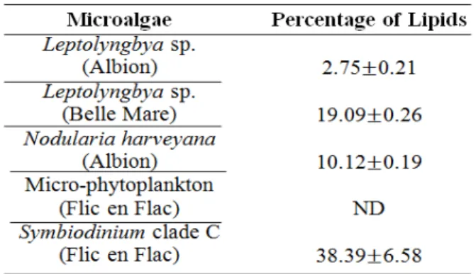
There were also differences recorded in the % lipid of the different microalgae (p<0.005) - among all, Symbiodinium clade C had the highest with (38.39±6.58%).
1H and 13C NMR analyses indicated the presence of the acyl glycerols
GRAPHICAL ABSTRACT
ARTICLE INFO ABSTRACT
Article history:
Received 17 February 2014 Received in revised form 1 April 2014 Accepted 1 April 2014
Available online 10 May 2014
Keywords:
Biodiesel Lipid Microalgae Mauritian waters
The aim of this study was to assess the lipid content and the subsequent potential of different microalgae present in the Mauritian marine water to produce biodiesel. The share of micro-phytoplankton species in the water column was determined. The cyanobacterial mats and endosymbiotic dinoflagellates were characterised morphologically and genetically using RFLP. The samples were quantified gravimetrically and analysed using 1H &13C NMR spectroscopy. Total micro-phytoplankton count amounted to 6.59±1.27x10⁵ cells L-1which was dominated by diatoms (95.2%), followed by dinoflagellates (2.9%) and cyanobacteria (1.9%). The cyanobacterial mats were identified as Leptolyngbya sp. and Nodularia harveyana, and the RFLP characterised the endosymbiotic dinoflagellates as the Symbiodinium clade C. The highest amount of lipid was recorded in the Symbiodinium clade C (38.39±6.58%). 1H and 13C NMR analyses indicated the presence of acyl glycerols. An attempt to synthesise biodiesel by alkaline trans-esterification reaction was also performed and the presence of biodiesel was detected using the Fourier Transform Infrared Spectroscopy. The Infrared analysis yielded peaks at around 1738cm-1 and 1200cm-1 characteristic of the carbonyl and ether groups respectively, indicating the presence of biodiesel.
© 2014 BRTeam. All rights reserved.
1. Introduction
In the twenty-first century, the atmospheric carbon dioxide level is 30% higher than the pre-industrial era (NOAA Earth System Research Laboratory 2013; Bernstein et al. 2007; Hofmann et al. 2009) sharing a direct relationship with the hike in fossil fuel burning since 1950 (US Energy Information Administration, 2012). In 2011, the global transport sector had a 28% energy share and accounted for almost a quarter of the world’s carbon dioxide
emissions (US Energy Information Administration 2012). Carbon dioxide is the major greenhouse gas contributing to global warming and ocean acidification; hence, triggering concern worldwide (Bernstein et al., 2007). The situational irony is that mankind is exploiting a dwindling global oil reserve (OPEC, 2012) at the expense of the environment-causing energy insecurity and climate catastrophes. Biodiesel is the ultimate solution for replacing the petroleum-centered transports industry (Chisti, 2008), subsequently, reducing the sector’s carbon dioxide emissions (Bernstein et al.
et al. / Biofuel Research Journal 2 (2014) 58- 64
2007). In addition to its greater energy currency than bioethanol (Chisti, 2008), it also conforms to the current diesel engines (Wang et al. 2000). However, the biofuel industry is being subjected to controversies including food insecurity due to the divergence of staple crops which are being used for biofuel production (Tenenbaum, 2008). Choosing a proper feedstock is therefore crucial. Lipids obtained from non-edible feedstocks are popular because they do not compete with the food market. Also, the prohibitive cost of edibleoils prevents their use in Biodiesel preparation. This is while nonedible oils are affordable for Biodiesel production (Karmee & Chadha, 2005). Nonedible oils which have been used for biodiesel production include Jatropha and Pongamia (Karmee et al. 2004; 2006). Microalgae, being anonedible lipid source, is another potential candidate for biodiesel production as it does not compete with food commodities (Gouveia, 2011) and has a high lipid content which is usually between 20-50% (Chisti, 2007). Studies have focused mostly on eukaryotic species such as Botryococcus braunii, Chlorella sp., Chlamydomonas reinhardtii and Nannocloropsis sp. because of their relatively higher lipid content (Scott et al. 2010). Cyanobacteria – a microalgae prokaryote – is also gaining momentum in the biodiesel production arena with respect to its fast growth rate and lipid content (Quintana et al. 2011).
The aim of this study was to gain preliminary data on the potential of different microalgae - micro-phytoplankton, filamentous cyanobacteria and endosymbiotic dinoflagellates - found in the Mauritian waters in order to identify prospective biodiesel feedstocks. The focus was on the determination of the moisture content as well as lipid content. An attempt to synthesise biodiesel from the microalgae’s lipids was also carried out.
2. Materials & methods
2.1. Collection and Identification of Samples
The microalgae which were collected from the Mauritian lagoons were cyanobacteria, micro-phytoplankton and endo-symbiotic dinoflagellates. The collection period was from September to December 2012 as illustrated in Table 1.
Table 1
Samples collection sites and the sampling periods.
The cyanobacterial mats were collected live and stored in a cooler bag along with some seawater. The micro-phytoplankton was collected as described by Sadally and coworkers. (2011). As for the Symbiodinium sp., part of the coral Fungia repanda was collected and the endosymbiotic dinoflagellates were water-picked and centrifuged at 5000rpm for 5 minutes.
The morphology and microscopic structures observed under a light microscope were used to identify the cyanobacterial mats while the micro-phyotoplankton was counted and identified following the methods described by Sadally et al. (2011). The endosymbiotic dinoflagellates were identified with respect to their cnidarian host and the clade by performing a Restriction Fragment Polymorphism (RFLP) using the restriction enzymes Taq1 and
Sau3A. The microalgae were stored below -20°C for further experimental procedures.
2.2. Determination of Moisture Content
The moisture content of the respective microalgae was determined gravimetrically. They were weighed, using the electronic mass balance -METTLER TOLEDO B303-S, before and after drying in a drying cabinet at
80°C. The dried cyanobacterial mats were ground and stored in opaque plastic bottles at 4°C for lipid extraction.
2.3. Lipid Extraction
Total lipids were extracted by a modified Folch method with chloroform: methanol (1:1, v/v) (Ryckebosch et al. 2011) and the volume of organic solvent was determined according to the principles used by Halim et al. (2011). The volume of organic solvent was calculated for each microalgae species using the following equations:
Where Vt is the total volume of solvent (ml) and Sw is the amount of dried sample (mg)
Where Vs is the volume of choloroform: methanol mixture (ml).
Where Vw is the volume of water (ml)
2.4. Analysis of Total Lipids
13C & 1H NMR spectroscopy were performed to establish the lipid profile
of the different samples. A few drops of the lipids that had been dissolved in the extracting solvent were added to an NMR capillary tube followed by deuterated chloroform using a Pasteur pipette. The NMR spectrum was calculated at 250 MHz for both 13C & 1H using the NMR machine 250 MHz
Bruker.
2.5. Biodiesel Synthesis
Biodiesel was only synthesized with the cyanobacterial mats’ lipid extracts through the trans-esterification reaction as described by Hossain et al. (2008). For a brief period, 18.5 mMol sodium methoxide in methanol was mixed with the lipid extracts in a quickfit flask which was then placed on the electric shaker MODEL G25 for 1.5hours at 300rpm.
2.6. Biodiesel Purification
The trans-esterified mixture was then transferred to a corning tube and left to settle for about 16 hours. After the settling period, the corning tube was centrifuged at 2000rpm for 10 minutes as described by Prommuak et al. (2011). An attempt was made to purify the upper layer through water-wash using 60°C distilled water as suggested by Nakpong & Wootthikanokkhan (2010) and dry-wash using silica gel. Both washes were unsuccessful.
2.7. Biodiesel Analysis
The presence of biodiesel was determined by using FT-IR spectroscopy. A few drops of the transesterified mixture were placed on the FTIR -BRUKER ALPHA - crystal. Absorbant measurements were carried out using 32 scans.
2.8. Statistical Analysis
A one-way analysis of variance (ANOVA) and a post-hoc Tukey’s test with a = 0.05 were performed to statistically evaluate the results using MINITAB 15.
3. Results and discussions
3.1. Identification
Figure 1 illustrates the share of micro-phytoplanktons in the water column collected at Flic en Flac.
Fig.1. Percentage of Micro-phytoplankton
The total micro-phytoplankton density amounted to (6.59±1.27) × 105
cells/L with the diatoms bearing the biggest share (95.2%) while the cyanobacteria had the smallest (1.9%). The RFLP analysis of the endosymbiotic dinoflagellates, as depicted in Figure 2, showed that the latter was the Symbiodinium clade C.
RFLP pattern of Clade C Symbiodinium from Fungia repanda. Lanes 1&2: restriction digests using Taq1; Lanes 3&4: restriction digests using Sau3A.
The morphology and microscopic structures of the cyanobacterial mats have been illustrated in Figure 3 and the latter were identified as
Leptolyngbya sp. (Charpy et al., 2010) and Nodulariaharveyana (Lyra et al., 2005) respectively.
3.2. Moisture Content
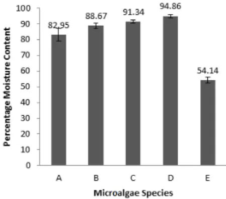
As depicted in Figure 4, the moisture content of the microalgae under study was highest in micro-phytoplankton and lowest in the Symbiodinium
clade C (P<0.05).
Fig.4.. Moisture content of the microalgae species (mean± SD; n=3). A: Leptolyngbya sp. (Albion), B: Leptolyngbya sp. (Belle Mare), C: Nodularia harveyana, D: Micro-phytoplankton & E: Symbiodinium clade C.
The moisture content of the Leptolyngbya sp. at Albion was not significantly different from that of Leptolyngbya sp. collected at Belle Mare. Furthermore, no significant differences were observed between Leptolyngbya
sp. (Belle Mare), Nodularia harveyana (Albion), Nodularia harveyana
(Albion) and micro-phytoplankton. The moisture content tallied with that of other literature (Patil et al. 2008). This particular trait is attributed to the poikilohydric-characteristic of the microalgae (Campbell et al., 2004); consequently, high energy input is required to remove the water molecules. Nonetheless, the removal of water is mandatory as water molecules around the cells and within the cells act as barriers against the organic solvent (Mercer & Amerta, 2011). No additional pre-treatments were deemed to be necessary due to their negligible effects (Rittmann, 2008), preceding lipid extraction.
3.3. Lipid Extracts
The lipid extracts of the different microalgae exhibited different colors as illustrated in Figure 5.
Fig.3. (a) Mat of Leptolyngbya sp. (b) Filaments of Leptolyngbya sp. with single trichomes (c) Mat of Nodularia harveyana (d) Filamentous structures of Nodularia
–
Fig.5. Pictures in the boxes depict the aqueous extracts’ colours while the others illustrate the pigments’ colours of the extracts of the different microalgae. *Aqueous extract for the micro-phytoplankton was colourless. A: Leptolyngbya sp. (Albion), B:
Leptolyngbya sp. (Belle Mare), C: Nodularia harveyana, D: Micro-phytoplankton &E: et al. / Biofuel Research Journal 2 (2014) 58- 64
Beetul
60
The aqueous extract of the Leptolyngbya sp. collected at different locations varied significantly-reddish brown (Albion) and pale green (Belle Mare). However, the pigments observed following evaporation of the organic solvent showed that both have similar pigments but with different ratios. On the other hand, the aqueous extract of Nodularia harveyana was green containing brownish/green pigments. The micro-phytoplankton yielded a colorless aqueous extract with white pigments while the Symbiodinium clade C gave a yellow aqueous extract with greenish yellow pigments. It was observed that the lipid extracts were of different colours as illustrated in Figure 5. Algae have different classes of photosynthetic pigments namely chlorophyll, carotenoids comprising of carotene and xantophylls, and phycobilins while phycobilin pigments are the main light harvesting pigments in cyanobacteria. Among the different classes of photosynthetic pigments, the chlorophylls and carotenes are the fat soluble molecules which can be extracted from the thylakoid membranes using organic solvents such as methanol (Robertson, 2011). For the cyanobacteria, the pigments can be identified mainly as chlorophyll and beta carotene. However, it may be that some of the phycobilin pigments tagged along with the lipids during the extraction as in the case of Nodularia harveyana. Simis et al. (2012) showed that phycocyanin, which might be mixed with phycoerythrocyanin, is the most abundant photosynthetic accessory pigment. Apart from the chlorophyll pigments, Leptolyngbya spp. contain rhodopsin which can be observed in Figure 5 as a red coloration. Finally, Daigo et al. (2008) observed the presence of zeaxanthin in the Symbiodinium genus; hence, explaining the characteristic yellowish colour obtained along with chlorophyll pigments Daigo et al. (2008). It has been reported that both rhodopsin and zeaxanthin can be extracted using organic solvents (Darszon et al., 1978; Chen et al., 2005) which implies that the red pigments obtained in Leptolyngbya spp. and the yellow colour observed in the lipid extract of the Symbiodinium genus are due to rhodopsin and zeaxanthin respectively.
3.4. Total Lipid Content
The quantification of the total lipid content has been illustrated in Table 2. Symbiodinium clade C had relatively high total lipid content compared to the cyanobacterial mats (P<0.05).
Table 2
Percentage of Lipid (mean ± SD; n=5; except Nodularia harveyana: n=4 & Symbiodinium clade C: n=3) *ND: Not Determined
Moreover, the total lipid content of the cyanobacterial mats collected at different locations was significantly different from each other. The lipid content of microalgae depends strongly on the storage structure of the organism (Rittmann, 2008), the enzymes involved (Wada & Sato, 2010) as well as the environmental conditions (Karatay & Donmez, 2011; Sakthivel et al., 2011). On the one hand, eukaryotic algae accumulate important amounts of lipids as reserves in the form of triacylglycerols and diacylglycerol for the membrane structures (Wada & Sato, 2010) while cyanobacteria accumulate lipids in their thylakoid membranes only (Rittmann, 2008), agreeing with the theory that cyanobacterial genomes lack the gene coding for the triacylglycerol synthesis (NREL 2012). On the other hand, the difference in the percentage of lipid among the different cyanobacterial mats may be explained by the enzymes present in cyanobacterial cells which differ from one species to another (Wada & Sato, 2010). The environmental factors such
as the pH and nitrate content should not be overlooked as studies have shown that these two factors influence the accumulation of lipids in the cyanobacteria (Karatay & Donmez, 2011; Sakthivel et al., 2011).
Symbiodinium clade C produced a relatively huge lipid content due to its mutualistic relationship with corals for faster calcification (Davy et al., 2012). The main energy store of corals is lipids – metabolites synthesized from the primary products obtained from the photosynthetically-fixed carbon translocated from the dinoflagellates symbionts (Davy et al., 2012). Kellogg and Patton (1983) showed that primary metabolites are not the only ones which are transferred to the coral; the dinoflagellates also release fat droplets suggesting that lipids are also translocated to the corals. Such activities account for the high metabolic rate of dinoflagellates as they have to cater for their own energy supply as well as that of their hosts. Consequently, they have to accumulate a significant amount of lipids as well as other products during and following photosynthesis. Nevertheless, the moisture content of the dinoflagellates was relatively very low because those organisms have to provide metabolites for two bodies at once leaving less space for moisture accumulation in the cells. Yet, it should be noted that such characteristics may differ in the isolated dinoflagellates (Sutton & Hoegh-Guldberg, 1990).
3.5 1H & 13C NMR Analysis
The proton NMR analysis yielded the results which have been illustrated in Figure 6.
Fig.6.1H NMR analysis. The singlet between 2.0ppm ppm and 2.5ppm is for the methyl in the
acetoxy group (COOR-CH3). The two singlets overlapping peaks between 3.0ppm and 3.5ppm
is characteristics of phospholipids. The peaks between 4.5ppm and 5.0ppm show the presence of glycerol methyl protons.
It was observed that the peaks were moved downfield due to the hydrogen bonds formed between the lipids and the methanol in which they were dissolved for storage. 4.3ppm is the benchmark for the presence of acyglycerols (Adosraku et al., 1994). However, due to the formation of inappropriate hydrogen bonds, that particular peak moved downfield – between 4.5ppm and 5ppm. The NMR spectra show that the Leptolyngbya sp. (Belle Mare) and Symbiodinium clade C have the highest amount of acyglycerols in the prokaryotes and eukaryotes arena respectively. Also, the lipid profile of the micro-phytotplankton is similar irrespective of the site of collection in the lagoon.
Figure 7 illustrates the 13C Lipid profile of the eukaryotic (Symbiodinium
clade C) and prokaryotic (Leptolyngbya sp.) microalgae. The resonance for
ether (–C-O) at chemical shifts 57.8ppm, 62.3ppm and 70.6ppm may indicate
the presence of sulfoquinovosyldiacylglycerol, phosphatidylgycerol and acylglycerols which are usually present in microalgae (Wada & Sato, 2010).
Fig.7.13C NMR spectrum of Symbiodinium clade C (Flic-en-Flac) and Lepolyngbya sp. (Belle
Mare). The resonance for the methyl group and methylene group are between the range of 0ppm to 30ppm and 15ppm to 55ppm respectively. It should be noted that the peak at 49.2ppm was not counted as a carbon atom in the lipid sample as this peak codes for methanol. The resonance for ether (–C-O) is between 55ppm and 90ppm. The peaks 183.5ppm and 185.2 ppm indicates the presence of carbonyl group found in esters confirming the presence of lipids.
The high number of carbon is crucial in the biodiesel industry as the longer and more saturated are the fatty acid chain the higher will be the cetane number. The cetane number is a standard for the measurement of ignition delay time of an engine and a larger cetane number for a diesel fuel leads to a shorter ignition delay (Knothe, 2006).
3.6. Biodiesel Analysis
As mentioned in the methodology section, both the water-wash and the dry-wash were unsuccessful; consequently, the biodiesel volume could not be quantified. However, during the washing attempt, a golden yellow solution, as illustrated in Figure 8, was obtained indicating the presence of biodiesel.
Fig.8.The brownish colour in (a) indicates the presence of the biodiesel synthesised during the trans-esterification reaction. Due to the minute amount produced, the latter appear as pigments but when methanol is added to it a golden yellow colour is obtained confirming the presence of biodiesel.
The FT IR analysis carried out indicated the presence of biodiesel for
Leptolyngbya sp. (Belle Mare) and Nodularia harveyana (Albion) lipids only. As illustrated in Figure 9, peaks at 1738.30cm-1 for C=O and 1171.49cm-1
and 1249.31cm-1 for –C-O were obtained for the Leptolynbya sp. (Belle
Mare). As for the Nodularia harveyana, peaks at 1737.84cm-1 for C=O and
1189.43cm-1 for –C-O were obtained (Knothe, 2006).
The alkali-catalysed process was carried out for the trans-esterification step in this study. Following the settling time, an important amount of glycerine deposit was observed which was of a semi-solid composition. Gerpen et al. (2004) suggested that the amount of catalyst to be used should be 1% of the extracted lipids’ mass but according to Hossain et al. (2008), the amount of catalyst used was sometimes 200 times higher than the expected amount generating an excessive number of water molecules which are toxic to triacylglycerols and diacylglycerols. The amount of lipid extracts being very small, the water molecules were enough to hydrolyse most of the acylglycerols in some cases, accounting for the semi-solid glycerine observed; but this may also be explained by the highly saturated lipids which might have been present (Gerpen et al., 2004). It should be noted that the amount of glycerine obtained was not considered since the qualitative data would have been deceptive comprising of the excessive sodium hydroxide salt.
Following glycerine and biodiesel separation, purification is mandatory to bring down the pH to 7. During the washing process, two phases must be observed in the separating funnel because biodiesel has a lower density than water – 0.93g/L (Sander & Murthy, 2010). Yet, during the water-wash no two phases were observed. This is because of the small amount of biodiesel obtained following the transesterification step. The relationship between oil and biodiesel is a 1:1 ratio provided that only triacylglycerol, diacylglycerol or monoacylglycerol are present. The average lipid content of the
Leptolyngbya sp. collected at Albion was 0.056g. Assuming that the oil had no free fatty acids, the amount of biodiesel obtained for the latter should be around 58.16µl (Gerpen et al., 2004). However, due to the high hydrolysis of the acylglycerol, the volume of biodiesel was expected to be much lower ruling out the utility of the water-wash for this study. A dry wash was also attempted but to no avail. While drying out, all methanols evaporated leaving the pigment-like substance. The golden yellow pigments clearly indicated the presence of a small quantity of biodiesel. This is not surprising because Daroch et al. (2013) reported that many studies failed to produce biodiesel from microalgae as the alkali-catalysed reaction failed to convert the lipids into biodiesel efficiently.
Since, quantification was hard to achieve, detecting the presence of biodiesel was done by using FTIR following the evaporation of the excess methanol in the biodiesel mixture. The presence of fatty acid methyl esters were detected only for the Leptolyngbya sp. collected at Belle Mare and the
Nodularia harveyana. According to the equation established by (Gerpen et al., 2004) the volumes of biodiesel which should have been obtained for these species were 170.57±1.74µl and 64.66±1.45µl respectively. The species’ lipids were more resistant to hydrolysis than the one collected at Albion.
The biodiesel also known as fatty acid methyl esters have the ester bond which is linked to a –CH3. The esters’ bonds IR spectral region normally have
peaks in the range of 1730-1750 cm-1 and 1000-1300 cm-1 for C=O and –C-O
respectively. However, it should be noted that acylglycerols have the same bonds as well. The IR spectral region at around 1200cm-1 for –O-CH
3
distinguishes between the fatty acid methyl esters and acylglycerols – presence of peak at 1200 cm-1 indicating the presence of fatty acid methyl
esters (Knothe, 2006). The peaks at 1249.31 cm-1 and 1738.30 cm-1 indicated
the presence of fatty acid methyl esters in the biodiesel mixture synthesized from the lipid extract of the Leptolyngbya sp. collected at Belle Mare while the peaks at 1737.84cm-1 and 1189.43cm-1 indicated the presence of fatty acid
methyl esters found in that of Nodularia harveyana.
Among the cyanobacterial mats used in this study, the Leptolyngbya sp. collected at Belle Mare had the highest lipid content of 19.09±0.26% and the hypothetical biodiesel yield was 170.57±1.74µl. A Singh and Gu (2010) analysis showed that the biodiesel yield from the 30% oil-microalgae was 58, 700 l/ha while rapeseed and canola, jatropha, karanj, corn, soybeen, peanut, coconut and oil palm yielded 1190 l/ha, 1892 l/ha, 2590 l/ha, 1721 l/ha, 446 l/ha, 1059 l/ha, 2689 l/ha and 5959 l/ha respectively. If a simple proportion is applied to our study comparing the biodiesel yields, it can be speculated that since 30% oil-microalgae produces 58, 700 l/ha, a 19% oil-microalgae can produce 37, 177 l/ha which is still higher than the other oleaginous crops.
et al. / Biofuel Research Journal 2 (2014) 58- 64 Beetul
4. Conclusions
In this study, the potential of microalgae found in Mauritian waters for biodiesel production was investigated. The share of micro-phytoplankton species was determined. Total micro-phytoplankton count amounted to 6.59±1.27x10⁵ cells L-1 which was dominated by diatoms (95.2%), followed
by dinoflagellates (2.9%) and cyanobacteria (1.9%). The cyanobacterial mats were identified as Leptolyngbya sp. and Nodularia harveyana, and the RFLP characterised the endosymbiotic dinoflagellates as the Symbiodinium clade C. The moisture content obtained was highest in micro-phytoplankton (94.86±0.99%) and lowest in the Symbiodinium clade C (54.14±1.97%). Differences in the % lipid of the different microalgae were also recorded (p<0.005) with Symbiodinium clade C having the highest (38.39±6.58%). 1H
and 13C NMR analyses indicated the presence of the acyl glycerols and the
Infrared analysis confirmed the presence of biodiesel. The preliminary data obtained in this study indicates the capacity of the different microalgae found in Mauritian waters for acting as a potential nonedible feedstock for the production of Biodiesel.
Acknowledgements
The authors wish to thank the Faculties of Agriculture and Science at the University of Mauritius for supporting this work. The precious help provided by the technical staff of the Department of Agriculture and Food Science of the Faculty of Agriculture and the Departments of Biological Sciences and Chemistry of the Faculty of Science is gratefully acknowledged.
References
Adosraku RK, Choi GTY, Constantinou-Kokotos V, Anderson MM, Gibbons WA. 1994. NMR lipid profiles of cells, tissues, and body fluids: proton NMR analysis of human erythrocyte lipids. Journal of lipid Research 35:1925 – 1931.
Bernstein L, Bosch P, Canziani 0, Chen Z, Christ R, Davidson O, Hare W, Huq S, Karoly D, Kattsov V, Kundzewicz Z, Liu J, Lohmann U, Manning M, Matsuno T, Menne B, Metz B, Mirza M, Nicholls N, Nurse L, Pachauri R, Palutikof J, Parry M, Qin D, Ravindranath N, Reisinger A, Ren J, Riahi K, Rosenzweig C, Rusticucci M, Schneider S, Sokona Y, Solomon S, Stott P, Stouffer R, Sugiyama T, Swart R, Tirpak D, Vogel C, Yohe G. 2007. Climate Change 2007: Synthesis Report – Summary for Policymakers. Intergovernmental Panel on Climate Change.
http://www.ipcc.ch/pdf/assessment-report/ar4/syr/ar4_syr.pdf (accessed on 12th October 2013).
Campbell A, Cooke P, Cass K, Earl K. 2004. Plant and Animal Evolution.
New Zealand, The University of Waikato.
http://sci.waikato.ac.nz/evolution/aboutus.shtml (accessed on 2nd April 2013)
Charpy L, Palinska Ka, Casareto B, Langlade Mj, Suzuki Y, Abed Rmm, Golubic S. 2010. Dinitrogen-Fixing Cyanobacteria in Microbial Mats of Two Shallow Coral Reef Ecosystems. Microb Ecol 59:174-186.
Chen F, Li H, Wong R N, Jiang Y. 2005. Isolation and purification of the bioactive carotenoid zeaxanthin from the microalga Microcystis aeruginosa by high-speed counter-current chromatography. Journal of Chromatography A 1064(2):183-186
Chisti Y. 2007. Biodiesel from microalgae. Biotechnology Advances 25:294-306.
Chisti Y. 2008. Biodiesel from microalgae beats bioethanol. Trends in Biotechnology 26(3):126-131.
Daigo K, Nakano Y, Casareto Be, Suzuki Y, Shioi Y. 2008. High performance Liquid Chromatographic Analysis of Photosynthetic Pigments in Corals: An Existence of a Variety of Epizoic, Endozoic and Endolithic Algae. 11th International Coral reef Symposium, 7-11 July 2008 Fort Lauderdale, FL, USA; 123-127
Daroch M, Geng S, Wang G. 2013. Recent advances in liquid biofuel production from algal feedstocks. Applied Energy 102:1371-1381. Darszon A, Philipp M, Zarco J, Montal M. 1978. Rhodopsin-phospholipi
complexes in apolar solvents: Formation and properties. The Journal of Membrane Biology 43(1):71-90.
Davy SK, Allemand D, Weis VM. 2012. Cell Biology of Cnidarian Dinoflagellate Symbiosis. Microbiology and Molecular Biology Reviews 76(2):229-261.
Gerpen JV, Shanks B, Pruszko R, Clements D, Knothe G. 2004. Biodiesel Production Technology. USA: NREL (SR-510-36244).
http://www.nrel.gov/docs/fy04osti/36244.pdf (accessed on 10th December 2012)
Gouveia L, 2011. Microalgae as Feedstock for Biofuels. In: Springer ed. Microalgae as Feedstock for Biofuels. New York: Springer Berlin Heidelberg, p 1-69.
Halim R, Gladman B, Danquah MK, Webley PA. 2011. Oil extraction from microalgae for biodiesel production. Bioresource Technology 102:178-185. Hofmann D J, Butler H J, Tans PP. 2009. A new look at atmospheric carbon
dioxide. Atmospheric Environment 43:2084-2086.
Hossain ABM, Shalleh A, Boyce AN, Naqiuddin M. 2008. Biodiesel fuel production from algae as renewable energy. American Journal of Biochemistry and Biotechnology 4(3):250-254.
Karatay SE, Donmez G. 2011. Microbial oil production from thermophile cyanobacteria for biodiesel production. Applied Energy 88(11):3632-3635. Karmee SK, Mahesh P, Ravi R, Chadha A. 2004. Kinetic study of the base-catalyzed transesterification of monoglycerides from pongamia oil. Journal of the American Oil Chemists' Society 81(5): 425-430
Karmee SK, Chadha A. 2005. Preparation of biodiesel from crude oil of Pongamia piñata. Bioresource Technology. 96(13): 1425-1429
Karmee SK, Chandna D, Ravi R, Chadha A. 2006. Kinetics of base-catalysed transesterification of triglycerides from Pongama oil. Journal of the American Oil Chemists’ Society. 83(10): 873-877.
Kellogg, RB., Patton, JS. 1983. Lipid droplets, medium of energy exchange in the symbiotic anemone Condylactis gigantea: a model coral polyp. Marine Biology 75(2-3); 137-149
Knothe G. 2006. Analyzing Biodiesel: Standards and Other Methods. JAOCS 83:823-833.
Lyra C, Laamanen M, Lehtimaki JM, Surakka A, Sivonen K. 2005. Benthic cyanobacteria of the genus Nodularia are non-toxic, with gas vacuoles, able to glide and genetically more diverse than planktonic Nodularia. International Journal of Systematic Microbiology 55:555-568.
Mercer P, Amerta RE. 2011. Development in oil extraction from microalgae. European Journal of Lipid Science and Technology 113(5):539-547. Nakpong P, Wootthikanokkhan S. 2010. Roselle (Hibiscus sabdariffa L.) oil
as an alternative feedstock for biodiesel production in Thailand. Fuel 89:1806-1811.
Noaa Earth System Research Laboratory. 2013. Atmospheric CO2. Mauna Loa. http://co2now.org/images/stories/data/co2-mlo-monthly-noaa-esrl.pdf
(accessed on 12th October 2013)
NREL. 2012. NREL Creates New Pathways for Producing Biofuels and
Acids from Cyanobacteria. USA, NREL.
http://www.nrel.gov/docs/fy13osti/55974.pdf (accessed on 9th March 2013)
Organization of The Petroleum Exporting Countries. 2012. World Oil Outlook 2012. Austria: OPEC.
Patil V, Tran K, Giselrod HR. 2008. Towards Sustainable Production of Biofuels from Microalgae. International Journal of Molecular Sciences 9:1188-1195.
Prommuak C, Pavasant P, Quitain AT, Goto M, Shotipruk A, 2011. Microalgal Lipid Extraction and Evaluation of Single-Step Biodiesel Production. Engineering Journal 16(5):158-166.
Quintana N, Van Der Kooy F, Van De Rhee MD, Voshol G P, Verpoorte R. 2011. Renewable energy from Cyanobacteria : energy production optimization by metabolic pathway engineering. Appl Microbiol Biotechnol 91:471-490.
Rittmann BE. 2008. Opportunities for Renewable Bioenergy Using Microorganisms. Biotechnology and Bioengineering 100 (2):203-212. Robertson D, 2011. Algal Pigments. USA, Clark University.
http://www.clarku.edu/faculty/robertson/ (accessed on 30th March 2013). Ryckebosch E, Muylaert K, Foubert I. 2011. Optimization of an Analytical
Procedure for Extraction of Lipids from Microalgae. Journal of the American Oil Chemists’ Society 89(2):189-198.
Sadally SB, Bhagooli R, Taleb-Hossenkhan N. 2011. Micro-Phytoplankton Distribution and Biomass at Two Lagoons around Mauritius Island. University of Mauritius Research Journal 18A:54-87.
Sander K, Murthy GS. 2010. Life cycle analysis of algae biodiesel. Int J Life Cyle Assess 15:704-714.
Sakthivel R, Elumalai S, Arif MM. 2011. Microalgae lipid research, past, present: A critical review for biodiesel production, in the future. Journal of Experimental Sciences 2(10):29-49.
Scott SA, Davey MP, Dennis JS, Horst I, Howe CJ, Lea-Smith D, Smith AG. 2010. Biodiesel from algae: challenges and prospects. Current Opinion in Biotechnology 21(3):277-286.
Simis SGH, Huot Y, Babin M, Seppala J, Metsamaa L. 2012. Optimization of variable fluorescence measurements of phytoplankton communities with cyanobacteria. Photosynthesis Research 112 (1):13-30.
Singh J, Gu S. 2010. Commercialization of potential of microalgae for biofuels production. Renewable and Sustainable Energy Reviews 14:2596-2610.
Sutton DC, Hoegh-Guldberg O. 1990. Host-Zooxanthella Interactions in Four Temperate Marine Invertebrate Symbioses: Assessment of Effect of Host Extracts on Symbionts. The Biological Bulletin 178:175-186.
Tenenbaum DJ. 2008. Food vs. Fuel. Environmental Health Perspectives 116(6):254-257.
US Energy Information Administration. 2012. Annual Energy Review 2011. United States: Energy Information Administration
Wada H, Sato N. 2010. Lipid Biosynthesis and its Regulation in Cyanobacteria. In: Wada H, Murata N, ed. Lipids in Photosynthesis. Netherlands: Springer Netherlands, p 157-177.
Wang WG, Lyons DW, Clark NN, Gautam M. 2000. Emissions from Nine Heavy Trucks Fueled by Diesel and Biodiesel Blend without Engine Modification. Environ. Sci. Technol. 34:933-939.
et al. / Biofuel Research Journal 2 (2014) 58- 64 Beetul