Ar
ti
cle
J. Braz. Chem. Soc., Vol. 21, No. 5, 927-933, 2010. Printed in Brazil - ©2010 Sociedade Brasileira de Química 0103 - 5053 $6.00+0.00
*e-mail: [email protected]
Triterpenoid Saponins from
Lippia alba
(Mill.) N. E. Brown
Mareni R. Farias,*,a Roberto Pértile,a Melissa M. Correa,a Maria Tereza R. de Almeida,b
Jorge A. Palermob and Eloir P. Schenkela
aPrograma de Pós-graduação em Farmácia, Universidade Federal de Santa Catarina, Campus
Universitário Trindade, 88040-900 Florianópolis-SC, Brazil and Departamento de Ciências Farmacêuticas, Universidade Federal de Santa Catarina, 88040-900 Florianópolis-SC, Brazil
bDepartamento de Química Orgánica, Facultad de Ciencias Exactas y Naturales, Universidad de
Buenos Aires, Ciudad Universitaria Pab.2, C1428EGA, Buenos Aires, Argentina
Das folhas de Lippia alba foram isoladas duas saponinas. As estruturas destas saponinas foram estabelecidas empregando métodos espectroscópicos, principalmente RMN mono e bi-dimensional e espectrometria de massas. Estes novos compostos foram caracterizados como ácido 3-O-β-D-glucopiranosil-28-O-(α-L-rhamnopiranosil-(1→3)-β-D-xilopiranosil-(1→ 4)-α-L-rhamnopiranosil-(1→2)-α-L-arabinopiranosil)-16α,23-di-hidróxi-olean-12-en-28-óico, designada Lippiasaponina I (2) e como ácido 3-O-β-D-glucopiranosil-28-O-(α -L-rhamnopiranosil-(1→3)-β-D-xilopiranosil-(1→4)-α-L-rhamnopiranosil-(1→3)-α-L-arabinopiranosil)-16α ,23-di-hidróxi-olean-12-en-28-óico, designada Lippiasaponina II (3).
Two saponins were isolated from the leaves of Lippia alba. Their structures were established using one- and two-dimensional NMR spectroscopy and mass spectrometry. These new compounds were elucidated as 3-O-β-D-glucopyranosyl-28-O-(α-L-rhamnopyranosyl-(1→3)-β -D-xylopyranosyl-(1→4)-α-L-rhamnopyranosyl-(1→2)-α-L-arabinopyranosyl)-16α ,23-dihydroxy-o-lean-12-en-28-oic acid, named as Lippiasaponin I (2) and as 3-O-β-D-glucopyranosyl-28-O -(α-L-rhamnopyranosyl-(1→3)-β-D-xylopyranosyl-(1→4)-α-L-rhamnopyranosyl-(1→3)-α -L-arabinopyranosyl)-16α,23-dihydroxy-olean-12-en-28-oic acid, named Lippiasaponin II (3).
Keywords:Lippia alba, verbenaceae, saponins
Introduction
Lippia alba (Mill.) N. E. Brown (Verbenaceae) is a shrub widely distributed throughout South America and it is popularly known as ‘cidreira’ or ‘false melissa’, designation derived from other medicinal plants (Melissa oficinalis L.and Cymbopogon citratus (DC) Stapf.) also used in the popular medicine in cases of respiratory distress. Traditionally the tea from its leaves is largely utilized in popular medicine from all Brazilian regions as a tranquilizer and also in gastrointestinal and respiratory disorders. From the pharmacological point of view, antifungal activity was reported for hydroalcoholic extracts.1,2 Inhibition of HSV-1 (strain 29R/acyclovir resistant) was reported for the n-butanol fraction, and antipoliovirus activity for the ethyl acetate fraction.3 In addition, sedative and myorelaxant
effects in vivo were reported for the hydroalcoholic extracts4 and antiulcerogenic activity was described for the leaves’ infusion.5
Most chemical studies of L. alba are related to the essential oil composition and at least three chemotypes have been proposed based on the volatile chemical composition of its leaves.6 For the aerial parts, the presence of lavonoids,7 iridoid and phenylethanoid glycosides8 was reported. For a detailed review see Pascual et al.9 and references therein quoted.
In the present work, we report the isolation and structural characterization of two new saponins. To the best of our knowledge, the presence of saponins in Lippia alba has not been previously reported. The complete hydrogen and carbon assignments of the new compounds was accomplished using 2D NMR experiments including 1H, 1H-COSY, RCT, 13C, DEPT 90 and 135, HSQC, HMQC
Triterpenoid Saponins from Lippia alba (Mill.) N. E. Brown J. Braz. Chem. Soc.
928
Results and Discussion
Solvent partition and chromatographic procedures allowed the isolation of the main triterpenoid saponins from the aerial parts of L. alba:Lippiasaponin I (2) and Lippiasaponin II (3). Basic hydrolysis of a mixture of 2
and 3 afforded only one prosapogenin (1).
Compound 1
The 13C NMR spectrum of the prosapogenin (1)showed 36 signals, whereas the DEPT spectrum revealed 6 methyls, 11 methylenes, 11 methines and 8 quaternary carbon atoms. Six carbons could clearly assigned to the sugar moiety, identiied as β-glucopyranose by the NMR signals of the anomeric position (d 13C 105.8; d 1H 4.39).
The 1H NMR and 13C NMR spectrum of the prosapogenin
1 in CD3OD displayed characteristic signals of a triterpene aglycone derived from oleanolic acid, showing a triplet at d 5.29, which correlated in the HSQC spectrum with an oleinic carbon doublet at d 123.4, assigned to C-12. Six three-hydrogen singlets at d0.70, d0.98, d0.79, d1.38, d 0.87 and d0.96 could be assigned to the C-24, C-25, C-26, C-27, C-29 and C-30 methyl groups respectively. A signal at d 181.3 in the 13C NMR spectrum was assigned to the carboxilic acid at C-28.10
Three oxygenated carbons were observed at d 83.5 (CH), d 64.9 (CH2) and d 75.3 (CH). The former was assigned as glycosidated C-3, conirmed by the HMBC correlation between the anomeric hydrogen at d 4.39 and the carbon at d 83.5. The oxygenated carbon at d 65.0 (CH2), in principle could be assigned to C-23 or C-24.10
This hydroxymethylene group was assigned to C-23 considering the 13C NMR chemical shifs of the methyl groups. It is reported that a hydroxymethylene group at the 4α-position (C-23) provokes a shielding for the 4β-methyl group (C-24) to ca.d 11-13.11 The shielded methyl group (d 13.4, s) was assignable to the 4β-methyl group (C-24), thus, this hydroxyl was linked at C-23 (d 65.0).
The remaining oxygenated carbon at d 75.3 (CH) was linked to the hydrogen at d 4.45 (m). The COSY spectrum showed that this hydrogen was coupled to hydrogens at d 1.35 and d 1.85, assigned to C-15 by HMBC correlations, thus unambiguously locating this hydroxyl group at C-16. The 16α-configuration was evident from the small J values of H-16 (broad multiplet at d 4.45) in the 1H NMR spectrum, characteristic of an equatorial hydrogen.
Therefore, the structure of 1 was elucidated as 3-O-β -D-glucopyranosyl-16α-23-dihydroxy-olean-12-en-28-oic acid. Total carbon and hydrogen assignments are shown on Table 1.
Compound 2
The FAB MS (positive ion mode) of compound 2
displayed a quasi-molecular ion peak at m/z 1229 [M + Na+] suggesting a molecular formula of C58H94O26. In addition, a fragment ion at m/z 1050 indicated the loss of an hexose moiety.
The 13C NMR spectrum of 2 showed 58 signals, whereas the DEPT spectrum revealed 8 methyls, 13 methylenes, 29 methines and 8 quaternary carbon atoms. The comparison of the 1H and 13C data (Table 1) indicated the same partial structure of compound 1. The major differences were observed at C-28 (d 175.6) and the presence of five anomeric carbons instead of one. The shielding of C-28 was attributed to glycosylation of the carboxyl group.10
The 13C NMR signals of the four additional anomeric carbons were located at d 94.0, d 101.4, d 102.5 and d 106.5 and correlated to anomeric hydrogens at d 5.60, d 5.03, d5.13 and d 4.53 respectively in the HSQC experiments (Table 1).
HMBC, COSY, RCT, and 2D J-Resolved spectra were used for the complete assignment of the resonances of each monosaccharide moiety, starting from the anomeric hydrogens.
The analysis of these spectra revealed the presence of a β-D-xylose (JH-1, H-2 = 7.7Hz), an α-L-arabinose in a predominant 1C
4 conformation ( 3J
H-1, H-2 = 3.8Hz) 12 and
Farias et al. 929 Vol. 21, No. 5, 2010
Table 1.13C NMR and 1H NMR data of Prosapogenin, Lippiasaponin I and Lippiasaponin II (in CD 3OD)
Prosapogenin (1) Lippiasaponin I (2) Lippiasaponin II (3)
Attribution DEPT dC dH (J in Hz) dC dH (J in Hz) dC dH (J in Hz)
Aglicone
1 CH2 39.6 0.98 m
1.62 m
39.6 1.61 m
1.65 m
39.6 0.96 m
1.62 m
2 CH2 26.4 1.74 m
1.95 m
26.3 1.90 m
1.90 m
26.4 1.75 m
1.93 m
3 CH 83.5 3.63 m* 83.6 3.64 m* 83.8 3.66 dd (5.4, 12.0)
4 C 43.9 - 43.5 - 43.9
-5 CH 48.3 1.23 m 48.4 1.23 d (6.2) 48.5 1.20 d (4.2)
6 CH2 18.9 1.37 m
1.48 m
19.0 1.37 m
1.47 m
19.2 1.37 m
1.47 m
7 CH2 33.8 1.28 m
1.64 m
33.7 1.37 m
1.61 m
33.7 1.48 m
1.58 m
8 C 40.6 - 40.7 - 40.8
-9 CH 48.2 1.69 m 48.1 1.67 m 48.1 1.67 m
10 C 37.7 - 37.6 - 37.7
-11 CH2 24.5 1.90 m
1.90 m
24.5 1.90 m
1.90 m
24.5 1.90 m
1.90 m
12 CH 123.4 5.29 br t 123.7 5.34 t (3.4) 123.5 5.32 t (3.5)
13 C 145.2 - 145.0 - 144.8
-14 C 42.8 - 42.3 - 42.8
-15 CH2 36.2 1.35 m
1.85 m
36.4 1.15 m
1.39 m
36.6 1.47 m
1.67 m
16 CH 75.3 4.45 m 74.6 4.48 br t 74.5 4.46 s
17 C 49.6 - 50.0 - 50.3
-18 CH 42.1 3.00 dd (4.0, 12.0) 42.1 3.02 dd (3.9, 12.0) 42.2 2.96 dd (4.0, 12.0)
19 CH2 47.7 1.02 m
2.28 t (12.0)
47.6 1.04 dd (12.0, 3.9) 2.27 t (12.0)
47.9 1.05 dd (4.0, 12.0) 2.28 t (12.0)
20 C 31.4 - 32.0 - 31.3
-21 CH2 36.6 1.14 m
1.93 m
36.4 1.43 dd (3.9, 12.0) 1.75 m
36.6 1.16 m
1.93 m
22 CH2 32.7 1.76 m
1.90 m
31.9 1.90 m
1.90 m
32.1 1.89 m
1.92 m
23 CH2 65.0 3.27 m
3.62 m
65.2 3.36 d (12.0) 3.62 d (12.0)
65.4 3.30 m
3.64 d (12.0)
24 CH3 13.4 0.70 s 13.5 0.72 s 13.6 0.74 s
25 CH3 16.6 0.98 s 16.6 0.99 s 16.7 0.99 s
26 CH3 17.8 0.79 s 18.0 0.77 s 17.8 0.77 s
27 CH3 27.3 1.38 s 27.3 1.37 s 27.2 1.35 s
28 C 181.3 -- 175.6 - 177.1
-29 CH3 33.4 0.87 m 33.3 0.87 s 33.3 0.87 s
30 CH3 24.9 0.96 s 25.1 0.96 s 24.9 0.94 s
3-O-sugar
Gly
1 CH 105.8 4.39 d (7.7) 105.7 4.40 d (7.7) 105.7 4.39 d (7.7)
2 CH 75.7 3.16 t (8.2) 75.6 3.17 t (8.0) 75.7 3.16 t (8.8)
3 CH 78.4 3.34 m* 78.3 3.35 t (8.0) 78.3 3.33 t (8.8)
4 CH 71.6 3.27 m* 71.6 3.29 m 71.6 3.28 t (8.8)
5 CH 77.8 3.27 m* 77.7 3.27 m 77.8 3.26 m
6 CH2 62.8 3.67 m*
3.83 dd (3.2, 10.0)
62.7 3.70 m
3.81 dd (3.5, 12.0)
Triterpenoid Saponins from Lippia alba (Mill.) N. E. Brown J. Braz. Chem. Soc.
930
acid, and named Lippiasaponin I. These sugar assignments are in accordance with literature data.14,15
Compound 3
As in the case of compound 2, the 13C NMR spectrum of
3 showed 58 signals, whereas the DEPT spectrum revealed 8 methyls, 13 methylenes, 29 methines and 8 quaternary carbon atoms (Table 1).
A comparative analysis of 1H and 13C NMR, and COSY spectra of 3,showed great similarities in the chemical shift of both compounds, but some differences in the resonances of the arabinose unit linked at C-28. All 13C signals of this arabinose were deshielding shifted (Table 1). Besides, the J of H-1 Ara (d 5.43) changed notably from 3.8Hz (2) to 5.2Hz (3).
The analysis of the COSY, RCT and 2D J-Resolved spectra of compound 3 revealed as in compound 2, the
28-O-sugars
Ara
1 CH 94.0 5.60 d (3.8) 95.5 5.43 d (5.2)
2 CH 75.8 3.77 t (3.8) 77.1 3.54 t (9.5 )
3 CH 71.2 3.86 m 76.6 3.55 m
4 CH 67.0 3.82 dd (3.5, 9.0) 70.9 3.52 m
5 CH2 63.8 3.50 m*
3.89 dd (9.0, 12.0)
66.7 3.28 d (12.0) 3.91 m
Rha I
1 CH 101.4 5.03 d (1.4) 101.3 5.28 d (1.6)
2 CH 72.1 3.85 m 71.9 3.91 dd (1.6, 3.4)
3 CH 72.4 3.84 m 72.4 3.81 dd (3.4, 9.0)
4 CH 83.4 3.55 t (9.0) 84.3 3.52 t (9.0)
5 CH 69.0 3.68 m 69.0 3.70 m
6 CH3 18.1 1.29 d (6.2) 18.4 1.31 d (6.2)
Xyl
1 CH 106.5 4.53 d (7.7) 107.0 4.48 d (7.7)
2 CH 76.2 3.34 t (8.6) 76.4 3.37 t (8.8 )
3 CH 84.1 3.45 t (8.6) 84.3 3.45 t (8.8)
4 CH 69.9 3.53 m 69.9 3.52 m
5 CH2 67.1 3.18 t (8.3)
3.82 m
67.2 3.21 dd (9.0, 12.0) 3.86 dd (5.5, 12.0)
Rha II
1 CH 102.5 5.13 d (1.6) 102.6 5.13 d (1.6)
2 CH 72.3 3.94 dd (1.6, 3.5) 72.3 3.95 dd (1.6, 3.4)
3 CH 72.2 3.71 dd (3.5, 9.2) 72.2 3.71 dd (3.4, 8.8)
4 CH 74.0 3.38 t (9.2) 74.0 3.37 t (9.5)
5 CH 70.0 3.99 dd (9.2, 7.0) 70.0 3.99 dd (5.4, 9.5)
6 CH3 17.9 1.23 d (7.0) 17.9 1.23 d (6.2)
* overlapping; br = broad.
Table 1. continuation
presence of a β-D-xylose (J H-1, H-2 = 7.7 Hz), a β-D-glucose (JH-1, H-2 = 7.7 Hz) and two α-rhamnoses (JH-1, H-2 = 1.6 Hz and JH-1, H-2 = 1.6 Hz). The main difference between 2 and
3 was observed in the α-L-arabinose unit, which in 3 was in a predominant 4C
1 conformation ( 3J
H-1, H-2 = 5.2 Hz). 12
For the tetraglycosidic chain linked to the carboxylic C-28 of the aglycone, the HMBC showed cross-peaks between H-1 (d 5.13) of a terminal rhamnose and C-3 (d 84.3) of xylose, between H-1 of xylose (d 4.48) and C-4 (d84.3) of an inner rhamnose, between H-1 (d 5.28) of an inner rhamnose and C-3 (76.6) of arabinose and between H-1 (d 5.43) of arabinose and C-28 (d 177.1). These data indicated the linkage between C-3 of arabinose and C-1 of the inner rhamnose, pointing to the main structural difference between Lippiasaponins I (2) and II (3). A value for the 3J
H-1–H-2 coupling of 3.8 Hz (Ara-H1) was observed in Lippiasaponin I. This data indicated a 1C
Farias et al. 931 Vol. 21, No. 5, 2010
Figure 1. Structures of the prosapogenin (1), lippiasaponin I (2) and lippiasaponin II (3). O
O HO
HO HO
OH HOH2C
OH O O O O OH HO O OH HO O
H3C O OH HO O O OH HO HO
H3C
3
28
6
1
lippiasaponin I (2)
2 3 1 6 5 4 1 2 3 5 4 6 5 1 2 3 4 1 3 4 5 5 12 16 O O HO HO HO
OH HOH2C
OH O O 3 28 6 1
lippiasaponin II (3)
2 3 1 5 4 5 12 16 O O HO HO HO
OH HOH2C
OH O O 3 28 6 1 5 12 16 O OH HO O
H3C
O OH HO O O OH HO HO
H3C
6 5 4 1 2 3 6 5 1 2 3 4 1 2 3 4 5 O OH O HO
Triterpenoid Saponins from Lippia alba (Mill.) N. E. Brown J. Braz. Chem. Soc.
932
in Lippiasaponin II a value of 5.2 Hz (Ara-H1) which suggests an α-L-arabinopyranoside in 4C
1 conformation. 12
The problem of the coniguration and conformation of arabinopyranoses in esters of hindered triterpene carboxylic acids has been discussed at length using 1H and 13C NMR arguments.12,16 Particularly puzzling is the fact that in
α-L-arabinosyl ester the 3J
H-1-H-2 couplings vary from 2.8 to 6.2 Hz depending on the equilibrium between 1C
4 and 4C
1 conformations.
The α-L-arabinopyranoside absolute stereochemistry was determined by comparison with methyl α -L-arabinopyranoside.17 The C-1 of arabinopyranose in esters of triterpene carboxylic acids differs from C-1 of methyl α-L-arabinopyranoside, because the priority order changes with the atomic number of atoms, therefore, the α-L-arabinopyranoside is S/R/S/S. In the molecular model of compound 2 we observed that the dihedral angle between Ara-H1 and Ara-H2 is aproximately 80-90o on
α-L-arabinopyranoside chair 1C
4 conformation, which explains the small J, in accordance with Karplus curve. The 1C
4 conformation on Lippiasaponin I (2) can be explained by O-Rha substituition at C-2 in Ara, which increases the population of 1C
4 conformation.
12 Additional proof was
obtained through the observation of a W coupling between H-3 (d 3.86) of arabinose and H-5 (d 3.50).
In the molecular model of α-L-arabinopyranoside chair 4C
1 conformation the dihedral angle between Ara-H1 and Ara-H2 is aproximately 180o, which points to a high value for the coupling constant. The predominance of 4C
1 conformation on Lippiasaponin II (3) can be explained by O-Rha substituition at C-3 in Ara, sincethe C-2 is hindered by the proximity of the O-substituent at position 1.
Therefore, compound 3 was identiied as 3-O-β -D-glucopyranosyl-28-O-(α-L-rhamnopyranosyl-(1→3)-β -D-xylopyranosyl-(1→4)-α-L-rhamnopyranosyl-(1→3)-α -L-arabinopyranosyl)-16α,23-dihydroxy-olean-12-en-28-oic acid, and named Lippiasaponin II. To the best of our knowledge, this is the irst report of the natural occurrence of 2 and 3.
Experimental
General procedures
NMR experiments were performed on a Bruker AM-500 (AM-500 MHz) spectrometer. NMR data were reported as d values, and referenced to the residual signal of the solvent (CD3OD). The MS experiments were performed on a UltrOTOF-Q, Bruker Daltonics, Billerica, MA spectrometer (positive ion mode). Silica gel 60 (0.063-0.200 mm, Merck) and silica gel 60 (0.040-0.063 mm,
Merck) were used for column chromatography. TLC was performed on precoated silica gel 60 F254 plates (Merck). HPLC separations were performed on a Shimadzu (FRC-10A) liquid chromatographer equipped with a UV-Vis detector using a preparative ODS column (column A, Shim-pack 20 x 250 mm, Shimadzu; detector: 254 nm; low rate: 5 mL min-1.
Plant material
Aerial parts of Lippia alba were collected in Cacupé, Florianópolis, Brazil, in April 2003. A voucher specimen (FLOR-31267) is deposited at the herbarium ICN (Federal University of Santa Catarina, Brazil).
Extraction and isolation
Material was dried and the leaves were separated from the stems and lowers. Leaves (840 g) were ground to powder and macerated two times with 96% ethanol. The alcohol extract (89.2 g) was concentrated, suspended in water, and then partitioned successively with petroleum ether (boiling range 30-60 oC), CH
2Cl2, EtOAc and n-BuOH (150 mL, 6 times with each solvent). The n- BuOH-soluble fraction (2.8 g) was applied to a silica gel 60 column (0.063-0.200 mm) eluting with the organic phase of the mixture EtOAc:MeOH: isopropanol:H2O:HOAc (7:1:2:5:0.2) to give ive sub-fractions (1-5). Fraction 5 (265 mg) was further chromatographed over silica gel 60 ( 0 . 0 4 0 - 0 . 0 6 3 m m ) , e l u t i n g w i t h t h e m i x t u r e EtOH:EtOAc:H2O (120:60:5) to yield two sub-fractions (5.1-5.2). Fraction 5.1 (218 mg) was separated by preparative HPLC (column A) using CH3CN:H2O, 70:30 (v:v) as eluant to yield 2 (35 mg) and 3 (32 mg).
Acid hydrolysis of Lippiasaponin I (2)
Compound 2 (2 mg) was hydrolyzed in 2 mol L-1 TFA at 120 oC for 1 h 30 min. The hydrolyzed monosaccharides were derivatized to the acetylated 1-deoxy-1-(2-hydroxypropylamino) alditols following published procedures12 and analysed by GC using a Hewlett-Packard Ultra-2 column (50m x 0.2 mm, thickness of liquid phase 0.11 µm) and identiied by coinjection with authentic standards prepared in a similar way.
Prosapogenin (1)
Farias et al. 933 Vol. 21, No. 5, 2010
Lippiasaponin I (2)
White powder, [α]D25 = −30.43 (C = 0.013, MeOH); FAB MS m/z = 1229 (100%); 1050 (10%); 1H NMR and 13C NMR spectral data: see Table 1.
Lippiasaponin II (3)
White powder; 1H NMR and 13C NMR spectral data: Table 1.
Supplementary Information
Supplementary data are available free of charge at http://jbcs.sbq.org.br, as PDF ile.
Acknowledgments
We are indebted to Lic. Diego Navarro (Facultad de Ciencias Exactas y Naturales, Universidad de Buenos Aires) for his help with the preparation of sugar derivatives and GC analysis and to Prof. Norberto Peporine Lopes (Faculdade de Ciências Farmacêuticas, USP - Ribeirão Preto) for the mass spectral meseasurements.
References
1. Holetz, F. B.; Pessini, G. L.; Sanches, N. R.; Cortez, D. A. G.; Nakamura, C. V.; Dias Filho, B. P.; Mem. Inst. Oswaldo Cruz
2002, 97, 1027.
2. Duarte, M. C. T.; Figueira, G. M.; Sartoratto, A.; Rehder, V. L. G.; Delarmelina, C.; J. Ethnopharmacol.2005, 97, 305.
3. Andrighetti-Frohner, C. R; Sincero, T. C. M; Da Silva, A. C.; Savi, L. A.; Gaido, C. M.; Bettega, J. M. R.; Mancini, M.; De Almeida, M. T. R.; Barbosa, R. A.; Farias, M. R.; Barardi, C. R. M; Simões, C. M. O.; Fitoterapia 2005, 76, 374.
4. Zétola, M.; De Lima, T. C. M.; Sonaglio, D.; González-Ortega, G.; Limberger, R. P.; Petrovick, P. R.; Bassani, V. L.;
J. Ethnopharmacol.2002, 82, 207.
5. Pascual, M. E.; Slowing, K.; Carretero, E.; Villar, A.; Il Farmaco
2001, 56, 501.
6. Matos, F. J. A.; Rev. Bras. Farm.1996, 77, 137.
7. Barbosa, F. G.; Lima, M. A.; Silveira, E. R.; Magn. Reson. Chem.2005, 43, 334.
8. Barbosa, F. G.; Lima, M. A. S.; Braz-Filho, R., Silveira, E. R.;
Biochem. Syst. Ecol.2006, 34, 819.
9. Pascual, M. E.; Slowing, K.; Carretero, E.; Sanchez Mata, D.; Villar, A.; J. Ethnopharmacol. 2001, 76, 201.
10. Tan, N.; Zhou, J.; Zhao, S.; Phytochemistry 1999, 52, 153. 11. Zhang, Y.-J.; Yang, C.-R.; Phytochemistry1994, 36, 997. 12. Ishii, H.; Kitagawa, I.; Matsushita, K.; Shirakawa, K.; Tori,
K.; Tozyo, T.; Yoshikawa, M.; Yoshimura, Y.; Tetrahedron Lett.
1981, 22, 1529.
13. Cases, M. R.; Cerezo, A. S.; Stortz, C. A.; Carbohydr. Res.
1995, 269, 333.
14. Eskander, J.; Lavaud, C.; Pouny, I.; Soliman, H. S. M.; Abdel-Khalik, S. M.; Mahmoud, I. I.; Phytochemistry2006, 67, 1793. 15. Sahu, N. P.; Koike, K.; Jia, Z.; Nikaido, T.; Phytochemistry
1997, 44, 1145.
16. Massiot, G.; Lavaud, C.; Besson, V.; Men-Olivier, L. L.; Binst, G. V.; J. Agric. Food Chem.1991, 39, 78.
17. Taniguchi, T.; Monde, K.; Miura N.; Nishimura, S.-I.;
Tetrahedron Lett.2004, 45, 8451.
Su
pp
le
m
enta
ry
Inf
or
m
ati
on
J. Braz. Chem. Soc., Vol. 21, No. 5, S1-S9, 2010. Printed in Brazil - ©2010 Sociedade Brasileira de Química 0103 - 5053 $6.00+0.00
*e-mail: [email protected]
Triterpenoid Saponins from
Lippia alba
(Mill.) N. E. Brown
Mareni R. Farias,*,a Roberto Pértile,a Melissa M. Correa,a Maria Tereza R. de Almeida,b
Jorge A. Palermob and Eloir P. Schenkela
aPrograma de Pós-graduação em Farmácia, Universidade Federal de Santa Catarina - Campus
Universitário Trindade, 88040-900 Florianópolis-SC, Brasil and Departamento de Ciências Farmacêuticas, Universidade Federal de Santa Catarina, 88040-900 Florianópolis-SC, Brazil
bDepartamento de Química Orgánica, Facultad de Ciencias Exactas y Naturales, Universidad de
Buenos Aires, Ciudad Universitaria Pab.2, C1428EGA, Buenos Aires, Argentina
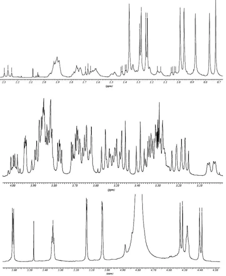
Figure S1. 1H NMR spectrum (500 MHz, CD
Triterpenoid Saponins from Lippia alba (Mill.) N. E. Brown J. Braz. Chem. Soc.
S2
Figure S2. Expansions of the 1H NMR spectrum (500 MHz, CD
Farias et al. S3 Vol. 21, No. 5, 2010
Figure S3. 2D J-Resolved spectrum (500 MHz, in CD3OD) of Lippiasaponin I (2).
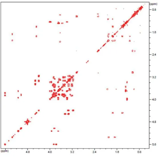
Figure S4. 1H-1H COSY spectrum (500 MHz, in CD
Triterpenoid Saponins from Lippia alba (Mill.) N. E. Brown J. Braz. Chem. Soc.
S4
Figure S5. 13C NMR spectrum (125 MHz, in CD
3OD) of Lippiasaponin I (2).
Farias et al. S5 Vol. 21, No. 5, 2010
Figure S7. RCT spectrum (500 MHz, in CD3OD) of Lippiasaponin I (2).
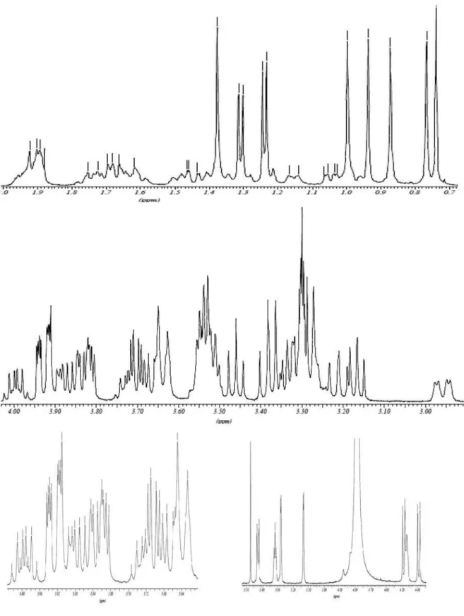
Figure S8. 1H NMR spectrum (500 MHz, CD
Triterpenoid Saponins from Lippia alba (Mill.) N. E. Brown J. Braz. Chem. Soc.
S6
Figure S9. Expansions of the 1H NMR spectrum (500 MHz, CD
Farias et al. S7 Vol. 21, No. 5, 2010
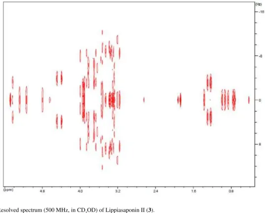
Figure S10. 2D J-Resolved spectrum (500 MHz, in CD3OD) of Lippiasaponin II (3).
Figure S11. 1H-1H COSY spectrum (500 MHz, in CD
Triterpenoid Saponins from Lippia alba (Mill.) N. E. Brown J. Braz. Chem. Soc.
S8
Figure S12. 13C NMR spectrum (125 MHz, in CD
3OD) of Lippiasaponin II (3).
Farias et al. S9 Vol. 21, No. 5, 2010