(1) Instituto Adolfo Lutz, São Paulo, SP, Brasil. (2) Instituto Adolfo Lutz, Campinas, SP, Brasil.
MOLECULAR EPIDEMIOLOGY OF A NOSOCOMIAL OUTBREAK DUE TO Enterobacter cloacae AND
Enterobacter agglomerans IN CAMPINAS, SÃO PAULO, BRAZIL
Célia R. GONÇALVES(1), Tania M.I.VAZ(1), Daniela LEITE(1), Beatriz PISANI(2), Marise SIMÕES(2), Maria Angela M. PRANDI(2), Marilu M.M. ROCHA(2), Paulo C. CESAR(3), Plinio TRABASSO(4), Angela von NOWAKONSKI(4) & Kinue IRINO(1)
SUMMARY
A total of 73 isolates (57 Enterobacter cloacae and 16 Enterobacter agglomerans), recovered during an outbreak of bacteremia
in the Campinas area, São Paulo, Brazil, were studied. Of these isolates, 61 were from parenteral nutrition solutions, 9 from blood cultures, 2 from a sealed bottle of parenteral nutrition solution, and one was of unknown origin. Of the 57 E. cloacae isolates, 54 were biotype 26, two were biotype 66 and one was non-typable. Of 39 E. cloacae isolates submitted to ribotyping, 87.2% showed the same
banding pattern after cleavage with EcoRI and BamHI. No important differences were observed in the antimicrobial susceptibility
patterns among E. cloacae isolates exhibiting the same biotype, serotype and ribotype. All E. agglomerans isolates, irrespective of
their origin, showed same patterns when cleaved with EcoRI and BamHI. The results of this investigation suggest an intrinsic
contamination of parenteral nutrition solutions and incriminate these products as a vehicle of infection in this outbreak.
KEYWORDS: Nosocomial outbreak; Enterobacter cloacae; Enterobacter agglomerans; Serotypes; Ribotyping .
INTRODUCTION
Enterobacter species are emerging as important human pathogens,
particularly among hospitalized patients5,14,33,37. Among 13 species currently recognized as members of the genus Enterobacter11,12, E.
cloacae and E. aerogenes are the most frequently isolated species
associated with human diseases1,10,17,19,30. E. agglomerans, commonly found in plants, soil, water and food stuffs, although rarely recognized as an agent of endogenous nosocomial infections6,8,14, can cause epidemics among hospitalized patients when associated with the use of contaminated intravenous products due to its ability to grow in commercial infusion fluids22,23.
Single source outbreaks of Enterobacter infections have been
frequently traced to contaminated intravenous products, blood products, distilled water and pressure monitoring devices2,7,22,24,25,31,32,34,35. A long-lasting epidemic caused by Enterobacter spp involving many hospitals and associated with the use of intrinsically contaminated intravenous solutions was reported in the 1970’s in the United States by MAKI et al.22. In March 24, 1997, one hospital of Campinas County, São Paulo State, Brazil, notified the Municipal Secretary of Health of an outbreak of bacteremia associated with intravenous therapy. According to the epidemiological investigation28 , this outbreak began between 21 and 22 March, and was associated with the use of a parenteral nutrition solution produced by one manufacturer and bearing the same batch number.
Enterobacter cloacae and Enterobacter agglomerans were isolated from
in-use infusion bags available in two of eight affected hospitals of Campinas County. Among 49 affected patients of this Municipality, 12 died (two adults and 10 newborns). Bacteremia was the typical clinical manifestation seen in the majority of patients receiving infusion bags after delivered by the manufacturer.
The purpose of this study was to investigate, by phenotypical and genotypic methods, the relatedness of E. cloacae and E. agglomerans
strains recovered during this outbreak.
MATERIAL AND METHODS
Bacterial strains. A total of 73 isolates, 57 of them Enterobacter cloacae, and 16 Enterobacter agglomerans, were studied. Of the 57 E. cloacae isolates, 7 were from blood cultures, 48 from solutions
administered to patients, one was from a sealed bottle and one was of unknown origin. Concerning the 16 E. agglomerans isolates, 2 were
Table 1
Origin and phenotypic and genotypic characteristics of enterobacteria isolated during the outbreak associated with the use of parenteral nutrition solution, March, 1997.
Patient Source Microorganism Biotype Serotype Ribotype Antimicrobial Resistance Pattern
1 PNS1 E. cloacae 26 O3 E1B1 CX CF AP GN
E. cloacae 66 NT2 E3B3 CX CF AP
2 PNS E. cloacae NT NT E5B5 CX CF AP
3 PNS E. cloacae 26 O3 E1B1 CX CF GN
4 PNS E. cloacae 26 O3 E1B1 CX CF AP GN
E. cloacae 26 R E1B1 CX CF GN
E. agglomerans -3 - E6B6 S
Blood E. cloacae 26 O3 E1B1 CX CF AP GN
E. agglomerans - - E6B6 S4
5 PNS E. cloacae 26 O3 E1B1 CX CF AP GN
E. cloacae 26 R5 E1B1 CX CF GN
E. cloacae 26 O3 - CX CF AP GN
E. agglomerans - - E6B6 S
6 PNS E. cloacae 26 O3 E1B1 CX CF AP GN
E. cloacae 26 R E1B1 CX CF AP GN
E. cloacae 26 O3 - CX CF AP GN
E. agglomerans - - E6B6 S
Blood E. cloacae 26 03 E1B1 CX CF AP GN
7 PNS E. cloacae 26 O3 E1B1 CX CL AP GN
E. cloacae 66 NT E4B4 Multiresistant6
E. cloacae 26 O3 - CX CF AP GN
E. agglomerans - - E6B6 Multiresistant7 8 PNS E. cloacae 26 O19 E2B2 Multiresistant 8
9 PNS E. cloacae 26 O3 E1B1 CX CF AP GN
E. cloacae 26 R - CX CF AP GN
E. cloacae 26 R - CX CF AP GN
E. agglomerans - - E6B6 S
Blood E. cloacae 26 O3 E1B1 CX CF AP GN
10 PNS E. cloacae 26 O3 E1B1 CX CF AP GN
E. cloacae 26 O3 - CX CF AP GN
E. cloacae 26 R - CX CF AP GN
11 Blood E. cloacae 26 R E1B1 CX CF AP GN
Patient Source Microorganism Biotype Serotype Ribotype Antimicrobial Resistance Pattern
13 PNS E. cloacae 26 O3 E1B1 CX CF AP GN
E. cloacae 26 O3 - CX CF AP GN
E. agglomerans - - E6B6 S
? E. cloacae 26 O3 E1B1 CX CF
14 PNS E. cloacae 26 O3 E1B1 CX CF AP GN
E. cloacae 26 R - CX CF AP GN
E. agglomerans - - E6B6 S
15 PNS E. cloacae 26 O3 E1B1 CX CF AP GN
E. cloacae 26 O3 - CX CF AP GN
E. cloacae 26 R - CX CF AP GN
16 PNS E. cloacae 26 O3 E1B1 CX CF AP GN
E. cloacae 26 O3 - CX CF AP GN
17 PNS E. cloacae 26 O3 E1B1 CX CF AP GN
18 PNS E. cloacae 26 O3 E1B1 CX CF AP GN
E. agglomerans - - E6B6 S
Blood E. agglomerans - - E6B6 S
19 Blood E. cloacae 26 O3 E1B1 CX CF AP GN
20 PNS E. cloacae 26 R E1B1 CX CF AP GN
E. cloacae 26 R - CX CF AP GN
E. agglomerans - - E6B6 S
21 PNS E. cloacae 26 O3 E1B1 CX CF AP GN
E. cloacae 26 O3 - CX CF AP GN
E. cloacae 26 O3 - CX CF AP GN
E. cloacae 26 R - CX CF AP GN
E. agglomerans - - E6B6 S
E. agglomerans - - E6B6 AP GN TT
E. agglomerans - - - S
22 PNS E. cloacae 26 O3 E1B1 CX CF AP GN
23 PNS E. cloacae 26 O3 E1B1 CX CF AP GN
24 PNS E. cloacae 26 O14 NT CX CF AP
25 PNS E. cloacae 26 O3 E1B1 CX CF AP GN
E. cloacae 26 O3 - CX CF AP GN
E. cloacae 26 R - CX CF AP GN
E. agglomerans - - E6B6 AP GN TT
26 PNS E. cloacae 26 O3 E1B1 CX CF AP GN
27 PNS E. cloacae 26 O3 E1B1 CX CF AP GN
28 Blood E. cloacae 26 O3 E1B1 CX CF AP GN
29 Blood E. cloacae 26 O3 E1B1 CX CF AP GN
Sealed Bottle E. cloacae 26 O3 E1B1 CX CF AP GN
E. agglomerans - - E6B6 S
Biochemical identification. All isolates were subjected to biochemical identification9, and all E. cloacae were biotyped as previously described27,36.
Serotyping. Nineteen E. cloacae isolates were serotyped in the
Central Public Health Laboratory, Laboratory of Hospital Infection, Colindale, London, UK. The remaining 38 E. cloacae were serotyped at
Instituto Adolfo Lutz, Central Public Health Laboratory, using O-antisera prepared with standard E. cloacae strains received from the Laboratory
of Hospital Infection, London, UK. O-serotyping was performed according to methods previously described15.
Antibiotic susceptibility testing. The susceptibility of all isolates to antimicrobial agents was determined by the standard disk diffusion method26 using the following commercial disks (Cecon, Centro de Controle e Produtos para Diagnósticos, S. Paulo, Brazil): amikacin (AM, 30µg), cefoperozone (CPZ, 75µg), cefotaxime (CTX, 30µg), imipenen (IPM, 10µg), ciprofloxacin (CIP, 5µg), tetracyclin (TT, 30µg), chloramphenicol (CLO, 30µg), sulfamethoxazole-trimethoprim (SUT, 1.25/23.75µg), ampicillin (AP, 10µg), cephalotin (CF, 30µg), gentamicin (GN, 10µg), and cephalexin (CX, 30µg).
Ribotyping. All isolates recovered from blood cultures were ribotyped as well as the isolates from sealed bottle. Among isolates from in-use parenteral nutrition solutions we selected at least one isolate of each bottle. Chromosomal DNA of E. cloacae and E. agglomerans was extracted and purified as previously described3. DNA samples (3 µg) were cleaved with the restriction enzymes EcoRI and BamHI according
to the reaction conditions recommended by the manufacturer (Pharmacia, LKB). DNA fragments were subjected to horizontal electrophoresis in 0.8% agarose (Sigma) in 0.04 M TRIS-acetate and 0.001 M EDTA buffer, and restriction fragments were transferred under vacuum (Vacugene, Pharmacia, LKB) to nylon membranes . The membranes were hybridized with the 16+23S cDNA probe transcribed by reverse transcriptase from
E. coli rRNA (Boerhinger Mannheim, Germany) and labelled with
digoxigenin according to POPOVIC et al.29. A Haemophilus aegyptius (strain # 3031) EcoRI DNA digest (fragment sizes of 1492 ,1713, 3228,
3789, 4960, 5575, 6334, and 17613 bp) was used as molecular marker. Fragment sizes were estimated with a computer program (DNASTAR Computer System for Molecular Biology and Genetics, London, UK).
RESULTS
Biotyping and serotyping. Of E. cloacae isolates 39 were assigned
to biotype 26 / serotype O3, 13 to biotype 26 / OR ( rough ), four belonged to other biotypes / serotypes and one was non-typable by biotyping or serotyping. The characteristics of all strains are summarized in Table 1.
Antimicrobial susceptibility patterns. The majority of E. cloacae
showed no important differences in the antimicrobial susceptibility patterns, being homogeneously resistant to ampicillin, cephalothin, cephalexin, and gentamicin. One isolate of biotype 26 / O19 and one of biotype 66 / NT were resistant to 9 drugs. Of 16 E. agglomerans isolates,
13 were susceptible to all tested drugs, 2 were resistant to ampicillin, gentamicin and tetracycline and one was resistant to ampicillin,
Ribotyping. DNA samples of 39 E. cloacae and 15 E. agglomerans
were digested with EcoRI and BamHI. All 34 E. cloacae isolates
belonging to biotype 26 and to serotype O3 or OR (rough isolates), irrespective of their origin (solution administered to patients, sealed bottle of parenteral nutrition solution and blood cultures) were homogeneously cleaved with Eco RI, generating 16 fragments ranging in size from 1.3 to 22.9 kp, and were assigned to pattern E1. These 34 isolates, which also showed a unique pattern (B1) with 8 bands ranging in size from 6.0 to 25.8 kp when cleaved with BamHI, were assigned to ribotype E1B1.
DNA sample (extraction was repeated twice) of one E. cloacae isolate
(biotype 26 / serotype O14) was sheared and was not digested. E. cloacae
isolates belonging to biotype 26 / serotype O19 (one isolate), biotype 66 / NT (two isolates) and NT / NT (one isolate) displayed patterns distinct from E1B1 when cleaved with EcoRI or BamHI and were classified as
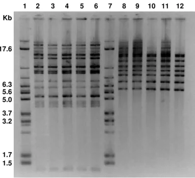
ribotypes E2B2, E3B3, E4B4, and E5B5, respectively. All E. agglomerans isolates when cleaved with EcoRI showed a unique pattern (E6) with 12 fragments ranging from 1.7 to 14.5 kp. The homogeneity of these isolates was confirmed with the second enzyme BamHI. All E. agglomerans isolates were classified as ribotype E6B6. Figure 1 shows
the banding patterns of E. cloacae and E. agglomerans recovered from
solutions administered to patients, from blood cultures and from one sealed bottle of parenteral nutrition solution, after digestion with EcoRI
and BamHI, respectively. Schematic representation of the banding
patterns of ribotypes of E. cloacae and E. agglomerans is shown in
Figure 2.
1 2 3 4 5 6 7 8 9 10 11 12
17.6
6.3 5.6 5.0
3.7 3.2
1.7 1.5 Kb
Fig. 1 - Banding patterns of E. cloacae belonging to biotype 26/serotype O3 (lanes 2 to 6) generated by EcoRI (pattern E1), and of E. agglomerans (lanes 8 to 12) after cleavage with
1 2 3 4 5 6 7 8 9 10 11 12 13 14 Kb
17.6
6.3 5.6 5.0
3.7 3.2
1.7 1.5
Fig. 2 - Schematic representation of the banding patterns E1 to E5 of E. cloacae obtained from DNA after digestion with EcoRI (lanes 2 to 6), and banding patterns B1 to B5 after digestion with
BamHI (lanes 9 to 13). Banding patterns of E. agglomerans from DNA after digestion with EcoRI (E6) and BamHI (B6) are represented in Lane 7 and 14. Lane 1 and 8, DNA molecular weight marker.
DISCUSSION
Phenotypic methods when used in combination could be sufficiently discriminatory and satisfactory to differentiate the majority of clinical isolates of E. cloacae13,36. Using conventional typing methods, we could differentiate a main group of 39 E. cloacae represented by isolates characterized as biotype 26 / serotype O3. E. cloacae serotype O3 strains
are the most frequently isolated serotype from human infections16,36 and are probably widely disseminated in the environment.
This group of phenotypically similar isolates was genetically homogeneous as determined by ribotyping. On the other hand, four isolates which belonged to distinct biotypes and serotypes were also heterogeneous when assayed by ribotyping.
Biochemical and genotypic similarities shared by E. cloacae isolated from a sealed bottle of parenteral nutrition solution and E. cloacae isolated
from solutions administered to patients point to the intrinsic contamination of these solutions, probably from environmental origin because of the antimicrobial susceptibilty pattern. Extrinsic contamination of the solutions administered to patients is unlikely, considering that nocomial strains of E. cloacae are usually multiresistant.
Multiple resistant E. cloacae strains belonging to some biotypes such
as biotype 26 and 66 are frequently associated with human infections probably because these strains are present in some hospital units such as intensive care units37. It was not surprising that such nosocomial pathogens could be isolated from some infusion bottles as was seen with Patient 7, where in addition to the intrinsic contamination, an extrinsic contamination by multiple resistant nosocomial E. cloacae
(biotype 66 / NT and ribotype E4B4) problably occurred. An extrinsic
Patient 8 from which it was isolated a multiple resistant E. cloacae strain belonging to biotype 26 / serotype O19, and ribotype E2B2.
The high genetic heterogeneity of E. agglomerans4,20,21 makes it difficult to establish a satisfactory system for its identification. According to FARMER11, only cultures that are yellow pigmented and triple decarboxylase-negative strains are usually identified as E. agglomerans.
All of our isolates that were triple decarboxylase-negative, although none was yellow pigmented, were identified as E. agglomerans. The lack of
phenotypic methods to discriminate strains and to support similarities among strains of E. agglomerans is a drawback in epidemiological studies. In this study the relatedness of E. agglomerans strains isolated
from solutions administered to patients, from blood cultures and from a sealed bottle of parenteral nutrition solution could be only confirmed by ribotyping. Both EcoRI and BamHI provided a precise evidence of strains
relatedness.
Ribotyping proved to be an extremely useful method to confirm the identity of our isolates as other investigators had already reported13,18,35,38. In our sample, most of E. cloacae isolates were assigned to the same
ribotype (E1B1) which assesses the genetic similarity of the isolates of the main group identified as biotype 26/serotype O3 or 26/OR of distinct origins.
Our results suggest that E. cloacae and E. agglomerans were present
RESUMO
Epidemiologia molecular de um surto de bacteriemia por
Enterobacter cloacae e Enterobacter agglomerans ocorrido na
região de Campinas, S. Paulo, Brasil
Foram estudadas um total de 73 cepas (57 de E. cloacae e 16 E. agglomerans), isoladas durante um surto de bacteriemia ocorrido na
região de Campinas, S. Paulo. Entre estas cepas, 61 foram isoladas de solução de nutrição parenteral, 9 de sangue, 2 de bolsa fechada de solução de nutrição parenteral e uma era de origem desconhecida. Entre as 57 cepas de E. cloacae, a maioria das cepas foram do biotipo 26/sorotipo
O3 (39 cepas) e do biotipo 26/OR (13). Entre as 39 cepas de E. cloacae
ribotipadas, 87,2% apresentaram o mesmo padrão de bandas com EcoRI
e BamHI. Cepas de E. cloacae pertencentes ao mesmo biotipo, sorotipo
e ribotipo não apresentaram diferenças significativas em relação ao padrão de sensibilidade aos agentes antimicrobianos. Todas as cepas de E. agglomerans, independente da origem, pertenciam ao mesmo ribotipo
após a clivagem com EcoRI e BamHI. Os resultados obtidos sugerem
uma contaminação intrínseca das soluções de nutrição parenteral, incriminando-as como o veículo de transmissão dos agentes etiológicos do surto.
ACKNOWLEDGEMENT
We thankDr T.L. Pitt, Laboratory of Hospital Infection, PHLS Central Public Health Laboratory, Colindale, London, UK, for typing our 19 E. cloacae isolates and for sending us the serotype strains for E. cloacae.
REFERENCES
1. ARPIN, C.; ROGUES, A.M.; GACHIE, J.P. et al. - Epidemiological study of an outbreak due to multidrug-resistant Enterobacter aerogenes in a medical intensive care unit.
J. clin. Microbiol., 34: 2163-2169, 1996.
2. BENNETT, S.N.; McNEIL, M.M.; BLAND, L.A. et al. - Postoperative infections traced to contamination of an intravenous anesthetic, propofol. New Engl. J. Med., 333: 147-154, 1995.
3. BRENNER, D.J.; McWHORTER, A.C.; KNUDSON, J.K. & STEIGERWALT, A.G.
-Escherichia vulneris: a new species of Enterobacteriaceae associated with human wounds. J. clin. Microbiol., 15: 1133-1146, 1982.
4. BRENNER, D.J.; FANNING, G.R.; KNUTSON, J.K.L. et al. - Attempts to classify Herbicola Group-Enterobacter agglomerans strains by deoxyribonucleic acid hybridization and phenotypic tests. Int. J. system. Bact., 34: 45-55, 1984. 5. BODEY, G.P.; ELTING, L.S. & RODRIGUES, S. - Bacteremia caused by Enterobacter:
15 years of experience in a cancer hospital. Rev. infect. Dis., 13: 550-558, 1991. 6. BURCHARD, K.W.; BARRAL, D.T.; REED, M. & SLOTMAN, G.J. - Enterobacter
bacteremia in surgical patients. Surgery, 100: 857-861, 1986.
7. BUCHHOTZ, D.H.; YOUNG, V.M.; FRIEDMAN, R.R. et al. - Bacterial proliferation in platelet products stored at room temperature: transfusion-induced Enterobacter sepsis.
New Engl. J. Med., 285: 429-433, 1971.
8. CHOW, J.W.; FINE, M.J.; SHALAES, D.M. et al. - Enterobacter bacteremia: clinical
9. EWING, W.H. - Edwards and Ewing’s identification of Enterobacteriaceae. 4. ed. New York, Elsevier Science, 1986.
10. FALKINER, F. R. - Enterobacter in hospital. J. Hosp. Infect., 20: 137-140, 1992. 11. FARMER III, J.J. - Biochemical identification of new species and biogroups of
Enterobacteriaceae isolated from clinical specimens. J. clin. Microbiol., 21: 46-76, 1985.
12. FARMER III, J.J. - Enterobacteriaceae: introduction and identification. In: MURRAY, P.R.; BARON, J.; TENOVER, F.C. & YOLKEN, R.H., ed. Manual of clinical microbiology. 6.ed. Washington, American Society of Microbiology, 1995. p.438-449.
13. GARAIZAR, J.; KAUFMANN, E. & PITT, T.L. - Comparison of ribotyping with conventional methods for the type identification of Enterobacter cloacae. J. clin. Microbiol., 29: 1303-1307, 1991.
14. GALLAGHER, P.G. - Enterobacter bacteremia in pediatric patients. Rev. infect. Dis., 12: 808-812, 1990.
15. GASTON, M.A.; BUCHER, C. & PITT, T.L. - O serotyping scheme for Enterobacter cloacae. J. clin. Microbiol., 18: 1079-1083, 1983.
16. GASTON, M.A.; CREES-MORRIS, J. & PITT, T.L. - Serotypes and biochemical profiles of British hospital strains of Enterobacter cloacae in relation to site of infection and antibiotic susceptibility. J. Hosp. Infect., 10: 17-27, 1987.
17. GHELDRE, Y.; MAES, N.; ROST, F. et al. - Molecular epidemiology of an outbreak of
multidrug-resistant Enterobacter aerogenes infections and in vivo emergence of imipenem resistance. J. clin. Microbiol., 35: 152-160, 1997.
18. GRATTARD, F.; POZZETTO, B.; BERTHELOT, P. et al. - Arbitrarily primed PCR,
ribotyping, and plasmid pattern analysis applied to investigation of a nosocomial outbreak due to Enterobacter cloacae in a neonatal intensive care unit. J. clin. Microbiol., 32: 596-602, 1994.
19. JALALUDDIN, S.; DEVASTER, J.M.; SCHEEN, R. et al. - Molecular epidemiological study of nosocomial Enterobacter aerogenes isolates in a Belgium hospital. J. clin. Microbiol., 36: 1846-1852, 1998.
20. LINDH, E.; KJAELDGAARD, P.; FREDERIKSEN, W. & URSING, J. - Phenotypical properties of Enterobacter agglomerans (Pantoea agglomerans) from human, animal
and plant sources. Acta path. microbiol. immunol. scand. Sect. B , 99: 347-352, 1991.
21. LIND, E & URSING, J. - Clinical strains of Enterobacter agglomerans (synonyms: Erwinia herbicola, Erwinia milletiae) identified by DNA-DNA hybridization. Acta path. microbiol. immunol. scand. Sect. B, 94: 205-213, 1986.
22. MAKI, D.G.; RHAME, F.S.; MACKEL, D.C. & BENNETT, J.V. - Nationwide epidemic of septicemia caused by contaminated intravenous products. I. Epidemiologic and clinical features. Amer. J. Med., 60: 471-485, 1976.
23. MAKI, D.G. & MARTIN, W.T. - Nationwide epidemic of septicemia caused by contaminated intravenous products. IV. Growth of microbial pathogens in fluids for intravenous infusion. J. infect. Dis., 131: 267-272, 1975.
24. MAYHALL, C.G.; LAMB, V.A .; GAYLE Jr., W.E. & HAYNES Jr., B.W. - Enterobacter cloacae septicemia in a burn center: epidemiology and control of an outbreak. J. infect. Dis., 139: 166-171, 1979.
25. MIRZA, G.E.; KARAKÜCÜK, S.; DOGANAY, M. & CAGLAYANGIL, A. -Postoperative endophthalmitis caused by Enterobacter species. J. Hosp. Infect., 26: 167-172, 1994.
-27. OLD, D.C. - Biotyping of Enterobacter cloacae. J. clin. Path., 35: 875-878, 1982. 28. PREFEITURA MUNICIPAL DE CAMPINAS. SECRETARIA MUNICIPAL DE
SAÚDE – Investigação de surto de bacteriemia/sepsis relacionado à administração de nutrição parenteral prolongada (NP) contaminada com a bactéria Enterobacter
sp. Comissão de Investigação – Portaria SMS N 05/97. Abril/Maio de 1997. 29. POPOVIC, T.; BOPP, C.A. & WACHSMUTH, K. - Epidemiologic application of a
standardized ribotype scheme for Vibrio cholerae O1. J. clin. Microbiol., 31: 2474-2482, 1993.
30. SANDERS Jr., W.E. & SANDERS, C. - Enterobacter spp: pathogens poised to flourish at the turn of the century. Clin. microbiol. Rev., 10: 220-241, 1997.
31. STRUELENS, M.J.; ROST, F.; DEPLANO, A. et al. - Pseudomonas aeruginosa and Enterobacteriaceae bacteremia after biliary endoscopy: an outbreak investigation using DNA macrorestriction analysis. Amer. J. Med., 95: 489-498, 1993. 32. THOMAS, A.; LALITHA, M.K.; JESUDASON, M.V. & JOHN, S. - Transducer related
Enterobacter cloacae sepsis in postoperative cardiothoracic patients. J. Hosp. Infect., 25: 211-214, 1993.
33. VAN NIEROP, W.J.; DUSE, A.G.; STEWART, R.G. et al. - Molecular epidemiology of
an outbreak of Enterobacter cloacae in the neonatal intensive care unit of a provincial hospital in Gauteng, South Africa. J. clin. Microbiol., 36: 3085-3087, 1998. 34. WAGNER, S.J.; FRIEDMAN, L. & DODD, R.Y. - Transfusion-associated bacterial sepsis.
Clin. microbiol. Rev., 7: 290-302, 1994.
35. WANG, C.C.; CHU, M.L.; HO, L.J. & HWANG, R.C. - Analysis of plasmid pattern in paediatric intensive care unit outbreaks of nosocomial infection due to Enterobacter cloacae. J. Hosp. Infect., 19: 33-40, 1991.
36. WEISCHER, M.; KOLMOS, H.J.; KAUFMANN, M.E. & ROSDAHL, V.T. - Biotyping, phage typing, and O-serotyping of clinical isolates of Enterobacter cloacae. Acta path. microbiol. immunol. scand. Sect. B, 101: 838-844, 1993.
37. WEISCHER, M. & KOLMOS, H.J.- Retrospective 6-year study of enterobacter bacteraemia in a Danish university hospital. J. Hosp. Infect., 20: 15-24, 1992. 38. WEISCHER, M. & KOLMOS, H.J. - Ribotyping of selected isolates of Enterobacter
cloacae and clinical data related to biotype, phage type, O-serotype, and ribotype.
Acta path. microbiol. immunol. scand. Sect. B, 101: 879-886, 1993. Received: 03 September 1999
SUMMARY OF THESIS*
CAVALHEIRO, Norma de Paula – Análise dos sorotipos do VHC identificados em pacientes da cidade de São Paulo, através de método imunoenzimático. São Paulo, 1999. (Dissertação de Mestrado - Faculdade de Medicina da Universidade de São Paulo).
ANALYSIS OF SEROTYPES OF HCV IN PATIENTS FROM THE CITY OF SÃO PAULO, BY MEANS OF A
ENZYME-IMMUNOASSAY METHOD
With the objective of analyzing the prevalence of the different types of Hepatitis C Virus (HCV) in a population of chronic carriers of HCV, through a serologic method (MUREX HCV Serotyping Assay), 219 patients were studied who showed a positive polymerase chain reaction. This sera were submitted to immunoenzymatic tests for the detection of antibodies in relation to HCV types 1,2,3,4,5 and 6. The samples were diluted and incubated in the presence of heterologous competing peptides, with microwells coated with serotype-specific antigens of HCV. Of the 219 patients, it was possible to detect the HCV serotype in 166, revealing a sensitivity of 75.8%. The results showed a predominance of type 1 (70.0%) in our medium, followed by type 3 (22.3%) and type 2 (4.2%). Serotypes 4 and 5 were present in 1.8% of the patients, but always associated with serotype 1. These samples, in spite of fulfilling the prerequisites of validity for testing, showed a very high optical density reading for all types of viruses tested, including positive and negative controls. The possibility of cross reactions in these cases should be considered. Confirmation by genotyping and a more detailed investigation on the origin and mode of acquisition of the HCV of these patients should be researched. Type 6 was not confirmed in any of the samples tested and probably was not present in this particular collection. The epidemiological parameters evaluated were: age, sex and means of