The solution structure of the soluble form of the lipid-modi
fi
ed azurin
from
Neisseria gonorrhoeae
, the electron donor of cytochrome
c
peroxidase
Cláudia S. Nóbrega
a, Ivo H. Saraiva
b, Cíntia Carreira
a, Bart Devreese
c,
Manolis Matzapetakis
b, So
fi
a R. Pauleta
a,⁎
aUCIBIO, REQUIMTE, Departamento de Química, Faculdade de Ciências e Tecnologia, Universidade Nova de Lisboa, Campus da Caparica, 2829-516 Caparica, Portugal bInstituto de Tecnologia Química e Biológica António Xavier, Universidade Nova de Lisboa, Av. da República, 2780-157 Oeiras, Portugal
cLaboratory of Protein Biochemistry and Biomolecular Engineering, Ghent University, K.L. Ledeganckstraat 35, B-9000 Ghent, Belgium
a b s t r a c t
a r t i c l e
i n f o
Article history:
Received 27 July 2015
Received in revised form 25 October 2015 Accepted 13 November 2015
Available online 14 November 2015
Neisseria gonorrhoeaecolonizes the genitourinary track, and in these environments, especially in the female host, the bacteria are subjected to low levels of oxygen, and reactive oxygen and nitrosyl species. Here, the biochemical characterization ofN. gonorrhoeaeLaz is presented, as well as, the solution structure of its soluble domain deter-mined by NMR.N. gonorrhoeaeLaz is a type 1 copper protein of the azurin-family based on its spectroscopic prop-erties and structure, with a redox potential of 277 ± 5 mV, at pH 7.0, that behaves as a monomer in solution. The globular Laz soluble domain adopts the Greek-key motif, with the copper center located at one end of theβ-barrel coordinated by Gly48, His49, Cys113, His118 and Met122, in a distorted trigonal geometry. The edge of the His118 imidazole ring is water exposed, in a surface that is proposed to be involved in the interaction with its redox partners. The heterologously expressed Laz was shown to be a competent electron donor to
N. gonorrhoeaecytochromecperoxidase. This is an evidence for its involvement in the mechanism of protection against hydrogen peroxide generated by neighboring lactobacilli in the host environment.
© 2015 Elsevier B.V. All rights reserved.
Keywords: Neisseria
Copper protein Azurin Laz
Cytochromecperoxidase Solution NMR structure
1. Introduction
During infection, pathogenic microorganisms are frequently ex-posed to oxidative stress due to reactive oxygen species (ROS) generat-ed by host defense mechanisms. Some of the most common ROS found in biological systems include superoxide anion (O2−
), hydrogen perox-ide (H2O2) and hydroxyl radical (HO)[1]. However, there are other sources of ROS, such as the bacteria's own metabolic process, exposure to factors within host environment, such as metal ions, or commensal organisms that generate oxidants. Pathogenic bacteria from the species Neisseria gonorrhoeaeandNeisseria meningitidis, that cause gonorrhea and meningitis, respectively, are constantly exposed to ROS from these various sources and, therefore, have developed numerous defense mechanisms to cope with oxidative stress[2]. These mechanisms are es-sential to cell survival, since ROS can damage DNA, proteins and cell membranes.
One of these defense mechanisms is based on cytochromec peroxi-dases. These enzymes arec-type heme containing periplasmic enzymes that catalyze the reduction of hydrogen peroxide to water, usingc-type
cytochromes or small type 1 copper proteins, of the respiratory chain, as electron donors[3,4].
An example of such an electron donor is azurin, a member of a family of copper-containing, water soluble, low molecular weight redox pro-teins called cupredoxins, which function as electron shuttles in the elec-tron transfer chain of several microorganisms, including some well-known pathogens, such asPseudomonas(Ps.)aeruginosa. These proteins have received great attention due to their unusual spectroscopy and electronic structure in the oxidized copper(II) form, as well as, their ability to transfer electrons rapidly to several enzymes[5,6].
Ps. aeruginosaazurin has been shown to be involved in the protec-tion against oxidative stress, since a mutant lacking this protein was shown to be very sensitive to ROS[1,7]. In another pathogenic bac-terium,N. gonorrhoeae, a mutant strain lacking the gene coding for a cupredoxin, the lipid-modified azurin (Laz), was found to be much more sensitive to hydrogen peroxide but not to superoxide, unlike the Ps. aeruginosaazurin mutant, resulting in reduced survival in human ectocervical epithelial cells[2].
TheNeisserialipid-modified azurin has a high sequence homology to other copper proteins from the azurin family[8,9], though it differs significantly from other members of this family in that it contains a N-terminal domain of 39 amino acids that encodes the H.8 epitope (common to pathogenicNeisseria), in which there arefive imperfect ⁎ Corresponding author.
E-mail address:[email protected](S.R. Pauleta).
http://dx.doi.org/10.1016/j.bbabio.2015.11.006
0005-2728/© 2015 Elsevier B.V. All rights reserved.
Contents lists available atScienceDirect
Biochimica et Biophysica Acta
affinity for oxygen, and can also use nitrite as an alternative electron ac-ceptor, as they present an incomplete denitrification chain, composed by a copper nitrite reductase (AniA) and nitric oxide reductase NorB[14]. InPs. aeruginosa, azurin can donate electrons to enzymes of the de-nitrification pathway, and thus, the role forNeisseriaLaz as electron donor to AniA had been proposed, since these two proteins share the same cellular location, as being tethered to the outer membrane[10]. Nevertheless, in the case ofN. gonorrhoeae, it was shown recently that the membrane bound cytochromec5and CcoP are essential as electrons shuttles to AniA[13]. Therefore, the question remains as to the identity of the redox partner(s) of Laz inN. gonorrhoeae.
Based on primary sequence homology, Laz and azurin belong to the same cupredoxin family, the azurin-family[15]. These small redox pro-teins have distinct spectroscopic properties, with a strong absorption band at around 600 nm (with anεof 3–6 mM−1cm−1), which has been assigned to a Scys–Cu charge transfer band, and a redox potential higher than the one found in most inorganic copper complexes[16]. In cupredoxins, the copper atom adopts a distorted tetrahedral or bipy-ramidal geometry, with three of the four/five copper coordinating resi-dues located at the C-terminus (Cys, His and Met) and the other(s) at the N-terminus. In the case of azurin, thefifth copper ligand is an oxy-gen from the carbonyl peptide bond of a glycine that is coordinating the copper atom, making the geometry more bipyramidal[15]. The structure ofN. gonorrhoeaeLaz incorporating Zn instead of Cu was re-cently determined by X-ray at 1.9 Å resolution (PDB ID 3AY2)[17]. In this work, we proceeded to solve the solution structure with Cu so as to evaluate any differences between Zn-Laz and the native like form es-pecially since Zn(II) prefers a quadrangular planar geometry.
This work focuses also on the biochemical characterization ofN. gonorrhoeaeLaz, as well as, on the determination of the solution struc-ture of its cupredoxin domain. Results obtained show that Laz can do-nate electrons to cytochromecperoxidase from the same organism, which might explain the involvement of this cupredoxin in the defense against oxidative stress.
2. Materials and methods
2.1. Protein purification
The gene encoding the soluble part of Laz was cloned into a pET vec-tor and the protein was heterologously produced inEscherichia coli
Copper (I) content was determined using a modified version of the method of Hanna et al.[19], which is based on the formation of a com-plex between CuIand 2, 2′-biquinoline in an acetic acid medium. All so-lutions were prepared fresh in deionized water. A sample of Laz (100μL, containing 10–30 nmol of protein) or a standard solution of copper ac-etate was reduced by adding 300μL of 20 mM sodium ascorbate (in 0.1 M sodium phosphate at pH 6.0) and incubated for 30 min. To this, 600μL of a 2,2′-biquinoline solution (0.5 mg/mL) prepared in glacial acetic acid was added, and the solution was incubated for 10 min prior to the measurement of the absorbance at 546 nm. The concentra-tion of CuIpresent in each sample was determined using the slope of the calibration curve prepared with copper(II) acetate. The extinction
coef-ficient obtained (6.3 mM−1 cm−1
) was identical to that described by Hanna et al.[19]. The method used to quantify the total protein content was the BCA method (Sigma)[20], according to the manufacturer's in-structions. The standard protein used was bovine serum albumin.
The extinction coefficient was determined taking into account the copper concentration of the sample, as a contamination with another copper protein is unlikely to occur, and it is rather difficult to have a pure protein sample (usually considered as pure when it isN95% purity).
2.2.2. Determination of molecular mass
The molecular mass of the purified Laz was determined by electrospray ionization mass spectrometry (ESI-MS). The apparent molecular mass of Laz was estimated by molecular-exclusion chromatog-raphy, using a Superdex 75 10/300 GL (GE Healthcare). The column was equilibrated with 50 mM Tris–HCl, pH 7.6 with or without 150 mM NaCl. Samples of oxidized Laz (1 nmol) were prepared in the running buffer. A calibration curve was prepared using the Gel Filtration Calibration Kit Low Molecular Weight (GE Healthcare) in the same running buffer, according to the manufacturer's instructions. The chromatograms to-gether with the analysis of this data are shown as Supplementary mate-rial (Fig. S1 and Fig. S2).
2.2.3. Spectroscopic characterization
The visible-spectra were recorded on a Shimadzu UV-1800 spectro-photometer using 1 cm quartz cells. Laz samples were oxidized with a solution of potassium ferricyanide and reduced with a solution of sodi-um ascorbate.
The Laz sample for EPR was 0.3 mM in 20 mM phosphate buffer, pH 7.0. The EPR spectra of Laz were recorded on a X-band Bruker EMX
spectrometer equipped with a rectangular cavity (model ER 4102T) and an Oxford Instruments continuous liquid heliumflow cryostat. Experi-mental conditions are described in the legend of the spectrum. The simulation of the spectrum was performed using WINEPR SimFonia software version 1.2, from Bruker.
2.2.4. Redox potential
Voltammetric measurements were performed on a potentiostat AUTOLAB/PSTAT 12 (ECO Chemie, Utrecht, The Netherlands). Data were collected and analyzed using GPES software package (ECO Chemie). The electrode potential values reported here were referred to the standard hydrogen electrode (SHE). Voltammetric experiments were conducted at room temperature using a three-electrode confi gu-ration cell with a saturated Ag/AgCl reference electrode (+ 197 mV vs SHE at room temperature), a platinum wire as the counter electrode and an edge plane pyrolytic graphite (PGE) as the working electrode. Nitrogen gas was purged through the solution for at least 15 min to re-move any dissolved oxygen before each experiment.
Before each experiment, the PGE surface was polished by hand on a polishing cloth using a 1 and 0.3μm alumina slurry, sonicated for 5 min and rinsed well with Milli-Q water. The electrode was prepared by dropping a 3μL drop of working solution containing Laz (145μM) and left to dry at room temperature for 1 h. The scan-rate dependence of the current was measure between 5 and 150 mV/s, in a solution con-taining 100 mM phosphate buffer pH 7.0 and 100 mM NaCl.
The pH dependence of the redox potential was determined in the pH range from 5.5 to 8.5, in a solution containing 20 mM of acetate, 2-morpholinoethanesulfonic acid, 2-[4-(2-hydroxyethyl)piperazin-1-yl] ethanesulfonic acid and N-[tris(hydroxymethyl)methyl]-3-aminopropanesulfonic acid. The reduction potential was determined for each pH at 10 mV/s scan rate. The pH dependence of the reduc-tion potential was defined by one equilibrium and two pKavalues. The data wasfitted to Eq.1, according to the equilibrium shown in the inset ofFig. 4B.
EB0
¼ElpB0þ2:nF3RTlog10
1þ KoR Hþ
!
1þ KoO Hþ
! 0
B B B B @
1
C C C C A
ð1Þ
In the equation, E0′is the measured reduction potential,E lp °'
is the reduction potential at low pH, and KaO and KaR are the proton dissociation constants of the oxidized and reduced forms, respectively. A non-linear regressionfit (with nofixed parameters) in Excel was
used to estimate the parameters: Elp0
′= 294 ± 5 mV, pKaO= 6.8 ± 0.2 and pKaR= 7.9 ± 0.2.
2.2.5. Kinetic assays
The activity ofN. gonorrhoeaecytochromecperoxidase (NgCCP) with Laz as electron donor was monitored on an Agilent Diode Array spectrophotometer, by the increase in absorbance at 625 nm as result of Laz oxidation over time. Laz was reduced with 1 mM sodium ascor-bate and 10μM DAD for 30 min at room temperature, which were re-moved using a desalting column, NAP-5 (GE Healthcare), equilibrated with 10 mM MES, pH 6.0.
The E. coli periplasmatic fraction containing heterologously expressedNgCCP inE. coliBL21(DE3) was used directly in the assay (data not shown,NgCCP gene was inserted into pET22-b, and co-expressed with pEC86[21]). As a control experiment, a periplasmatic fraction of the sameE. colistrain co-transformed with pEC86 and pET22b, without the gene encodingNgCCP, and grown in same condi-tions was used. The periplasmic extract was obtained by 5 freeze-thaw cycles of the resuspended cells, followed by centrifugation at 48,000gin an Avanti J-26 XPI centrifuge (Beckman Coulter), during 10 min, at 4 °C.
The assay was performed at 25 °C in 10 mM MES pH 6.0 containing 10 mM NaCl and 1.0 mM CaCl2. The order of the assay additions was: buffer, Laz to thefinal concentration of 11μM, 0.02 or 0.004μg of total protein of periplasmatic fraction, and 100μM H2O2to initiate the assay. In the end, a small aliquot of potassium ferricyanide solution was added to fully oxidize and confirm that all Laz was oxidized in the assay.
2.3. NMR experiments and structure determination
The NMR samples were 1.5 mM Laz in 20 mM sodium phosphate, pH 7.0, 1 mM sodium azide, 5 mM sodium ascorbate and 10%2H2O. Sodium ascorbate was used to reduce the copper site of the protein, making it diamagnetic[18]. NMR data was collected, at 298 K, on a Bruker AvanceIII 800 MHz equipped with a TXI-HCN gradient probe and on a Bruker AvanceIII 600 MHz equipped with a TCI-cryoprobe. Res-onance assignments have already been reported[18]and deposited in the BioMagResBank (http://www.bmrm.wisc.edu) under BMRB acces-sion number 18636.
The NOE data from reduced Laz were obtained from the1H13C HSQC-NOESY experiment, acquired simultaneously with a 1H15N HSQC-NOESY experiment[22], with a mixing time of 120 ms.
ARIA2.3[23]and CNS1.21[24,25]were used for NOE assignment and structure calculation. TALOS-N[26]was used to predict dihedral Fig. 2.Schematic representation of the electron transfer chain ofNeisseria gonorrhoeaeand role of Laz and other cytochromes as electron acceptors/donors. The arrows represent electron transfer: black (established), blue (proposed here), dotted (proposed but not proven) and red (shown here). CcoN/CcoO/CcoP is cytochromecoxidasecbb3. Redox centers are represented
defined in the parameterfile (parallhdg5.3.pro). The Cu(I) atom mass and non-bonded parameters used were the ones present in the ion.top and ion.paramfiles from CNS1.21, respectively (see description of the
files in the Supplementary materials).
The restraints used for the structure calculations are summarized in
Table 1. The validation of the data was performed using CING[28]. The NMR data was deposited in the BioMagResBank (www.bmrb.wisc. edu) under accession number 18636 and the coordinates used for the ensemble of NMR structures have been deposited in the Protein Data Bank (http://www.rcsb.org/) under the PDB ID 2n0m.
As a remark, a disulfide bond was identified between C6 and C29 based on the NOE observed between the Hβprotons of these two resi-dues. This was added as a patch in the preparation of the calculation and not added as a restraint.
The RMSD between average NMR structure and the X-ray structure (PDB ID: 3AY2) was determined using the online tool SuperPose version 1.0 (http://wishart.biology.ualberta.ca/).
modified at the second cysteine residue with a diacylglyceride[9,10], and thus its cellular localization has been proposed to be the outer membrane with the azurin-like domain facing the inside of the periplasmic space[10,12], similarly to other gonococcal proteins, such as AniA, a copper containing nitrite reductase[29], and bacterial cytochromecperoxidase[30].
In order to obtain soluble protein for the structural characterization, the DNA sequence corresponding to this soluble azurin-like domain was cloned into a pET expression vector, with the signal peptide of Paracoccus pantotrophuspseudoazurin, which was previously shown to be well recognized by theE. coliSec-dependent pathway[31].
The expression conditions oflazgene inE. coliwere optimized, and in order to obtain Laz in the soluble periplasmic extract, the transformed cells had to be grown at 16 °C during 7 h, after induction with IPTG. Laz in the periplasmic extract was mostly in the reduced state, as upon ad-dition of some crystals of potassium ferricyanide, a bright blue color appears. The yield of the expression was 50 mg and 20 mg of purified Laz/L of rich and M9 medium, respectively.
The isolated Laz has a ratio of A625 nmof the oxidized to A278 nmof the untreated sample, of approximately 0.8 or higher, and could be consid-ered pure judged by its SDS-PAGE. The amount of copper per total pro-tein was determined to be 0.79 ± 0.04, as expected for a cupredoxin (with 1 Cu/protein).
The heterologously produced Laz has an apparent molecular mass of 21 ± 2 kDa or 25 ± 2 kDa in 50 mM Tris–HCl, pH 7.6, in the presence or absence of 150 mM NaCl, respectively (Figs. S1 and S2, Supplementary material). These values could indicate that Laz exhibits a monomer– dimer equilibrium (since the expected molecular mass from the prima-ry sequence is 13,633.3 kDa and the one determined by ESI-MS was 13,631 ± 1 Da). Nevertheless, backbone15N relaxation studies at the 800 MHz NMR spectrometer of the reduced form at low ionic strength (500μM Laz in 20 mM phosphate buffer, pH 7.0) gave an average R1 of 1.18 ± 0.05 s−1
and an R2 of 10.0 ± 0.1 s−1
. The resulting value of R2/R1 of 8.46 is consistent with a monomeric protein (τc of 6.5 ± 0.1 ns). In addition, the oxidized and reduced forms (at the same conditions) exhibited identical diffusion rates and R1 and R2 values, suggesting that the oxidized protein is also not a dimer under NMR conditions. Thus, the discrepancy between the two methods of molecular size determination could be attributed to the elongatedβ-barrel structure (vide infra) of Laz (higher Stokes volume) and to the need of a calibration curve with“standard proteins”in the size-exclusion chromatography.
3.2. Spectroscopic characterization
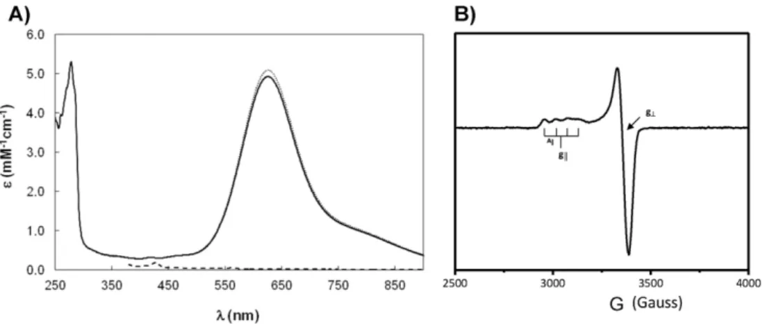
Laz visible spectrum, shown inFig. 3A, has a maximum absorption band at 625 nm. This absorption band has an extinction coefficient of 5.1 mM−1cm−1(considering the copper quanti
fication), and has a ligand-to-metal charge transfer character of Scys to Cu ion: CysS π→Cu2+(dx2−y2)[32], which is characteristic of the type 1 copper
proteins, and responsible for its intense blue color[33]. This other spec-troscopic feature of Laz is the small hyperfine coupling constant (A||) of Table 1
Summary of structural statistics obtained for the reducedNeisseriaLaz. The statistics pre-sented are for the 10 lowest energy structures after water refinement.
Number of distances and dihedral angle constraints
NOE-distance constraints 3345
Intra-residue 968
Sequential 699
Medium range (1b|i−j|b5) 373
Long range (|i−j|N4) 1305
Ambiguous 169
Dihedral angle constraints 65 Constraints statistics
r.m.s.d. of distance violations 0.154 ± 0.014 r.m.s.d. of dihedral violations 0.247 ± 0.059 Structure r.m.s.d.
Backbone 0.57 ± 0.07
Heavy atoms 0.93 ± 0.06
Structural quality Ramachandran (PROCHECK)
Most favored regions 86.1%
Allowed regions 13.4%
Generously allowed regions 0.4%
Disallowed regions 0.1%
WHATIF Z-scores
Backbone conformation −0.232 ± 0.355
2nd generation packing quality 7.349 ± 2.394 Ramachandran plot appearance −1.799 ± 0.514 χ1/χ2rotamer quality −3.056 ± 0.447
CING ROG analysis
Red 15%
Orange 22%
57 gauss[34,35], in the EPR spectrum of Laz (Fig. 3B), with g|| 2.266 and g┴2.048, which is due to the highly covalent character of the Cu2+– S(cys)πbond (vide infra, short distance between of Cu–S–Cys).
The wavelength of the maximum absorption in azurin and in Laz is redshifted by around 30 nm, when compared to other cupredoxins, which has been attributed to the existence of a disulfide bond between two N-terminalβ-strands (vide-infra)[16].
In fact, the maximum absorption band occurs at the same wavelength as the one observed for soluble azurins isolated from different microorganisms, such asPs. aeruginosa[36,37],Pseudomonas chlororaphis[38]andAlcaligenes denitrificans[39]. Likewise, the EPR parameters are very similar to ones of cupredoxins that belong to the azurin-family[38–40].
3.3. Redox properties of Laz
The electrochemical behavior of Laz formN. gonorrhoeaewas ana-lyzed at a PGE by proteinfilm voltammetry in a solution containing 100 mM phosphate pH 7.0 and 100 mM NaCl. Cyclic voltammograms obtained for different scan rates (5≤v≤150 mV/s) have quite symmet-rical oxidation and reduction peaks as can be observed inFig. 4A.
Analysis of the voltammograms shows that the peak currents (ip) vary linearly withv, indicative of a diffusionless one-electron redox
process with both the oxidized and the reduced forms being adsorbed. The ratio of the cathodic and anodic peak currents (ipa/ipc) are indepen-dent ofvand close to 1 for all scan rates. The width of half height (ΔEp,1/2) for anodic and cathodic peak has a value close to 90 mV.
Laz electrochemical behavior shows a reversible system of one elec-tron process and a formal potential could be calculated. For all scan rates, the average of E is constant within the experimental error and a formal reduction potentialE°′= 277 ± 5 mV was estimated at pH 7.0. The value of the estimated formal potential is similar to the one report-ed for azurin fromPs. aeruginosa(+ 267 mV) immobilized on a PGE
[41].
The pH dependence of the reduction potential (Fig. 4B) shows that this value decreases with pH. The variation observed can befitted with two pKa values, one for the reduced and another for the oxidized form of Laz, pKaO(6.8 ± 0.2) and pKaR(7.9 ± 0.2), which are similar to the ones determined forPs. aeruginosaazurin by electrochemistry using a PGE, (7.1 and 7.8, respectively)[42]. In the absence of additional biochemical data on Laz, these pKa values can be attributed to an ioniz-able group close to the copper center, probably a histidine residue due to the pKa values determined, as in the case ofPs. aeruginosaazurin (His35)[40,42]. In fact, this residue is conserved in the azurin family (in Laz it is His38) and is located in the second coordination sphere of the copper center[42]. In the case ofPs. aeruginosaazurin, the results Fig. 3.Spectroscopic properties ofNeisseria gonorrhoeaeLaz. A) UV–visible spectra of Laz in 20 mM phosphate buffer, pH 7.0. The as-isolated spectrum of Laz is shown as a continuous line, oxidized as dotted and the ascorbate reduced spectrum is shown as a dashed line. B) X-band EPR spectrum of 300μM Laz in 20 mM phosphate buffer, pH 7.0, 70 K, 25 dB, 5 G of modulation and 1 × 10⁵gain.
Fig. 4.Electrochemical behavior of Laz at a PGE and pH effect on the redox potential. A) Cyclic voltammograms (5≤v≤150 mV/s) at a PGE of 145μMNeisseria gonorrhoeaeLaz in 100 mM phosphate buffer, pH 7.0 and 100 mM NaCl. B) Effect of pH on Laz formal reduction potential. Experiments were performed and analyzed as described in Section2. Open andfilled circles are two independent titrations towards lower or higher pH values, respectively. The curve wasfitted with two pKavalues, 6.8 ± 0.2 and 7.9 ± 0.2, using Eq.(1). Inset: proton dissociation
The structure was determined using a total of around 3300 NOE-distance constraints (26 per residue, Fig. S5) and 65 dihedral angle constraints, with the complete statistics shown inTable 1.
It is worth mentioning that about 1300 of the total NOEs are long range distance restraints (39%), which results in afinal number of 10 long range NOEs per residue. The per-residue global displacement in the 10 lower energy structures to the mean structure (calculated after superimposition for minimal RMSD of all backbone atoms, Fig. S4) iden-tifies that apart from the 5first residues, the structure is well-defined (Fig. S5), with a backbone r.m.s.d below 0.6 Å, except for three more
flexible loops, 38–41, 77–80 and 105–107, that are located afterβ3 and beforeβ5 andβ7, respectively. The copper ligands and its surround-ings are also well-defined.
The Laz has the typical cupredoxin fold of the azurin-family[15], consisting of an eight-strandedβ-barrel formed by two anti-parallel β-sheets arranged in the Greek key motif (Fig. 5A), with a helix after β4 (residues 58–68). Sheet 1 is composed ofβ1 (residues 6–12),β3 (residues 32–39) andβ6 (residue 93–99) and sheet 2 composed ofβ2 (residues 21–27),β4 (residues 51–56),β5 (residues 84–86),β7 (resi-dues 108–113) andβ8 (residue 122–129). This very rigid well-packed hydrophobic core andβ-barrel structure explains the high number of long range NOEs per residue that were assigned.
Like in other azurins[43,44], there is a disulfide bond between cys-teine residues 6 and 29, which was identified by the typical chemical shift of the Cβof these residues for an oxidized cysteine and by the strong NOE observed between the Hβprotons of Cys6 and Cys29, in the earlier stages of the structure calculation. This disulfide bond con-nects the two N-terminalβ-strands (β1 andβ2). Just as a note, all the proline residues are in thecisconformation based on the chemical shift of their Cβand Cγ.
The copper center in Laz is coordinated by Nδ1His49 (2.1 ± 0.1 Å to Cu), SγCys113 (2.2 ± 0.1 Å to Cu), Nδ1His118 (2.1 Å ± 0.1 to Cu), car-bonyl O Gly48 (2.9 ± 0.1 Å to Cu), and SδMet122 (3.1 ± 0.1 Å to Cu), in a pseudotrigonal bipyramidal geometry (Fig. 5B). This geometry is similar
This fact is supportive of the entatic or rack-induced state hypothesis initially proposed by Vallee & Williams[47,48]. In this hypothesis the protein backbone exerts a strain on the copper atom, and thus the ge-ometry of the reduced and oxidized copper center is almost identical, minimizing the reorganization energy of the electron transfer process. As a consequence the electron transfer rates are enhanced, which ex-plains the high self-exchange rate constants found in cupredoxins, when compared with the ones of Cu(II)/Cu(I) small complexes, and their electronic properties.
As a remark, the structure reported here is similar to one determined for the Zn-Laz from the same organism using X-ray crystallography (PDB ID 3AY2)[17], with a backbone RMSD of 0.6 Å and an all atom RMSD of 1.2 Å.
The electrostatic surface of Laz presented inFig. 5C shows that the surface containing the surface exposed His118 edge, one of the copper ligands, is hydrophobic but without any specific charged patch. Most of these small redox proteins have patches of positive or negative charges, usually as a ring surrounding a hydrophobic region, which are proposed to be critical in the orientation of the small protein to-wards the partner, while the hydrophobic patch contributes to the sta-bilization of the active transient electron transfer complex and also to a high electron transfer rate[4,49].
The fact of Laz being tethered to the outer membrane through a
flexible linker will increase its local concentration, and thus the need for pre-orientation prior to electron transfer complex formation, in which charged patches would be instrumental, decreases. Moreover, the absence of a strong positive patch at the protein surface (not shown) might also be important to allow Laz to interact with its redox partner and not be associated with the membrane in unproductive orientations.
Moreover, the absence of clear charged patches together with the in-crease in its local concentration, might indicate that the electron com-plexes formed with Laz have a more dynamic nature, with several effective orientations. In case the redox partner is also tethered to the
Fig. 5.Structure and electrostatic surface potential ofNeisseria gonorrhoeaeLaz. In Panel A, the backbone is represented with the identified secondary structure as cartoon. Theα-helix is colored in red and theβ-sheets in blue, with the copper atom as a light blue sphere and His118 as stick. In Panel B, it is shown the type 1 copper center (orange sphere), coordinated by the side chains of His49, His118, Cys113, Met122 and oxygen of Gly48 carbonyl. In Panel C, the electrostatic surface potential is represented between−4 and 4 kT/e, and the image is a 90°
outer membrane (vide infra), the need for the two proteins to diffuse to-wards each other does not exist and thus the formation of a productive complex has a higher probability to occur.
3.5. Laz as electron donor to Neisseria cytochrome c peroxidase
Initially, Laz was proposed to be the electron donor to AniA, a copper nitrite reductase, as both proteins are outer membrane proteins[10], and in other microorganisms asPs. aeruginosa, azurin has a prominent role in electron shuttling in the periplasm during denitrification[50]. However, recently, it was shown that inN. gonorrhoeaea Laz mutant strain could reduce nitrite either under aerobic or microaerobic condi-tions at similar rate as the wild-type strain[13], and that cytochrome c5and CcoP, a dihemic and trihemic membrane bound cytochromes, re-spectively, are essential to maintain nitrite reduction by this microor-ganism[13](Fig. 2).
Although, it could still be speculated that Laz might donate electrons to AniA, but can only be reduced by one of those cytochromes, its role may be associated with electron shuttling to cytochromecperoxidase, which is also bound to the outer membrane. In fact, a Laz mutant N. gonorrhoeaestrain was shown to be more sensitive to hydrogen peroxide stress[2]than the wild-type strain.
In order to test the hypothesis that Laz functions as electron donor to cytochromecperoxidase, the periplasmic extract ofE. coli heterologous-ly expressingN. gonorrhoeaebacterial cytochromecperoxidase was prepared, and used to determine whether in its presence there was Laz oxidation upon addition of hydrogen peroxide. This oxidation rate corresponds to the electron transfer between reduced Laz and the en-zyme in the presence of hydrogen peroxide (Fig. 6). These electrons are required to maintain the catalytic cycle of cytochromecperoxidase during the reduction of the substrate.
In fact, a significant Laz oxidation rate is only observed in the presence of a periplasmic extract fromE. colicells that produce theN. gonorrhoeaebacterial peroxidase. Another assay was carried out, using in substitution to reduced Laz, reduced horse cytochrome c, but no oxidation of Laz was detected using the same periplasmic extracts (data not shown). This is an excellent evidence that Laz can donate electron to the bacterial cytochrome
cperoxidase fromN. gonorrhoeae. The complete kinetic and struc-tural characterization of this interaction is being carried out.
4. Conclusions
The soluble domain of Laz, a lipid-bound copper protein, of N. gonorrhoeae, was heterologously produced in the periplasm ofE. coli. This protein has all the spectroscopic properties, as well as, the fold of a cupredoxin of the azurin-family. The analysis of the electrostat-ic surface of Laz indelectrostat-icates the presence of a hydrophobelectrostat-ic patch around the exposed histidine edge that coordinates the copper atom and is pro-posed to be the electron entry/exit, but the presence of charged patches that could be required for the orientation of Laz towards the redox partner(s) is not observed. This distinct property might be a direct con-sequence of the tethering of this protein to the outer membrane and the evolutionary pressure of the environment to whichN. gonorrhoeaeis subjected: cannot freely diffuse in the periplasm and the presence of variable ionic strength that would have hindered the interaction, if it would be governed by electrostatic forces. In addition, the absence of a positive charged patch avoids its association with the membrane and increases the probability of producing productive encounter complexes with cytochromecperoxidase.
Here, it is proposed that one of the redox partners of Laz is cytochromecperoxidase, that catalyses the reduction of hydrogen per-oxide to water. The activity of this enzyme is vital for the survival of N. gonorrhoeaein the host upon exposure to oxidative stress during in-fection. Thus, the formation of this complex explains why a Laz mutant strain ofNeisseria gonorrhoeaeis more sensitive to hydrogen peroxide
[2].
Transparency document
ThisTransparency documentassociated with this article can be found, in online version.
Acknowledgments
We thank Fundação para a Ciência e Tecnologia (FCT) for thefi -nancial support provided to SRP (PTDC/BIA-PRO/109796/2009), CSN (SFRH/BD/87878/2012) and IHS (SFRH/BPD/84404/2012), and that support the 600 MHz and 800 MHz NMR spectrometers that are part of the National NMR Network (RECI/BBB-BQB/0230/2012).
Appendix A. Supplementary data
Supplementary data to this article can be found online athttp://dx. doi.org/10.1016/j.bbabio.2015.11.006.
References
[1] K.L. Seib, H.J. Wu, S.P. Kidd, M.A. Apicella, M.P. Jennings, A.G. McEwan, Defenses against oxidative stress inNeisseria gonorrhoeae: a system tailored for a challenging environment, Microbiol. Mol. Biol. Rev. 70 (2006) 344–361.
[2] H.J. Wu, K.L. Seib, J.L. Edwards, M.A. Apicella, A.G. McEwan, M.P. Jennings, Azurin of pathogenicNeisseriaspp. is involved in defense against hydrogen peroxide and sur-vival within cervical epithelial cells, Infect. Immun. 73 (2005) 8444–8448.
[3] J.M. Atack, D.J. Kelly, Structure, mechanism and physiological roles of bacterial cyto-chrome c peroxidases, Adv. Microb. Physiol. 52 (2007) 73–106.
[4] G.W. Pettigrew, A. Echalier, S.R. Pauleta, Structure and mechanism in the bacterial dihaem cytochrome c peroxidases, J. Inorg. Biochem. 100 (2006) 551–567.
[5] M.P. McLaughlin, M. Retegan, E. Bill, T.M. Payne, H.S. Shafaat, S. Pena, J. Sudhamsu, A.A. Ensign, B.R. Crane, F. Neese, P.L. Holland, Azurin as a protein scaffold for a low-coordinate nonheme iron site with a small-molecule binding pocket, J. Am. Chem. Soc. 134 (2012) 19746–19757.
[6] M.E. Zaballa, L.A. Abriata, A. Donaire, A.J. Vila, Flexibility of the metal-binding region in apo-cupredoxins, Proc. Natl. Acad. Sci. U. S. A. 109 (2012) 9254–9259.
[7] A. Chaudhari, A.M. Fialho, D. Ratner, P. Gupta, C.S. Hong, S. Kahali, T. Yamada, K. Haldar, S. Murphy, W. Cho, V.S. Chauhan, T.K. Das Gupta, A.M. Chakrabarty, AzurinPlasmodium falciparum malaria and HIV/AIDS: inhibition of parasitic and viral growth by Azurin, Cell Cycle 5 (2006) 1642–1648.
Fig. 6.Neisseria gonorrhoeaeLaz is a competent electron donor to cytochromecperoxidase from the same organism. Laz oxidation rate in the presence of 0.02μg protein (full line) or 0.004μg of protein (dashed line) from the periplasmic extractE. coli/NgCCP/pEC86, and 0.02μg of periplasmic extractE. coli/pET22b/pEC86 (dotted line), after addition of hydrogen peroxide. The assays were performed as described in Section2, and the initial rates were determined to be 0.86 ± 0.08, 0.12 ± 0.01 and ~0μM Laz/s, respectively (after subtraction of the initial oxidation rate in the presence of the periplasmic extract). Time was adjusted to the addition of H2O2, as time 0 s (a complete kinetic trace is
Microbiol. 17 (2015) 2114–2132.
[14] K.R. Barth, V.M. Isabella, V.L. Clark, Biochemical and genomic analysis of the
denitri-fication pathway within the genusNeisseria, Microbiology 155 (2009) 4093–4103.
[15]S. Najmudin, S.R. Pauleta, I. Moura, M.J. Romao, The 1.4 A resolution structure of
Paracoccus pantotrophuspseudoazurin, Acta Crystallogr. Sect. F: Struct. Biol. Cryst. Commun. 66 (2010) 627–635.
[16] E.I. Solomon, M.J. Baldwin, M.D. Lowery, Electronic-structures of active-sites in cop-per proteins—contributions to reactivity, Chem. Rev. 92 (1992) 521–542.
[17] W. Hashimoto, A. Ochiai, C.S. Hong, K. Murata, A.M. Chakrabarty, Structural studies on Laz, a promiscuous anticancer neisserial protein, Bioengineered 6 (2015) 141–148.
[18] C.S. Nobrega, M. Matzapetakis, S.R. Pauleta, (1)H, (1)(3)C and (1)(5)N resonance as-signment of the soluble form of the lipid-modified Azurin from Neisseria gonorrhoeae, Biomol. NMR Assign. 7 (2013) 311–314.
[19]P.M. Hanna, R. Tamilarasan, D.R. McMillin, Cu(I) analysis of blue copper proteins, Biochem. J. 256 (1988) 1001–1004.
[20]P.K. Smith, R.I. Krohn, G.T. Hermanson, A.K. Mallia, F.H. Gartner, M.D. Provenzano, E.K. Fujimoto, N.M. Goeke, B.J. Olson, D.C. Klenk, Measurement of protein using bicinchoninic acid, Anal. Biochem. 150 (1985) 76–85.
[21] E. Arslan, H. Schulz, R. Zufferey, P. Kunzler, L. Thony-Meyer, Overproduction of the
Bradyrhizobium japonicumc-type cytochrome subunits of the cbb3 oxidase in
Escherichia coli, Biochem. Biophys. Res. Commun. 251 (1998) 744–747.
[22]S.M. Pascal, R. Muhandiram, T. Yamazaki, J.D. Forman-Kay, L.E. Kay, Simultaneous acquisition of N-15-edited and C-13-edited NOW spectra of proteins dissolved in H2O, J. Magn. Reson. B 103 (1994) 197–201.
[23]W. Rieping, M. Habeck, B. Bardiaux, A. Bernard, T.E. Malliavin, M. Nilges, ARIA2: automated NOE assignment and data integration in NMR structure calculation, Bioinformatics 23 (2007) 381–382.
[24]A.T. Brunger, P.D. Adams, G.M. Clore, W.L. DeLano, P. Gros, R.W. Grosse-Kunstleve, J.S. Jiang, J. Kuszewski, M. Nilges, N.S. Pannu, R.J. Read, L.M. Rice, T. Simonson, G.L. Warren, Crystallography & NMR system: a new software suite for macromolecular structure determination, Acta Crystallogr. D Biol. Crystallogr. 54 (1998) 905–921.
[25]A.T. Brunger, Version 1.2 of the crystallography and NMR system, Nat. Protoc. 2 (2007) 2728–2733.
[26] Y. Shen, A. Bax, Protein backbone and sidechain torsion angles predicted from NMR chemical shifts using artificial neural networks, J. Biomol. NMR 56 (2013) 227–241.
[27]M. Nilges, A. Bernard, B. Bardiaux, T. Malliavin, M. Habeck, W. Rieping, Accurate NMR structures through minimization of an extended hybrid energy, Structure 16 (2008) 1305–1312.
[28] J.F. Doreleijers, A.W. Sousa da Silva, E. Krieger, S.B. Nabuurs, C.A. Spronk, T.J. Stevens, W.F. Vranken, G. Vriend, G.W. Vuister, CING: an integrated residue-based structure validation program suite, J. Biomol. NMR 54 (2012) 267–283.
X-ray absorption spectroscopic studies of the blue copper site—metal and ligand K-edge studies to probe the origin of the Epr hyperfine splitting in plastocyanin, J. Am. Chem. Soc. 115 (1993) 767–776.
[36] C.M. Groeneveld, M.C. Feiters, S.S. Hasnain, J. van Rijn, J. Reedijk, G.W. Canters, The pH and redox-state dependence of the copper site in azurin fromPseudomonas aeruginosaas studied by EXAFS, Biochim. Biophys. Acta 873 (1986) 214–227.
[37] T. Sakurai, The alkaline transition of blue copper proteins,Cucumis sativus plastocy-anin and Pseudomonas aeruginosa azurin, FEBS Lett 580 (2006) 1729–1732.
[38] D. Pinho, S. Besson, C.D. Brondino, E. Pereira, B. de Castro, I. Moura, Two azurins with un-usual redox and spectroscopic properties isolated from thePseudomonas chlororaphis
strains DSM 50083T and DSM 50135, J. Inorg. Biochem. 98 (2004) 276–286.
[39]E.W. Ainscough, A.G. Bingham, A.M. Brodie, W.R. Ellis, H.B. Gray, T.M. Loehr, J.E. Plowman, G.E. Norris, E.N. Baker, Spectrochemical studies on the blue copper pro-tein azurin fromAlcaligenes denitrificans, Biochemistry 26 (1987) 71–82.
[40] T. Pascher, B.G. Karlsson, M. Nordling, B.G. Malmstrom, T. Vanngard, Reduction po-tentials and their pH dependence in site-directed-mutant forms of azurin from Pseu-domonas aeruginosa, Eur. J. Biochem. 212 (1993) 289–296.
[41]J. Hirst, F.A. Armstrong, Fast-scan cyclic voltammetry of proteinfilms on pyrolytic graphite edge electrodes: characteristics of electron exchange, Anal. Chem. 70 (1998) 5062–5071.
[42] L.J.C. Jeuken, L.J. Wisson, F.A. Armstrong, The kinetics of a weakly electron-coupled proton transfer in azurin, Inorg. Chim. Acta 331 (2002) 216–223.
[43] H. Nar, A. Messerschmidt, R. Huber, M. van de Kamp, G.W. Canters, Crystal structure analysis of oxidizedPseudomonas aeruginosaazurin at pH 5.5 and pH 9.0. A pH-induced conformational transition involves a peptide bondflip, J. Mol. Biol. 221 (1991) 765–772.
[44] E.N. Baker, Structure of azurin fromAlcaligenes denitrificansrefinement at 1.8 A res-olution and comparison of the two crystallographically independent molecules, J. Mol. Biol. 203 (1988) 1071–1095.
[45]K. Paraskevopoulos, M. Sundararajan, R. Surendran, M.A. Hough, R.R. EadyI, I.H. Hillier, S.S. Hasnain, Active site structures and the redox properties of blue copper proteins: atomic resolution structure of azurin II and electronic structure calcula-tions of azurin, plastocyanin and stellacyanin, Dalton Trans (2006) 3067–3076.
[47]B.L. Vallee, R.J. Williams, Metalloenzymes: the entatic nature of their active sites, Proc. Natl. Acad. Sci. U. S. A. 59 (1968) 498–505.
[48] L.A. Abriata, A.J. Vila, M. Dal Peraro, Molecular dynamics simulations of apocupredoxins: insights into the formation and stabilization of copper sites under entatic control, J. Biol. Inorg. Chem. 19 (2014) 565–575.
[49] J. Schilder, M. Ubbink, Formation of transient protein complexes, Curr. Opin. Struct. Biol. 23 (2013) 911–918.