AIMS Genetics, 4 (3): 166-191 DOI: 10.3934/genet.2017.3.166 Received: 13 March 2017 Accepted: 30 June 2017 Published: 11 August 2017 http://www.aimspress.com/journal/Genetics
Review
The use of genotoxicity biomarkers in molecular epidemiology:
applications in environmental, occupational and dietary studies
Carina Ladeira1,2,3,* and Lenka Smajdova4
1 Environment and Health Research Group, Escola Superior de Tecnologia da Saúde de
Lisboa-Instituto Politécnico de Lisboa (ESTeSL–IPL), Av. D. João II, Lote 4.69.01, 1990-096 Lisboa, Portugal
2 Grupo de Investigação em Genética e Metabolismo, Escola Superior de Tecnologia da Saúde de
Lisboa-Instituto Politécnico de Lisboa (ESTeSL–IPL), Av. D. João II, Lote 4.69.01, 1990-096 Lisboa, Portugal
3 Centro de Investigação em Saúde Pública-Escola Nacional de Saúde Pública, (CISP-ENSP),
Universidade Nova de Lisboa, Portugal
4 Faculty of Social Sciences, London Metropolitan University, London, United Kingdom
* Correspondence: Email: carina.ladeira@estesl.ipl.pt; Tel: +35-121-898-0445; Fax: +35-121-898-0460.
Keywords: molecular epidemiology; biomarkers; genotoxicity; micronuclei; comet assay;
environment; occupation; diet
1. Introduction
Genetic factors are clearly important in terms of influencing individual susceptibility to carcinogens; however, external factors represent the greatest opportunity for primary prevention. By ‘external factors’ we mean those related with environment—a broad scope, including all non-genetic factors such as diet, lifestyle and infectious agents. In a more specific approach, environmental factors include natural or man-made agents encountered by humans in their daily life, upon which they have no or limited personal control. The most important ‘environmental’ exposures, defined in this strict sense, include outdoor and indoor air pollution and soil and drinking water contamination [1]. In a more specific environmental niche are the occupational settings. People who work in certain jobs may have a higher risk of cancer due to exposure to some chemicals, radiation, or other aspects of their work (ergonomics, complex networks of safety risks, and many and varied psychosocial factors). Activities such as agriculture, painting, and industry are examples where workers can handle certain chemicals or be exposed to hazardous agents that can increase the risk of developing cancer [2]. Diet is also included in environment, particularly in lifestyle, and recognition of its importance has increased in recent decades, since it is a factor linked to some types of cancer [3,4]. The molecular epidemiology approach, measuring molecular or cellular biomarkers as indicators of disease risk or exposure to causative or preventive factors, has applications in studies of environmental and occupational exposure, disease etiology, nutrition, lifestyle and others [5], particularly in biomonitoring of populations.
This review aims to demonstrate the importance of genotoxicity biomarkers, such as those provided by cytokinesis blocked micronucleus assay and comet assay, as molecular epidemiology tools in human biomonitoring studies. With this approach, it is possible to detect, and therefore, prevent disease, specifically cancer in a wide variety of exposures—environmental, occupational and from diet.
2. Molecular Epidemiology
Classical epidemiology has historically been the hallmark approach to demonstrate associations between exposure to hazardous substances and development of disease; however, inter-individual variation, i.e., genetic/individual susceptibility, did not have a place in this equation. The development of molecular biology and its use as a potential tool in epidemiological studies strengthened the identification of diseases associated with environmental exposures related to lifestyle, occupation, or ambient pollution. In molecular epidemiology, laboratory methods are employed to document the molecular basis and preclinical effects of environmental carcinogenesis [6-9].
epidemiology studies are incorporating panels of biomarkers relevant to exposure, preclinical effects and susceptibility, using blood and exfoliated cells, tissues and body fluids. These biomarkers are now being widely used in cross-sectional, retrospective, prospective and nested case-control epidemiologic studies, with the aim of improving our understanding of the causes of specific human cancers [5,11].
It is well established that maintaining the integrity of the genome is essential for normal cell function and any disruption in the process can lead to either cell death or cancer development [12], and so the majority of the available biomarkers used in molecular epidemiology studies are related to agents that cause DNA damage and are mutagenic [5,13]. Major gains in cancer prevention should stem from theoretically important strategies, namely regulations, public education programs, health surveillance, behavior modification, and chemoprevention programs and other interventions that adequately protect these groups from environmental carcinogens [10,14].
3. Biomarkers of Genotoxicity
Traditionally, biomarkers are defined as biomarkers of exposure, effect and individual susceptibility. For the purpose of this review, we will focus on biomarkers of effect. A biomarker can be any substance, structure or process that can be monitored in tissues or fluids and that predicts or influences health; or that assesses the incidence or biological behavior of a disease, but is not a measure of disease, disorder or health condition itself [15,16]. Ideally, biomarkers should be accessible (non-invasive), non-destructive, easy and cheap to measure [17,18].
One of the criteria for establishing associations between an exposure and disease is biological plausibility. In this context, biomarkers may contribute by illuminating some of the carcinogenic steps linked to a particular risk factor. This is possibly an undervalued area where biomarkers can make significant contributions to cancer epidemiology. If a particular chemical exposure from ambient air is associated with increased risk, the additional information that exposed individuals have higher levels of DNA damage would add support to the exposure-disease association [19].
Biomarkers of effect offer the opportunity to provide scientific confirmation of proposed exposure-disease pathways inhuman populations, since they can be elicited as a result of interaction of the biological system with the environment [20,21]. The increasing demand for information about health risks derived from exposure to complex mixtures calls for the identification of biomarkers to evaluate genotoxic effects associated with occupational and environmental exposure to chemicals, and other potential sources of damage. An important group of effect biomarkers are genotoxicity biomarkers, which have been developed in vitro (cells and cell lines), in vivo (animals) and ex vivo (cells from humans). Cytogenetic biomarkers are the most frequently used endpoints in human biomonitoring studies, and are extensively used to assess the impact of environmental, occupational and other factors in genetic (in)stability [20-22]. Among the wide range of cytogenetic biomarkers, micronuclei in lymphocytes provide a promising approach to assess health risks [23].
mutagenic and/or carcinogenic chemicals, and in studies of chemoprevention of cancer (antioxidants) and evaluation of malignant transformation of preneoplastic lesions associated with oral squamous cell carcinoma [25-33].
3.1. Cytokinesis Blocked Micronucleus (CBMN) Assay
Living organisms may be exposed to mutagenic substances that cause cellular damage, which may be induced by chemical, physical or biological agents that affect DNA, chromosome replication and gene transcription, causing abnormalities that may lead to cancer and cell death [34].
The cytokinesis-blocked micronucleus (CBMN) assay is a comprehensive system for measuring DNA damage, cytostasis and cytotoxicity-DNA damage events scored specifically in once-divided binucleated cells. It is a method for assessing DNA damage caused by xenobiotics, allowing detection of effects caused by clastogenic agents (that provoke chromosome breakage) and aneugenic agents (abnormal chromosome segregation associated with loss) [34-38]. Other endpoints that can be measured are nucleoplasmic bridges (NPB), a biomarker of DNA misrepair and/or telomere end-fusions, and nuclear buds (NBUD), a biomarker of elimination of amplified DNA and/or DNA repair complexes [29,39].
The CBMN assay is regularly used as an in vitro test in genotoxicity testing (OECD 487) and it is the preferred method in human biomonitoring studies to detect cytogenetic effects after exposure to genotoxic agents. It is regarded as an indicator of mutagen sensitivity, a biological dosimeter of ionizing radiation exposure, a measure of DNA-repair capacity and genomic stability, and a predictor of cancer susceptibility/risk [40,41]. In summary, it is defined as a robust assay for genetic damage with applications in ecotoxicology, nutrition, radiation sensitivity testing both for cancer risk assessment and optimization of radiotherapy; as well as these applications in biomonitoring of human populations, it is important for testing new pharmaceuticals and other chemicals. There are expectations regarding the future development of an automated system that can reliably score the various endpoints which are possible with the CBMN assay [29].
3.2. Comet Assay
The comet assay (otherwise called single-cell gel electrophoresis—SCGE) is a simple, sensitive method for detecting DNA-strand breaks. DNA strand breaks can originate from the direct modification of DNA by chemical agents or their metabolites; from the processes of DNA excision repair, replication, and recombination; or from the process of apoptosis. Direct breakage of the DNA strands occurs when reactive oxidative species (ROS) interact with DNA. Alkali-labile sites generated by loss of bases in the DNA, are converted to strand breaks by alkaline treatment (pH above 13.1) and so are also detected with the comet assay [42].
This assay was adapted to measure oxidized purines and oxidized pyrimidines by incubation of the nucleoids (the DNA structures remaining after lysis of agarose-embedded cells) with bacterial DNA repair enzymes [43] including formamidopyrimidine DNA glycosylase (Fpg), which recognizes the oxidized purine 8-oxoguanine, one of the most studied molecules regarding oxidative damage [34,43].
epidemiology, ecogenotoxicology (monitoring environmental pollution by studying sentinel organisms), research on oxidative damage as a factor in disease, monitoring oxidative stress in animals or human subjects resulting from exercise, or diet, or exposure to environmental agents as well as fundamental research in DNA damage and repair [9,44-46].
The congruence of results between the comet assay and other endpoints such as micronuclei or sister chromatid exchanges (SCE), has been one of the principal reasons to increase the use of the comet assay as a biomarker for hazard assessment, particularly in monitoring the effects of occupational hazards [47-52].
4. Human Genome-Environment Interaction—Biomonitoring as a Tool
The relative contribution of genetics versus the environment to human illness has been debated for decades, as the so-called gene-environment interaction. The importance of environmental exposures has been supported by geographic differences in incidence of disease, by variation in incidence trends over time, and by studies of disease patterns in immigrant populations [53].
Understanding risks to human health in the light of human genome-environment interactions is one of the most compelling challenges in environmental public health. With the sequencing of the human genome, renewed interest in understanding the role of the environment as a cause of human disease has emerged. Genes are expressed in response to the environment [54] and there are two kinds of susceptibility genes: those that predispose to disease without exposure to environmental factors and those that increase risk only by interaction with environmental agents [53]. Information about environmental risk factors should point to genes that might modify the risk, and identification of susceptibility genes should help identify previously unrecognized environmental risk factors [53].
Human biomonitoring has tremendous utility providing an efficient and cost-effective means of measuring human exposure to hazardous substances establishing evidence that both exposure and uptake have been taking place [55,56]. This approach considers all routes of uptake and all sources which are relevant, making it an ideal instrument for risk assessment and risk management. It can identify new chemical exposures, trends and changes in exposure, establish distribution of exposure among the general population, identify vulnerable groups and populations with higher exposures, and identify environmental risks at specific contaminated sites with relatively low expenditure [56]. More attention should be given to monitoring populations which are known to be exposed to hazardous environmental contaminants and to providing reliable health risk evaluation, since that information is useful for supporting regulations on protection of the environment [57].
5. Environmental Exposure
Nowadays people have to suffer the mutagenic and carcinogenic effects of many genotoxic agents in daily life and working environments due to changing lifestyles and innovations, for instance, chemical substances such as drugs, food additives, pesticides, and nanomaterials [58].
Anthropogenic pollution has become inherent to the modern environment. The global and rapid increase in technogenic stress in the biosphere raises the question about possible consequences for biota, including man, acknowledging that all forms of life are inter-connected and that human health is strongly linked to the ecosystem’s health [59]. Environmental chemicals and contaminants are ubiquitous, occurring in water, air, food and soil. While some chemicals are short-lived in the environment and may elicit no harmful effects in humans, other chemicals bioaccumulate or persist for a long time in the environment or the human body due to frequent exposure, potentially leading to adverse health effects [60].
A more integrated approach is needed to deal with the fact that adverse biological effects induced by exposure to complex pollutant mixtures are not easily interpreted from a set of chemical analyses. The toxic effect of different interacting pollutants can be either additive, synergistic or antagonistic [61]. Molecular epidemiology studies on populations environmentally or occupationally exposed to high levels of complex mixtures of urban air pollutants have revealed genotoxic effects in terms of increased incidence of DNA damage [5,62]. Atmospheric pollutants, such as carbon monoxide, ozone, nitrogen oxides, sulfur dioxide, polycyclic aromatic hydrocarbons, and particulate matter are examples of chemical agents that may lead to DNA damage [34] and pose a serious threat to the health and the well-being of humans. According to their physicochemical properties, for instance, polycyclic aromatic hydrocarbons (PAHs) are released into the environment from both natural and anthropogenic sources, and are highly mobile in the environment, allowing them to distribute across air, soil, and water, becoming effectively ubiquitous [63,64]. It is also of great importance to assess the risk of future health effects from accidental or occupational radiation exposure to humans in order to be able to take appropriate measures to protect exposed individuals [65]. Multidisciplinary approaches combining chemical, ecotoxicological and ecological data have been undertaken to develop effective methods for assessing the quality of the environment, identifying the extent of genetic changes that occur when organisms are exposed to chronic, low-level, anthropogenic pollutants in selected species, such as protozoa, dicotyledonous plants [61], Scots pine [59], invertebrate and vertebrate native marine species [66], and others.
Table 1. Studies of human populations related environmental exposures.
Risk factor/exposure Studied population/number of samples/sample
Genotoxicity biomarkers
Results Refs.
Air pollutants (CO, NO2, SO2, benzene, O3, PM10 and PM2.5)
Children (Northen Italy)/N = 181/exfoliated buccal cells
MN assay MN mean ± SD: 0.29 ± 0.13.
MN mean frequency of 0.29%: 2–3-fold higher than that considered as a “reference” value for children of this age.
[67]
Air pollutants:
domestic heating (SO2 and PM); traffic (NOx VOCs)
Children (suburban, urban-traffic sites in Turkey)/N = 1.841 summer; N = 1.497 winter/buccal epithelial cells
MN assay MN (‰) (mean ± SD) BEC with MN (‰) (mean ± SD) [68] Summer period 2.73 ± 1.98 2.28 ± 1.57
Winter period 1.87 ± 1.66 1.62 ± 1.33
p value 0.001 0.003
No statistical differences between summer and winter (p > 0.05) in suburban children.
Urban-traffic sites
MN (‰) (mean ± SD) BEC with MN (‰) (mean ± SD) Summer period 2.68 ± 1.99 2.68 ± 1.99
Winter period 1.64 ± 1.59 1.38 ± 1.15
p value 0.004 0.005
MN frequencies of urban-traffic children significantly higher in the summer than that of the winter (p < 0.05).
Formaldehyde,
nitrogen dioxide (NO2) in the air
Children 6–12 years old (living near chipboard-Viadana-Italy)/N = 413/oral mucosa cells
Comet assay MN assay
Children living near (<2 km) the chipboard industries—highest average exposure to formaldehyde.
[69]
Comet assay Mean
Tail intensity (%) 3.25
Tail lenght (µm) 11.69
Tail moment 0.20
Formaldehyde increase (0.20 μg/m3) associated with a 0.13% (95% CI: 0.03, 0.22%) higher comet tail intensity, 0.007 (95% CI: 0.001, 0.012) higher tail moment.
Micronuclei assay (%) MN: 0.12
NBUDs: 0.23
NO2 increase (2.13 μg/m3) was associated with a 16% relative increase (RR = 1.16; 95% CI: 1.06, 1.26) in NBUDs.
Heavy Metals: arsenic, chromium, lead, manganese, molybdenum, zinc
Adults (working in the Panasqueira mine or living in the same region)/N = 122/blood samples
Comet assay (% DNAT) MN assay
Controls Environmentally exposed
p-value [70]
Mean Mean
% DNAT 12.40 24.58 <0.001
MN (‰) 6.45 8.46 0.002
Heavy metals Adults (average age: 35.41) in 5 Bosnian regions with extensive mining, industrial activities/N = 104/blood samples
CBMN assay. Frequencies—range and mean ± SD [71]
Total number of MN in BN cells: 1.00–27.00‰ and 8.35 ± 5.38. MN: 0.10–2.50% and 0.83 ± 0.54.
NPB: 0.00–12.00‰ and 3.46 ± 2.89. NBUD: 0.00–10.00‰ and 2.40 ± 2.22.
MN frequency (%) in BN cells no statistically significant differences between any of the studied group as compared to the control group (p > 0.05).
NPBs differences were found to be statistically significant between 3 regions as compared to the controls (p < 0.05), and NBUDs in the local population of 1 region as compared to the control group (p < 0.05).
Herbicide (alachlor) N = 1 male (age 43)/N = 1
female (age 30)/mononuclear isolated
leukocytes
CBMN assay The induction of MN-BN in isolated lymphocytes was not statistically significant (p = 0.18) although one of the replicates at the highest concentration (20 μg mL−1) was much higher than the other replicate, leading to a higher, but not statistically significant difference.
[72]
Isolated blood lymphocytes
Alachlor [μg/mL] MN (per 1000)
0.0 6.0 ± 0.0
2.5 6.0 ± 2.1
5.0 5.5 ± 0.7
10.0 6.8 ± 0.4
20.0 10.3 ± 4.6
Isolated human lymphocytes treated for last 51 h of a 72 h culture period. Isolated human lymphocytes
Alachlor [μg/mL] MN in BN cells (per 1000)
0.0 3.8 ± 0.4
2.5 4.8 ± 3.2
5.0 4.5 ± 0.7
10.0 4.8 ± 1.8
20.0 Too few dividing cells
40.0 Too few dividing cells
4 h treatment with alachlor
Alachlor [μg/mL] MN in BN cells (per 1000)
0.0 6.5 ± 2.1
2.5 n.d.
5.0 n.d.
10.0 n.d.
20.0 4.5 ± 0.7
40.0 13.5 ± 3.5
Mobile phone radiation
Male adults (age 20–30)/N = 300 (150 high mobile users and 150 low mobile users)/buccal epithelial cells
MN assay Group I mean ± SD (0.77 ± 0.815). [73]
Group II mean ± SD (1.52 ± 1.176).
Significant increase in the mean MN count in group II in comparison to the group I (p-value < 0.0001).
In group II, the MN count in the side of mobile phone use was found to be statistically significantly elevated (1.52 ± 1.176) in comparison to the opposite side (0.90 ± 0.3992).
MN mean count was found to be significantly increased in non-head phone users (2.08 ± 1.291) in comparison to headphone users (0.96 ± 0.699).
Pesticides (complex mixtures):
carbamates, organophosphates, pyrethroids
N = 239 agricultural workers/N = 231 unexposed controls/lymphocytes of peripheral blood (PBL) and exfoliated cells of the oral mucosa
CBMN assay in PBL
MN assay
Mean ± SE [74]
BNMN Control 12.25 ± 0.60
Exposed 11.40 ± 0.49
MNL Control 13.82 ± 0.69
Exposed 12.55 ± 0.55
BCMN Control 1.06 ± 0.10
Exposed 1.03 ± 0.09
MNBC Control 1.18 ± 0.12
Exposed 1.12 ± 0.10
Pesticides
environmental exposure (through inhalation):
glyphosate, liquid formulations of cypermethrin,
chlorpyrifos
Children (age 4–14)/N = 50 pesticide spraying areas (Córdoba)/N = 25 children from the city of Río Cuarto (Córdoba), not exposed to pesticides/buccal mucosa cells
MN assay MN mean per 1000 cells Marcos Juárez: 5.20 ± 0.58 Río Cuarto: 3.36 ± 0.63
Genotoxicity is present in a group of children in Marcos Juárez was higher compared from to the Río Cuarto.
[75]
Pollution containing: cadmium, lead, p,p'-DDE,
hexachlorobenzene, PCBs, dioxin-like t,t'-muconic acid, 1-hydroxypyrene
Adult residents (age 50–65) from 9 areas with different types of pollution/N = 1583/peripheral blood cells
MN assay
Comet assay (% DNA)
MN mean % DNA mean [76]
Antwerp 7.30 1.69
Antwerp port 6.65 1.23
Fruit area 6.00 1.35
Olen 7.00 1.60
Ghent 7.25 2.03
Waste incinerators 8.60 2.24
Rural area 7.00 1.97
Within an industrial area DNA strand break levels were almost three times higher close to industrial installations than 5 kilometres upwind of the main industrial installations (p < 0.0001).
Overall significant differences between areas were still observed for oxidative DNA damage (p = 0.040) and for DNA-strand breaks (p < 0.001) and for MN (p = 0.11).
Polycyclic aromatic hydrocarbons
(PAHs) in the air
Children (age: 6–15)/5 groups of Tabasco-Mexico 5 groups/peripheral blood lymphocytes
Comet assay Exposed children Control group [77]
Tail lenght 14.21–42.14 12.25
Tail/head 0.97–2.83 0.63
PAHs and lead (Pb) Children (age: 5–14), 2 most polluted cities-Katowice, Sosnowice/N = 74/peripheral blood lymphocytes
MN assay MN mean: 4.44
Individual values reaching 17 MN cells per 1000 binucleated cells.
Positive significant correlation was found between PbB and MN levels (r = 0.347, p < 0.05).
[78]
Pyrethroid insecticide
Males (age: 25–30)/N = 5/peripheral blood samples /human hepatoblastoma derived cell line HepG2
Alkaline comet assay with FPG
Dose dependent increase of DNA damage in both cell types, positive correlations between DNA damage in lymphocytes (tail DNA, r = 0.982, p > 0.001 and tail lenght, tail DNA, r = 0.957, p > 0.001.
HepG2: tail DNA, r = 0.848, p < 0.05 and tail lenght, r = 0.848, p < 0.05.
6. Occupational Exposure
A wide range of chemicals that can act as environmental hazards, may also be exposure factors in specific occupational settings, and this is an extremely important consideration. For instance, besides the risks to the general public, atmospheric pollution can be considered an occupational health hazard to professional groups, such as traffic police or professional drivers working in urban areas [62], organic solvents [34, 80, 81], and others. Biomonitoring of exposure to toxic chemicals in the workplace is a fundamental tool to evaluate human health risks, supporting strategies to establish a safe work environment [82-85]. Table 2 summarizes some important occupational exposures, namely, antineoplastics [84], byproducts of petrol [85], formaldehyde [86], heavy metals [69,87,88], methyl bromide [89], organic solvents and smoke generated from biomass burning [34,80,81,90-92].
Occupational risk assessment may be defined as the qualitative and quantitative characterization of an occupational risk, i.e., the probability that an adverse health effect may result from human exposure to a toxic agent which is present in the occupational setting. It has three fundamental tools: environmental monitoring, health surveillance and biological monitoring. Risk assessment is meant to quantify the likelihood that a quantitatively defined occupational exposure of an individual (or group of individuals) to a chemical might result in adverse health effects [14,82].
Table 2. Studies of human populations related occupational exposures.
Risk
factor/exposure
Studied population/number of samples/sample
Genotoxicity biomarkers
Results Refs
. Antineoplastics Occupationally exposed
nurses N= 27/N = 111 non-exposed subjects/peripheral blood cells
CBMN assay MN lymphocytes mean ± SE (range) [84]
Controls: 2.09 ± 0.312 (0–15) Exposed: 10.11 ± 2.053 (1–58)
The occupationally exposed group showed significantly higher MN mean (p value < 0.001, Mann-Whitney test).
Benzene Gasoline station attendants (GSA) N = 43/controls N = 28/whole blood, buccal exfoliated cells
Comet assay in whole blood MN assay in buccal exfoliated cells
DNA damage index, significant increase in the damage score in the GSA group compared to controls (Mann-Whitney test, p < 0.001).
3.8-fold higher in the GSA group compared to controls (Mann-Whitney test, p < 0.001).
[81]
Benzene and atmospheric pollutants
Gas station attendants (GSA N = 43) taxi drivers (TD N = 34)/persons without known occupational exposures (NE N = 22)/buccal cells, blood
MN assay buccal cells
Comet assay blood
lymphocytes
Micronucleus assay [34]
In the MN assay, no significant difference was observed among the groups (p > 0.05). Frequency of abnormal cells (MN/1000 cells):
NE: 0.72 GSA: 2.70 TD: 1.30 Comet assay
Significant increase in DNA damage index (DI) in GSA and TD groups comparing to NE group (p < 0.001).
Byproducts of petrol and lead
Workers of car and battery repair garages N = 60/control group N = 80 workers who were not
MN assay MN mean (3000 cells per individual) Exposed: 8.22
Controls: 2.12
A significant difference (p < 0.001) was found between the exposed and the control.
[85]
exposed to byproducts of petrol and lead/exfoliated cells of buccal mucosa
Formaldehyde N = 46 workers occupationally exposed to formaldehyde (20–61 years old)/N = 85 unexposed individuals (20–53 years old)
CBMN assay in peripheral blood lymphocytes MN assay in buccal cells
MN in lymphocytes
NPB NBUD MN in buccal cells [86]
Mean Mean Mean Mean
Controls 0.81 0.18 0.07 0.16
Exposed 3.96 3.04 0.98 0.96
All genotoxicity biomarkers showed significant increases in exposed workers in comparison with controls (Mann-Whitney test, p < 0.002).
Heavy metals: arsenic, lead, chromium, ma-nganese, moly-bdenum, zinc
Adults (workers in the Panasqueira/N = 122/blood samples
Comet assay (% DNA) MN assay
Controls Occupationaly exposed p-value [69]
Mean Mean
% DNA 12.40 18.73 <0.001
MN (‰) 6.45 4.98 0.002
The occupationally exposed group showed significantly higher % DNA. Heavy metals
lead (Pb)
N = 90 male Pb recovery unit workers/N = 90 matched controls/peripheral blood lymphocytes, buccal exfoliated cells
Comet assay in PBL
MN assay
in buccal exfoliated
cells and PBL
Comet assay [87]
Comet tail lengh (μm)
Controls 8.15
Exposed 17.86
The results indicated that the exposed workers had a significantly higher mean comet tail length than that of controls (p < 0.05).
Micronucleus assay
MN frequency (‰) Buccal cells Lymphocytes
Controls 2.97 3.17
Exposed 4.66 6.46
Increased MN frequency in exposed subjects than in controls (p < 0.05). Heavy metals:
nickel
N = 204 male subjects (age: 18–50) in India/N = 102
Comet assay MN assay
Basal DNA damage (µm) MN frequency (%) [88]
Mean Range Mean Range
chromium welders employed in welding plants, durations of exposure (1–24 years)/N = 102 subjects-control group/blood lymphocytes, buccal epithelial cells
Control 8.94 4.14–17.10 0.32 0.00–0.80
Welders 23.05 17.24–35.62 1.30 0.12–2.89
The results indicated that the welders had a larger mean comet tail length than that of the controls (p < 0.001).
Welders showed a significant increase in micronucleated cells compared with controls (p < 0.001).
Methyl bromide
N = 31 Methyl bromide-exposed fumigation workers/n = 27 referents/blood
lymphocytes and oropharyngeal cells
Oropharyngeal MN assay (buccal cells) lymphocyte
MN assay (blood
lymphocytes)
MN assay (MN/1000 buccal cells) mean: [89]
Workers: 2.00 Referents: 1.31
Two-sided p-value = 0.08.
Kinetochore-negative micronucleated cells/1000 lymphocytes mean: Workers: 10.48
Referents: 10.41
Kinetochore-positive micronucleated cells/1000 lymphocytes mean: Workers: 10.81
Referents: 10.44
No statistically significant differences were observed between workers and referents for mean kinetochore-negative lymphocyte MN.
Organic solvent mixtures: acetone, 1-hexane, toluene, methylethylket one
N = 45 footwear industry workers: solvent based adhesive (SBA N = 29)/water solvent based adhesive (WSA N = 16)/N = 25 controls/blood, buccal cells
Comet assay CBMN assay
Control WBA SBA [90]
Comet assay (blood)
Damage index 3.44 ± 3.24 2.13 ± 2.45 8.35 ± 7.85 Damage frequency (%) 1.52 ± 1.31 0.78 ± 0.91 2.76 ± 1.99 Micronucleus test
MN (lymphocytes) 5.20 ± 2.33 3.88 ± 1.93 4.90 ± 2.34 NPB (lymphocytes) 3.00 ± 1.97 2.56 ± 2.53 3.69 ± 2.49 MN (exfoliated buccal cells) 0.62 ± 0.73 0.69 ± 0.87 1.15 ± 1.45
The Comet assay results showed that there was a significant increase in the mean damage index for the SBA (p < 0.001) group in comparison to the WBA group and control (p < 0.05). For the MN test in binucleated lymphocytes and exfoliated buccal cells, the 3 groups were not statistically different.
Smoke
generated by biomass
burning
N = 23 sugar cane workers/N = 30 control group/blood lymphocytes, buccal exfoliated cells
MN assay Micronucleus assay (MN/1000 cells) [91]
MN mean (lymphocytes) MN mean (buccal cells)
Controls 1.27 9.70
Cutters 8.22 22.75
The MN frequencies in lymphocytes were higher (p < 0.001) in the sugar cane workers compared with the control group.
A higher MN frequency in exfoliated cells was obtained in the group of sugar cane cutters compared with the controls (p < 0.001).
Toluene N = 34 male industrial painters, occupationally exposed to toluene/N = 27 control group subjects with no history of occupational exposure/blood
lymphocytes, buccal cells
Comet assay MN assay
Comet assay (DNA damage index): [80]
Controls: 39.4 Painters: 60.4
Significant increase in DNA damage index between painters and controls (p < 0.001). Micronucleus assay (MN/1000 cells)
Controls: 2.24 Painters: 2.74
No significant difference between painters and controls (p > 0.05). N = 34 women from
shoemaking plants (n = 16 plant A + n = 18 plant B)/N =
19 controls/blood mononuclear lymphocytes
Comet assay TM % TDNA [92]
Controls 5.37 ± 2.48 18.18 ± 6.26
Workers plant A 5.85 ± 2.43 19.49 ± 5.80
Workers plant B 6.09 ± 1.91 20.26 ± 4.35
Vehicle exhaust
N = 49 traffic police with outdoor activities
N = 36 indoor workers from university/lymphocytes
CBMN assay Mean ± S.D. 95% CI [62]
Controls 4.83 ± 1.84 4.20–5.46
Traffic police 7.06 ± 2.87 6.23–7.89
7. Diet
Dietary habits are recognized to be an important modifiable environmental factor influencing cancer risk and tumor development, and other diseases. Although some studies have estimated that about 30–40% of all cancers are related to dietary habits, the actual percentage is highly dependent on the foods consumed and the specific type of cancer [18,93,94]. Epidemiological studies on the role of environmental exposure to carcinogens in diet have identified specific cancers whose incidence is known to vary considerably among countries [89]; substantial increases in the risk of certain cancers are observed in populations migrating from low- to high-risk areas, and this suggests that international differences in cancer incidence can be attributed primarily to environmental or lifestyle rather than genetic factors [93,95]. Diet can influence cancer development in several ways, namely by direct action of carcinogens in food that can damage DNA, by dietary components that can change enzyme activity, or by inadequate intake of molecules involved in antioxidant protection, DNA synthesis, repair or methylation that can influence mutation rate or changes in gene expression [96], and others. It is important to note, however, that the role of dietary components with potential cancer chemopreventive activity is not the subject of this review [3].
Another perspective of diet related to cancer risk is unintended contamination, which can result from compounds used in agriculture (e.g., pesticides and herbicides in plant-based foods, and growth hormones or antibiotics used in animal farming), or food processing (e.g., preservatives, smoking) and food packaging (e.g., bisphenol A or phthalates). The latter are not known to directly cause cancer, but they may influence cancer risk in other ways—for example, by acting as hormone-like substances in the body [97]. Is important to note that heavy metals, such as cadmium or mercury, may enter the food chain, such as in fish, or they may enter through contamination or their natural presence in soil or water.
Many substances are added to foods to prolong shelf and storage life and to enhance color, flavor, and texture. The possible role of food additives in cancer risk is an area of great public interest [97]. Briefly, food additive is a substance not normally consumed as food by itself and not normally used as a typical ingredient of the food, whether or not it has nutritive value [98].
The presence of such chemical contaminants or other unwanted substances in food and feed is often unavoidable as some of these substances are ubiquitous in the environment. However, the collection of dietary intake data along with chemical analysis of biological samples allows human biomonitoring programs to identify chemical exposures that might be associated with diet [60].
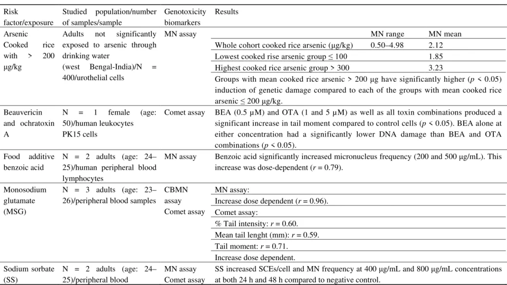
Table 3. Studies of human populations related dietary exposures.
Risk
factor/exposure
Studied population/number of samples/sample
Genotoxicity biomarkers
Results Refs.
Arsenic
Cooked rice with > 200
μg/kg
Adults not significantly exposed to arsenic through drinking water
(west Bengal-India)/N = 400/urothelial cells
MN assay MN range MN mean [101]
Whole cohort cooked rice arsenic (μg/kg) 0.50–4.98 2.12 Lowest cooked rise arsenic group ≤ 100 1.85 Highest cooked rice arsenic group > 300 3.23
Groups with mean cooked rice arsenic > 200 μg have significantly higher (p < 0.05) induction of genetic damage compared to each of the groups with mean cooked rice arsenic ≤ 200 μg/kg.
Beauvericin and ochratoxin A
N = 1 female (age: 50)/human leukocytes PK15 cells
Comet assay BEA (0.5 µM) and OTA (1 and 5 µM) as well as all toxin combinations produced a significant increase in tail moment compared to control cells (p < 0.05). BEA alone at either concentration had a significantly lower DNA damage than BEA and OTA combinations (p < 0.05).
[102]
Food additive benzoic acid
N = 2 adults (age: 24– 25)/human peripheral blood lymphocytes
MN assay Benzoic acid significantly increased micronucleus frequency (200 and 500 μg/mL). This increase was dose-dependent (r = 0.79).
[103]
Monosodium glutamate (MSG)
N = 3 adults (age: 23– 26)/peripheral blood samples
CBMN assay Comet assay
MN assay: [58]
Increase dose dependent (r = 0.96). Comet assay:
% Tail intensity: r = 0.60. Mean tail lenght (mm): r = 0.59. Tail moment: r = 0.71.
Increase dose dependent. Sodium sorbate
(SS)
N = 2 adults (age: 24– 25)/peripheral blood
MN assay Comet assay
SS increased SCEs/cell and MN frequency at 400 μg/mL and 800 μg/mL concentrations at both 24 h and 48 h compared to negative control.
[104]
Comet assay Average tail intensity (%)
Negative control (c = 0 μg/mL) 2.73
SS (c = 400 μg/mL) 10.91
SS (c = 8000 μg/mL) 5.97
SS is genotoxic to the human peripheral blood lymphocytes in vitro at the highest concentrations.
Synthetic food colorants
Sunset yellow FCF and brilliant blue FCF
N = 10 adults/blood samples. MN assay MN frequency was increased with increasing concentrations of sunset yellow and brilliant blue.
[105] Sunset yellow, significant increases in the MN rates were detected 30 mg/mL and 40 mg/mL of the concentrations (p < 0.05).
Brilliant blue, significant increases in the MN rates were detected 30 mg/mL and 40 mg/mL of the concentrations (p < 0.05).
Erythrosine (E127), tartrazine (E102),
ponceau 4R (E124), sunset yellow (E110), brilliant blue FCF (E133), fast green (E143),
carmoisine
(E122), and indigo carmine (E132)
N = 1 adult/blood samples. CBMN assay
Statistically significant increase in MN means induced by various food colors (multivariate analysis, p = 0.001 and pairwise comparisons, p < 0.05).
Control = 10
100µg/mL = 12 ± 0.7 200µg/mL = 12.8 ± 0.8 300µg/mL = 13.7 ± 0.7
Table 3 summarizes some important studies in diet field, namely the exposure to arsenic [101], mycotoxins as contaminants in food items [102], food additives [103,104], flavor enhancers [58], and synthetic food colorants [98,105].
For many other compounds for which the effects on cancer risk are not clear, there may be other good reasons to limit exposure. But at the levels that these are found in the food supply, lowering cancer risk is unlikely to be a major reason to justify this. There are moves to redefine maximum permissible limits for food colorants, instead of setting arbitrary limits for food additives in general; for instance in the case of colorants, each dye should have an individual limit based on well controlled genetic studies [98].
8. Conclusions
Human biomonitoring is a scientifically-developed approach for assessing human exposures to natural and synthetic compounds from the environment, occupation, and lifestyle, including diet [56]. It is the only available tool to integrate exposures from all sources and provide data for epidemiological studies of strengths of associations, dose response relations, etc.; however, it does not differentiate the exposure by source. Furthermore, human biomonitoring alone cannot provide information on how long a chemical has been in the body. Additional data collected from questionnaires, interviews and exposure assessment, combined with background knowledge, may provide valuable information regarding sources [21,60].
Although there has been growing recognition for the need to incorporate complex interactions between environmental exposures together with genetic factors, in order to fully understand cancer and diseases causation, since genetic instability is the startup point of carcinogenesis, there is growing recognition that environmental challenges not only interact with genes but may also modulate genetic effects and influence phenotypes [106]. An optimistic message is the fact that cancer development is not an inevitable consequence of the aging process per se, although there is a
partly avoidable increased likelihood of the requisite number of mutations occurred, and the human species is not inevitably destined to suffer a high incidence of cancer. This awareness has lent greater urgency to the search for more powerful tools for primary prevention, for early warning systems to identify causal environmental agents and flag risks well before a disease condition develops [5].
In conclusion, the potential benefits of biomarkers and molecular epidemiology in illness prevention justify a major commitment to the further development of human biomonitoring programs, the only available tool that combines exposure assessment from different sources and relates their effects, together with individual susceptibility, to the risk of disease.
Acknowledgements
The authors would like to acknowledge Professor Susana Viegas and Professor Carla Viegas for their contribution in conceiving the idea of this review and the CA15132 hCOMET COST Action– European Cooperation in Science and Technology.
Conflict of interest
References
1. Boffetta P, Nyberg F (2003) Contribution of environmental factors to cancer risk. Br Med Bull
68: 71-94.
2. Cancer Research UK, Cancer risk in the workplace. Cancer Research UK, 2016. Available from: http://www.cancerresearchuk.org/about-cancer/causes-of-cancer/cancer-risks-in-the-workplace.
3. Ladeira C, Gomes MC, Brito M (2014) Human nutrition, DNA damage and cancer: a review,
In: Mutagenesis: Exploring Novel Genes and Pathways. Wageningen: Wageningen Academic
Publishers, 73-104.
4. Key JT, Schatzkin A, Willett CW, et al. (2004) Diet, nutrition and the prevention of cancer.
Public Health Nutr 7: 187-200.
5. Perera FP, Weinstein IB (2000) Molecular epidemiology: recent advances and future directions.
Carcinogenesis 21: 517-524.
6. Portier CJ, Bell DA (1998) Genetic susceptibility: significance in risk assessment. Toxicol Lett
28: 185-189.
7. Vainio H (1998) Use of biomarkers—new frontiers in occupational toxicology and epidemiology. Toxicol Lett 102-103:581-589.
8. Bartsch H (2000) Studies on biomarkers in cancer etiology and prevention: a summary and challenge of 20 years of interdisciplinary research. Mutat Res, Rev Mutat Res 462: 255-279. 9. Dusinska M, Collins AR (2008) The comet assay in human biomonitoring: gene–environment
interactions. Mutagenesis 23: 191-205.
10. Perera FP (1996) Molecular epidemiology: insights into cancer susceptibility, risk assessment, and prevention. J Natl Cancer Inst 88: 496-509.
11. Au WW (2007) Usefulness of biomarkers in population studies: From exposure to susceptibility and to prediction of cancer. Int J Hyg Environ Health 210: 239-246.
12. El-Zein R, Vral A, Etzel CJ, et al. (2011) Cytokinesis-blocked micronucleus assay and cancer risk assessment. Mutagenesis 26:101-106.
13. Husgafvel-Pursiainen K (2002) Molecular biomarkers in studies on environmental cancer. J
Epidemiol Community Health 56(10):730-1.
14. Perera FP (2000) Molecular epidemiology: On the path to prevention? J Natl Cancer Inst 92:
602-612.
15. Goldstein B, Gibson J, Henderson R, et al. (1987) Biological markers in environmental health research. Environ Health Perspect 74: 3-9.
16. Fergurson L (2008) Biomarkers as endpoints in intervention studies. In: Wild, C., Vineis, P., Garte, S. Author, Molecular Epidemiology of Chronic Diseases, West Sussex: John Wiley &
Sons Ltd, 255-266.
17. Schulte P, Mazzuckelli LF (1991) Validation of biological markers for quantitative risk assessment. Environ Health Perspect 90: 239-246.
18. Davis CD, Milner JA (2007) Biomarkers for diet and cancer prevention research: potentials and challenges. Acta pharmacol Sin 28: 1262-1273.
20. Barrett JC, Vainio H, Peakall D, et al. (1997) 12th meeting of the scientific group on methodologies for the safety evaluation of chemicals: susceptibility to environmental hazards.
Environ Health Perspect 105: 699-737.
21. Ladeira C, Viegas S (2016) Human biomonitoring—An overview on biomarkers and their application in occupational and environmental health. Biomonitoring 3: 15-24.
22. Battershill JM, Burnett K, Bull S (2008) Factors affecting the incidence of genotoxicity biomarkers in peripheral blood lymphocytes: impact on design of biomonitoring studies.
Mutagenesis 23: 423-437.
23. Knudsen LE, Hansen AM (2007) Biomarkers of intermediate endpoints in environmental and occupational health. Int J Hygiene Environ Health 210: 461-470.
24. Cavallo D, Ursini CL, Rondinone B et al. (2009) Evaluation of a suitable DNA damage biomarker for human biomonitoring of exposed workers. Environmental and Molecular
Mutagenesis 50 (9):781–790.
25. Fenech M, Crott J, Turner J, et al. (1999) Necrosis, apoptosis, cytostasis and DNA damage in human lymphocytes measured simultaneously within the cytokinesis-block micronucleus assay: description of the method and results for hydrogen peroxide. Mutagenesis 14:605-612.
26. Majer BJ, Laky B, Knasmüller S, et al. (2001) Use of the micronucleus assay with exfoliated epithelial cells as a biomarker for monitoring individuals at elevated risk of genetic damage and in chemoprevention trials. Mutat Res 489: 147-172.
27. Burgaz S, Erdem O, Cakmak G, et al. (2002) Cytogenetic analysis of buccal cells from shoe-workers and pathology and anatomy laboratory shoe-workers exposed to n-hexane, toluene, methyl ethyl ketone and formaldehyde. Biomarkers 7: 151-161.
28. Proia NK (2006) Smoking and smokeless tobacco-associated human buccal cell mutations and their association with oral cancer—A Review. Cancer Epidemiol Biomarkers Prev 15:
1061-1077.
29. Fenech M (2007) Cytokinesis-block micronucleus cytome assay. Nat Protoc 2: 1084-1104.
30. Holland N, Bolognesi C, Kirschvolders M, et al. (2008) The micronucleus assay in human buccal cells as a tool for biomonitoring DNA damage: The HUMN project perspective on current status and knowledge gaps. Mutat Res 659: 93-108.
31. Thomas P, Fenech M (2011) Buccal micronucleus cytome assay. Methods Mol Biol 682: 235-248.
32. Cerqueira EMM, Meireles JRC (2012) The use of the micronucleus test to monitoring individuals at risk for oral cancer. In: The Research and Biology of Cancer, Hong Kong: Icon
Press Ltd, 1-26.
33. Kashyap B, Reddy PS (2012) Micronuclei assay of exfoliated oral buccal cells: means to assess the nuclear abnormalities in different diseases. J Cancer Res Ther 8: 184-191.
34. Göethel G, Brucker N, Moro AM, et al. (2014) Evaluation of genotoxicity in workers exposed to benzene and atmospheric pollutants. Mutat Res Genet Toxicol Environ Mutagen 770: 61-65.
35. Fenech M (1997) The advantages and disadvantages of the cytokinesis-block micronucleus method. Mutat Res 392: 11-18.
36. Fenech M (2000) The in vitro micronucleus technique. Mutat Res 455: 81-95.
38. Mateuca R, Lombaert N, Aka PV, et al. (2006) Chromosomal changes: induction, detection methods and applicability in human biomonitoring. Biochimie 88: 1515-1531.
39. Fenech M (2006) Cytokinesis-block micronucleus assay evolves into a ‘cytome’ assay of chromosomal instability, mitotic dysfunction and cell death. Mutat Res 600: 58-66.
40. Fenech M, Kirsch-Volders M, Natarajan AT, et al. (2011) Molecular mechanisms of micronucleus, nucleoplasmic bridge and nuclear bud formation in mammalian and human cells.
Mutagenesis 26: 125-132.
41. Speit G (2013) Does the recommended lymphocyte cytokinesis-block micronucleus assay forhuman biomonitoring actually detect DNA damage induced by occupational and environmental exposure to genotoxic chemicals? Mutagenesis 28: 375-380.
42. Moller P, Knudsen LE, Loft S, et al. (2000) The comet assay as a rapid test in biomonitoring occupational exposure to DNA-damaging agents and effect of confounding factors. Cancer
Epidemiol Biomarkers Prev 9: 1005-1015.
43. Collins A, Dusinska M (2009) Applications of the comet assay in human biomonitoring. In: Dhawan, A., Anderson, D., Author, The Comet Assay in Toxicology, Cambridge: Royal Society of Chemistry, 201-202.
44. Collins AR (2004) The comet assay for DNA damage and repair: principles, applications, and limitations. Molecular Biotechnol 26: 249-261.
45. Collins AR (2009) Investigating oxidative DNA damage and its repair using the comet assay.
Mutat Res 681: 24-32.
46. Azqueta A (2009) Detection of oxidised DNA using DNA repair enzymes. In: Dhawan, A., Anderson, D., Author, The Comet Assay in Toxicology, Cambridge: Royal Society of
Chemistry, 58-63.
47. Valverde M, Rojas E (2009) Environmental and occupational biomonitoring using the comet assay. Mutat Res 681: 93-109.
48. Valverde M, Rojas E (2009) The comet assay in human biomonitoring. In: Dhawan, A., Anderson, D., Author, The Comet Assay in Toxicology. Cambridge: Royal Society of
Chemistry, 227-251.
49. Digue L, Orsière T, De Méo M, et al. (1999) Evaluation of the genotoxic activity of paclitaxel by the in vitro micronucleus test in combination with fluorescent in situ hybridization of a DNA centromeric probe and the alkaline single cell gel electrophoresis technique (comet assay) in Human T-Lymphocytes. Environ Mol Mutagenesis 34: 269-278.
50. Hoffmann H, Speit G (2005) Assessment of DNA damage in peripheral blood of heavy smokers with the comet assay and the micronucleus test Mutat Res 581: 105-114.
51. Vasquez MZ (2010) Combining the in vivo comet and micronucleus assays: a practical approach to genotoxicity testing and data interpretation. Mutagenesis 25: 187-199.
52. Minozzo R, Deimling LI, Santos-Mello R (2010) Cytokinesis-blocked micronucleus cytome and comet assays in peripheral blood lymphocytes of workers exposed to lead considering folate and vitamin B12 status. Mutat Res/Genet Toxicol Environ Mutagen697: 24-32.
53. Olden K, Guthrie J (2001) Genomics: implications for toxicology. Mutat Res 473: 3-10.
54. Toscano WA, Oehlke KP (2005) Systems biology: new approaches to old environmental health problems. Int J Environ Res Public Health 2: 4-9.
56. Angerer J, Ewers U, Wilhelm M (2007) Human biomonitoring: state of the art. Int J Hygiene
Environ Health 210: 201-228.
57. Au WW, Cajas-Salazar N, Salama S (1998) Factors contributing to discrepancies in population monitoring studies. Mutat Res, Fundam Mol Mech Mutagen 400: 467-478.
58. Ataseven N, Yüzbaşıoğlu D, Keskin AÇ, et al. (2016) Genotoxicity of monosodium glutamate.
Food Chem Toxicol 91: 8-18.
59. Geras’kin SA, Kimb JK, Oudalova AA (2005) Bio-monitoring the genotoxicity of populations of Scots pine in the vicinity of a radioactive waste storage facility. Mutat Res 583: 55-66.
60. Choi J, Morck TA, Joas A, et al. (2015) Major national human biomonitoring programs in chemical exposure assessment. Environ Sci 2: 782-802.
61. Dagnino A, Bo T, Copetta A, et al. (2013) Development and application of an innovative expert decision support system to manage sediments and to assess environmental risk in freshwater ecosystems. Environ Int 60: 171-182.
62. Maffei F, Hrelia P, Angelini S, et al. (2005) Effects of environmental benzene: Micronucleus frequencies and haematological values in traffic police working in an urban area. Mutat Res 583: 1-11.
63. Kim K-H, Jahan SA, Kabir E (2013) A review of airborne polycyclic aromatic hydrocarbons (PAHs) and their human health effects. Environ Inter 60: 71-80.
64. Song XF, Chen ZY, Zang ZJ (2013) Investigation of polycyclic aromatic hydrocarbon level in blood and semen quality for residents in Pearl River Delta Region in China. Environ Int 60: 97-105.
65. Grawe ́ J, Biko J, Lorenz R, et al. (2005) Evaluation of the reticulocyte micronucleus assay in
patients treated with radioiodine for thyroid cancer. Mutat Res 583: 12-25.
66. Harvey JS, Lyons BP, Page TS, et al. (1999) An assessment of the genotoxic impact of the Sea Empress oil spill by the measurement of DNA adduct levels in selected invertebrate and vertebrate species. Mutat Res 441: 103-114.
67. Ceretti E, Feretti D, Viola GC, et al. (2014) DNA damage in buccal mucosa cells of pre-school children exposed to high levels of urban air pollutants. PLoS One 2: 1-9.
68. Demircigil GÇ, Erdem O, Gaga EO, et al. (2014) Cytogenetic biomonitoring of primary school children exposed to air pollutants: micronuclei analysis of buccal epithelial cells. Environ Sci
Pollut Res Int 21: 1197-1207.
69. Marcon A, Fracasso ME, Marchetti P, et al. (2014) Outdoor formaldehyde and NO2 exposures
and markers of genotoxicity in children living near chipboard industries. Environ Health
Perspect 122: 639-645.
70. Coelho P, García-Lestón J, Costa S, et al. (2013) Genotoxic effect of exposure to metal(loid)s. A molecular epidemiology survey of populations living and working in Panasqueira mine area, Portugal. Environ Int 60: 163-170.
71. Mesic A, Nefic H (2015) Assessment of the genotoxicity and cytotoxicity in environmentally exposed human populations to heavy metals using the cytokinesis-block micronucleus cytome assay. Environ Toxicol 30: 1331-1342.
73. Banerjee S, Singh NN, Sreedhar G, et al. (2016) Analysis of the genotoxic effects of mobile phone radiation using buccal micronucleus assay: A comparative evaluation. J Clin Diagn Res
10: 82-85.
74. Pastor S, Creus A, Parrón T, et al. (2003) Biomonitoring of four European populations occupationally exposed to pesticides: use of micronuclei as biomarkers. Mutagenesis 18: 249-258.
75. Bernardi N, Gentile N, Mañas F, et al. (2015) Assessment of the level of damage to the genetic material of children exposed to pesticides in the province of Córdoba. Arch Argent Pediatr 113:
126-131
76. De Coster S, Koppen G, Bracke M, et al. (2008) Pollutant effects on genotoxic parameters and tumor-associated protein levels in adults: A cross sectional study. Environ Health 7: 26.
77. Rodríguez TG, Aldeco RG, Alvarez HB, et al. (2008) Genotoxicity in child populations exposed to polycyclic aromatic hydrocarbons (PAHs) in the air from Tabasco, Mexico. Int J
Environ Res Public Health 5: 349-355.
78. Mielzyńska D, Siwińska E, Kapka L, et al. (2006) The influence of environmental exposure to
complex mixtures including PAHs and lead on genotoxic effects in children living in Upper Silesia, Poland. Mutagenesis 21: 295-304.
79. Nagya K, Rácz G, Matsumotoa T, et al. (2014) Evaluation of the genotoxicity of the pyrethroid insecticide Phenothrin. Mutat Res, Genet Toxicol Environ Mutagen 770: 1-5.
80. Moro AM, Brucker N, Charão M, et al. (2012) Evaluation of genotoxicity and oxidative damage in painters exposed to low levels of toluene. Mutat Res, Genet Toxicol Environ Mutagen 746:
42-48.
81. Moro AM, Charãoa MF, Brucker N, et al. (2013) Genotoxicity and oxidative stress in gasoline station attendants. Mutat Res, Genet Toxicol Environ Mutagen 754: 63-70.
82. Marco P, Priestly B, Buckett K (1998) Carcinogen risk assessment. Can we harmonise? Toxicol Lett 102-103: 241-246.
83. International Labour Organization (ILO). Chemical Exposure Limits. ILO 2011. Available from: http://www.ilo.org/safework/info/publications/WCMS_151534/lang--en/index.htm .
84. Ladeira C, Viegas S, Pádua M, et al. (2014) Assessment of genotoxic effects in nurses handling cytostatic drugs. J Toxicol Environ Health 77: 879-887.
85. Martino-Roth MG, Viégas J, Amaral M, et al. (2002) Evaluation of genotoxicity through micronuclei test in workers of car and battery repair garages. Genet Mol Biol 25: 495-500.
86. Ladeira C, Viegas S, Carolino E, et al. (2011) Genotoxicity biomarkers in occupational exposure to formaldehyde—The case of histopathology laboratories. Mutatn Res, Genet Toxicol
Environ Mutagen 721: 115-120.
87. Grover P, Rekhadevi PV, Danadevi K, et al. (2010) Genotoxicity evaluation in workers occupationally exposed to lead. Int J Hygiene Environ Health213: 99-106.
88. Danadevi K, Rozati R, Banu BS, et al. (2004) Genotoxic evaluation of welders occupationally exposed to chromium and nickel using the comet and micronucleus assays. Mutagenesis 19:
35-41.
89. Calvert GM, Talaska G, Mueller CA, et al. (1998) Genotoxicity in workers exposed to methyl bromide. Mutat Res, Genet Toxicol Environ Mutagen 417: 115-128.
91. Silveira HC, Schmidt-Carrijo M, Seidel EH, et al. (2013) Emissions generated by sugarcane burning promote genotoxicity in rural workers: A case study in Barretos, Brazil. Environ Health
12: 87.
92. Pitarque M, Vaglenov A, Nosko M, et al. (1999) Evaluation of DNA damage by the comet assay in shoe workers exposed to toluene and other organic solvents. Mutat Res 44: 115-127. 93. Strickland PT, Groopman JD (1995) Biomarkers for assessing environmental exposure to
carcinogens in the diet. Am J Clin Nutr 61: 710-720.
94. Sutandyo N (2010) Nutritional carcinogenesis. Acta Med Indones 42: 36-42.
95. Anand P, Kunnumakkara AB, Kunnumakara AB, et al. (2008) Cancer is a preventable disease that requires major lifestyle changes. Pharm Res 25: 2097-2116.
96. Willett W, Giovannucci E, et al. (2006) Epidemiology of diet and cancer risk. In: Shils, M.E., Shike, M. Author, Modern Nutrition in Health and Disease, Philadelphia: Lippincot Williams
and Wilkins, 1627.
97. The American Cancer Society, Food additives, safety, and organic foods. The American Cancer Society medical and editorial content team, 2012. Available from:
https://www.cancer.org/healthy/eat-healthy-get-active/acs-guidelines-nutrition-physical-activity-cancer-prevention/food-additives.html .
98. Swaroop VR, Dinesh RD, Vijayakumar T (2011) Genotoxicity of synthetic food colorants. J
Food Sci Eng 1: 128-134.
99. Duarte-Salles T, Mendez MA, Meltzer HM, et al. (2013) Dietary benzo(a)pyrene intake during pregnancy and birth weight: Associations modified by vitamin C intakes in the Norwegian mother and child cohort study (MoBa). Environ Int 60: 217-223.
100. Papadopoulou E, Caspersen IH, Kvalem HE (2013) Maternal dietary intake of dioxins and polychlorinated biphenyls and birth size in the Norwegian mother and child cohort study (MoBa). Environ Int 60: 209-216.
101. Banerjee M, Banerjee N, Bhattacharjee P, et al. (2013) High arsenic in rice is associated with elevated genotoxic effects in humans. Sci Rep 3: 1-8.
102. Klarić MS, Darabos D, Rozgaj R, et al. (2010) Beauvericin and ochratoxin A genotoxicity evaluated using the alkaline comet assay: Single and combined genotoxic action. Arch Toxicol
84: 641-650.
103. Yılmaz S, Ünal F, Yüzbaşıoğlu D (2009) The in vitro genotoxicity of benzoic acid in human peripheral blood lymphocytes. Cytotechnology 60: 55-61.
104. Mamur S, Yüzbaşıoğlu D, Unal F, et al. (2012) Genotoxicity of food preservative sodium sorbate in human lymphocytes in vitro. Cytotechnology 64: 553-562.
105. Kus E, Eroglu HE (2015) Genotoxic and cytotoxic effects of sunset yellow and brilliant blue, colorant food additives, on human blood lymphocytes, Pak J Pharm Sci 28: 227-230.
106. Spitz MR, Bondy ML (2010) The evolving discipline of molecular epidemiology of cancer.
Carcinogenesis 31: 127-134.
© 2017 Carina Ladeira et al., licensee AIMS Press. This is an open access article distributed under the terms of the Creative Commons Attribution License