Improved phylogeny of brown algae
Cystoseira (Fucales) from the
Atlantic-Mediterranean region based on
mitochondrial sequences
Carolina Bruno de Sousa1, Cymon J. Cox1, Luı´s Brito1, Maria Madalena Pavão1, Hugo Pereira1, Ana Ferreira2, Catarina Ginja3, Lenea Campino4, Ricardo Bermejo5,6, Manuela Parente7, João VarelaID1*
1 Centro de Ciências do Mar, Universidade do Algarve, Faro, Portugal, 2 Universidade dos Ac¸ores, Faculdade de Ciências e Tecnologia, Ponta Delgada, Ac¸ores, Portugal, 3 CIBIO-InBIO, Centro de Investigac¸ão em Biodiversidade e Recursos Gene´ticos, Universidade do Porto, Campus Agra´rio de Vairão, Vairão, Portugal, 4 Global Health and Tropical Medicine, Instituto de Higiene e Medicina Tropical,
Universidade Nova de Lisboa, Lisboa, Portugal, 5 Departamento de Biologı´a- A´ rea de Ecologı´a, Facultad de Ciencias del Mar y Ambientales, Universidad de Ca´diz, Puerto Real, Ca´ diz, Spain, 6 Irish Seaweed Research Group & Earth and Ocean Sciences Department, Ryan Institute and School of Natural Sciences, National University of Ireland, Galway, Ireland, 7 CIBIO-Ac¸ores, Centro de Investigac¸ão em Biodiversidade e Recursos Gene´ticos, InBIO Laborato´ rio Associado, Po´lo dos Ac¸ores, Departamento de Biologia, Universidade dos Ac¸ores, Ponta Delgada, Portugal
*jvarela@ualg.pt
Abstract
Cystoseira is a common brown algal genus widely distributed throughout the Atlantic and Mediterranean regions whose taxonomical assignment of specimens is often hampered by intra- and interspecific morphological variability. In this study, three mitochondrial regions, namely cytochrome oxidase subunit 1 (COI), 23S rDNA (23S), and 23S-tRNAVal intergenic spacer (mt-spacer) were used to analyse the phylogenetic relationships of 22 Cystoseira taxa (n = 93 samples). A total of 135 sequences (48 from COI, 43 from 23S and 44 from mt-spacer) were newly generated and analysed together with Cystoseira sequences (9 COI, 31 23S and 35 mt-spacer) from other authors. Phylogenetic analysis of these three markers identified 3 well-resolved clades and also corroborated the polyphyletic nature of the genus. The resolution of Cystoseira taxa within the three clades improves significantly when the inclusion of specimens of related genera was minimized. COI and mt-spacer markers resolved the phylogeny of some of the Cystoseira taxa, such as the C. baccata, C. foenicula-cea and C. usneoides. Furthermore, trends between phylogeny, embryonic development and available chemotaxonomic classifications were identified, showing that phylogenetic, chemical and morphological data should be taken into account to study the evolutionary relationships among the algae currently classified as Cystoseira. The resolution of Cysto-seira macroalgae into three well supported clades achieved here is relevant for a more accurate isolation and identification of natural compounds and the implementation of con-servation measures for target species.
a1111111111 a1111111111 a1111111111 a1111111111 a1111111111 OPEN ACCESS
Citation: Bruno de Sousa C, Cox CJ, Brito L, Pavão MM, Pereira H, Ferreira A, et al. (2019) Improved phylogeny of brown algae Cystoseira (Fucales) from the Atlantic-Mediterranean region based on mitochondrial sequences. PLoS ONE 14(1): e0210143.https://doi.org/10.1371/journal. pone.0210143
Editor: Suzannah Rutherford, Fred Hutchinson
Cancer Research Center, UNITED STATES
Received: September 28, 2017 Accepted: December 18, 2018 Published: January 30, 2019
Copyright:© 2019 Bruno de Sousa et al. This is an open access article distributed under the terms of theCreative Commons Attribution License, which permits unrestricted use, distribution, and reproduction in any medium, provided the original author and source are credited.
Data Availability Statement: All sequences files
are available from thehttps://www.ncbi.nlm.nih. gov/genbank/database (accession number(s) KF525356, KF525357, KF525359, KF525364, KF525365 and from MF767957 to MF768079).
Funding: CBS, HP and CG were supported by
Fundac¸ão para a Ciência e a Tecnologia (http:// www.fct.pt/index.phtml.pt) doctoral (SFRH/BD/ 78062/2011 and SFRH/BD/105541/2014, respectively) and contract (IF/00866/2014) grants.
Introduction
Cystoseira (Fucales, Heterokonta) brown algae are key elements of the marine seascape along
warm-temperate North African and European coasts [1–4]. They form marine forests with a complex three-dimensional structure and provide habitat for other algae, invertebrates and fish [5–8], playing a key role in the determination of biodiversity patterns and ecosystem func-tioning [6]. Currently, manyCystoseira taxa are undergoing a strong demographic decline
attributed to both local and global pressures [9–11]. Moreover, it has been suggested that this loss of biodiversity might be caused by the sensitivity of these macroalgae to increased water turbidity, eutrophication and pollution [12–14], as a consequence of the increasing anthropo-genic activity near the Atlantic and Mediterranean coastal areas [10,11]. Changes in the distri-butions and abundance of various species are also expected as a consequence of climate change [15,16]. Because of the ecological importance of assemblages dominated byCystoseira
and the deterioration of their populations during the past decades, the Mediterranean species of this genus are protected under the Barcelona Convention (Annex II, COM/2009/0585 FIN) and reforestation has been proposed as a management action to improve the conservation sta-tus of these macroalgae [14,17].
The importance of the genusCystoseira is further underscored by the observation that its
members produce several potentially bioactive metabolites such as terpenoids, fatty acids, tria-cylglycerols, steroids, phlorotannins, and polysaccharides [18,19]. Indeed, antioxidant, anti-inflammatory, antiproliferative, antifungal, antiviral, antibacterial and antiprotozoal activities have been reported to occur inCystoseira algae with increasing frequency [20–26]. This wide range of bioactivities detected in extracts of these algae might be explained by the bio- and chemical diversity of the genus [27,28].
The accuracy of the taxonomic identification of the biomass used for the isolation and iden-tification of natural compounds is, however, an important issue concerning the reproducibility and reliability of the results as well as for the implementation of conservation measures for the target macroalgae [29]. Taxonomic classification within the genusCystoseira is challenging
and controversial [30,31]. Erroneous taxonomical assignments are frequent due to the wide morphological variability ofCystoseira individuals, in addition to there being many species
that are still undergoing active speciation and hybridization [32–34]. This has become espe-cially apparent due to frequent conflicts between classification of specimens based on mor-phology and molecular data. Chemotaxonomic classifications based on the presence or absence of specific chemicals (e.g. meroterpenoids) have also been attempted [18,28,35–37]. In addition, analysis of the global chemical profile and the lipophilic composition of five Cysto-seira taxa from Brittany have been found to be in agreement with the phylogenetic
relation-ships established by the ITS2 region [37]. However, congruence between morphology, chemistry and molecular taxonomy at the species level is yet to be achieved [34], and the results obtained so far have not fully resolved theCystoseira phylogeny [37].
Several authors have previously attempted the elucidation of the relationships within this genus and with related genera using phylogenetic methods [34,38–40]. Analysis of Fucales (Phaeophyceae) Kylin based on large subunit (LSU) and small subunit (SSU) of the ribosomal DNA sequences led to the merging of the Cystoseiraceae De Toni and Sargassaceae Ku¨tzing families [41]. The mitochondrial 23S ribosomal subunit (23S) proved to be useful for defining genera in the Fucales [34] and in addition a set of 10 additional mitochondrial, plastid and nuclear markers has also been used to investigate the evolutionary history of brown algae at the ordinal level [42]. Other analysis including also organellar markers revealed that the genus
Cystoseira was composed of at least six distinct, but clearly polyphyletic, evolutionary lineages.
However, only 3 lineages (see below) were eventually classified as separated genera [34]. Based
RB holds a Training programme for Academic Staff fellowship of the Spanish Ministry of Education. This work was supported by Fundac¸ão para a Ciência e a Tecnologia (http://www.fct.pt/index. phtml.pt), projects CCMAR/Multi/04326/2013 and PEst-OE/EQB/LA0023/2011. The funders had no role in study design, data collection and analysis, decision to publish, or preparation of the manuscript.
Competing interests: The authors have declared
on morphologic, embryonic development characters and genetic data, several members of the genus were reclassified as belonging to the generaSirophysalis (Tropical Indo-West-Pacific), Polycladia (eastern Indian Ocean) and Stephanocystis (North Pacific) [34]. All otherCystoseira
taxa, despite forming at least three separate Northeastern Atlantic-endemic clades, retained the original classification. Currently, the genusCystoseira encompasses approximately 40 taxa,
the majority of which occurs in the Mediterranean and Atlantic-Mediterranean regions [43,
44]. However, to date, full infrageneric resolution of the genus and their position among related Sargassaceae genera has not been established. Therefore, the taxonomy of the Cysto-seira species is still unclear.
The mitochondrial gene coding for cytochrome oxidase subunit 1 (COI) is a well-known molecular tool used for the identification of different metazoan taxa [45–47]. Although the COI gene was used in the study of red [48] and brown algae [49–51], the utility of this marker for the infrageneric identification ofCystoseira individuals has not been evaluated so far. With
the purpose of improving the resolution of theCystoseira species identification and clarify
their phylogenetic relationships, a comprehensive study combining sequence information on the cytochrome oxidase subunit 1 (COI), 23S rDNA (23S), and 23S-tRNAVal intergenic spacer (mt-spacer) was undertaken. The results of this study confirm the polyphyly of the genus, which was resolved into 3 well supported clades by using sequence information on the pro-tein-coding COI gene.
Material and methods
Ethics statement
We state that no specific permissions were required for the taxa sampled in this work. The samples were taken from public sea places and not from any national park or protected area. The Portuguese Foundation for Science and Technology approved this type of research by sup-porting our research projects CCMAR/Multi/04326/2013 and PEst-E/EQB/LA0023/2011.
Sampling
Overall, this study includes 93 samples ofCystoseira and 210 sequences belonging to 31 species
of the Sargassaceae family (Cystoseira: 22 taxa; Bifurcaria: 1 species; Polycladia: 2 species; Siro-physalis: 1 species; Stephanocystis: 4 species and Turbinaria: 1 species). A detailed list of
sam-ples and sequence information is provided in the supporting information (S1 Table). However, it should be noted that, in this study, “Cystoseira” is a term of convenience, which includes all taxa previously classified as belonging to this genus and which have not been rede-fined by Draisma et al. [31].
Fifty-nine samples ofCystoseira sp. (n = 55) and Bifurcaria bifurcata (n = 4) were collected
along the Atlantic and Mediterranean coasts (Fig 1), and mtDNA markers were specifically amplified. The samples, collected by the authors or kindly provided by expert colleagues, were morphologically classified using the taxonomic characteristics following Go´mez-Garreta et al. [38] and Cormaci et al. [52]. Guiry and Guiry [44] was used as an additional reference for taxo-nomic validity. After washing with tap water, biomass was silica-dried and stored at room tem-perature for DNA extraction. Vouchers of the studied specimens were deposited in the herbarium of the University of Algarve (https://www.ualg.pt/pt/content/algu)—Index herbar-iorum code: ALGU. Additional vouchers were deposited in the herbarium of the Marine Bio-technology Group of the Centre of the Marine Sciences (MarBiotech / CCMAR).
AdditionalCystoseira sequences (9 COI, 31 23S and 35 mt-spacer) and other Sargassaceae
species (3 COI, 8 23S and 8 mt-spacer) publicly available in the GenBank database at the National Center for Biotechnology Information (NCBI) were included in the analysis [53] (Fig
1). Additionally, sequences from 4 species of the Fucaceae family (4 COI, 4 23S and 4 mt-spacer) were also obtained from GenBank and used as outgroup.
DNA extraction, amplification and sequencing
Genomic DNA was extracted from 5–10 mg of the silica gel-dried algal tissue using the method described by Doyle and Doyle [54]. The primers for amplification of the COI and 23S fragments were described by Lane et al. [55] and Draisma et al. [34], respectively. Primer pairs for amplification of the mt-spacer fragment were designed specifically for this study. Primer information, such as locus names, nucleotide sequences, and references are provided in
Table 1.
Mitochondrial 23S and mt-spacer were PCR-amplified in a final volume of 20.5μL reac-tions containing 5μL of genomic DNA (~10 ng/mL), 4 μL 5×PCR Buffer, 4 μL dNTP mix (1 mM of each dNTP), 2μL 25 mM MgCl2, 0.6μL Taq DNA polymerase (GoTaq DNA Polymer-ase, Promega), 0.5μL of 10 μM 23S forward (mt23S-FB) and reverse (mt23S-RB) primers or 0.25μL of 10μM mt-spacer forward (mt-spacer-F) and reverse (mt-spacer-R) primers. COI
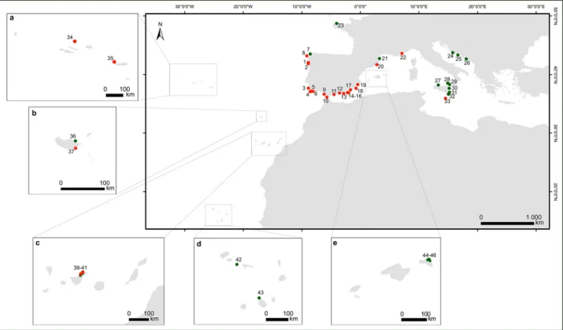
Fig 1. Geographical distribution of theCystoseira samples used in this study. Green dots represent GenBank sequences and the red dots data obtained in this study.
The boxes show the archipelagos ofaMadeira,bAzores,cCanary,dCape Verde andeBalearics. Locations and sampling number marked as red dots are:1Moledo (n = 1);
2
Areosa (n = 2);3Odeceixe (n = 1);4Manuel Lourenc¸o (n = 4);5Olhos de A´ gua (n = 9);6Arrifes (n = 1);7A Coruña (n = 4);8Santa Mariña (n = 2);9Santibañez (n = 2);
10El Mirlo (
n = 3);11Calaburras (
n = 1);12Herradura (
n = 1);13Guardias Viejas (
n = 1);14La isleta del Moro (
n = 2);15El Playazo (
n = 2);16Las Negras (
n = 1);17La Serena
(n = 1);18Cabo de Palos (n = 1);19Santa Pola (n = 1);20Blanes (n = 3);21Cote Vermeille (
n = 2);22Pointe I´lette (
n = 1);23Santec (
n = 2);24Prvic Island (
n = 1);25Brac
Island (n = 1);26Dubrovnik city arean = 1);27Capo Gallo (n = 1);28Aeolian Island (n = 2);29Capo Milazzo (n = 1);30S. Maria la Scala (n = 3);31Marzameni (n = 4);
32
Capo Passero (n = 2);33Xghajra (n = 1);34Carapacho (n = 1);35Ponta dos Mosteiros (n = 2);36Porto da Cruz (n = 1);37Canic¸o (n = 2);38Bajamar (n = 1);39Mesa del Mar (n = 4);40Punta del Hidalgo (
n = 6);41Tacoronte (
n = 1);42Branco island (
n = 1);43Tarrafal Bay (
n = 1);44Cala Viola de Llevant (n = 1);45La Llosa d’en Patro Pere
(n = 1);46Illots de Tirant (
n = 1);47Cala Mica (
n = 2);48Illa d’es Porros (
n = 1). For further information about the location of sample points, please refer to theS1 Table. https://doi.org/10.1371/journal.pone.0210143.g001
amplifications were perfomed in a 12-μL mix containing 2 μL of genomic DNA, 1.25 μL 5×PCR Buffer, 0.6 μL dNTP mix (1 mM of each dNTP), 1.25 μL 25 mM MgCl2, 0.1 μL Taq DNA polymerase, 0.25μL of 10 μM COI forward (GazF2) and reverse (GazR2) primers. Amplifications were performed using an Applied Biosystems 2720 Thermal Cycler with the following conditions: 95˚C for 6 min; 10 cycles of 95˚C for 30 s, 64˚C (decreasing 0.5˚C per cycle) for 30 s, 72˚C for 60 s; 35 cycles of 95˚C for 30 s, 59˚C for 30 s, 72˚C for 60 s; and a final elongation step of 10 min at 72˚C for the 23S and mt-spacer fragments; for COI, samples were incubated at 95˚C for 2 minutes; 5 cycles of 95˚C for 30 s, 45˚C for 30 s and 72˚C for 1 min; 35 cycles of 95˚C for 30 s, 46.5˚C for 30s and 72˚C for 1 min; and a 72˚C elongation step for 7 min. PCR amplicons were screened for specific fragment size on 2% agarose gel electrophore-sis and subsequently purified using a EZNA MicroElute Cycle-Pure Kit (Omega Bio-Tek, USA) purification kit. Amplified fragments were sequenced using the Sanger method at the Molecular Biology Core Laboratory, Centre of Marine Sciences (Algarve University, Faro), in an 3130XL Genetic Analyzer (Applied Biosystems) using PCR primers in cycle sequencing reactions.
Sequence validation and genetic diversity
New sequences generated from amplicons obtained from both strands were compared with GenBank data using BLASTn [56] to determine whether the biological source was a Sargassa-ceae alga. GenBank accession numbers of the sequences are indicated inS1 Table. Sequences were also organized in two datasets: one including only sequences from individuals of the
Cystoseira genus, and the other comprising the same data plus those from the Sargassaceae
and Fucales families.
The 23S and mt-spacer sequences were aligned with the CLC Sequence Viewer V.7.6.1 (Quiagen), using the default settings. For COI, sequences were aligned with transAlign soft-ware [57] using ClustalW multiple sequence alignment [58]. Alignments were further inspected with CLC Sequence Viewer V.7.6.1 and manually improved before a final curation step with Gblocks v.0.91b software [59] available at the Phylogeny.fr web service [60]. Gap positions within the final blocks option were allowed and a maximum of 8 contiguous non-conserved positions were considered with a minimum block length of 5 nucleotides (nt). The concatenated matrix was obtained using Seaview v.4.5.3 [61]. The number of polymorphic and phylogenetically informative sites of the aligned sequences were estimated for each marker using DnaSP v.5.10.1 software [62]. Haplotype identification was carried out for each mito-chondrial marker using this software.
Evolutionary divergence and phylogenetic relationships
Genetic distance analysis was used to investigate inter- and intraspecific evolutionary diver-gence betweenCystoseira sequences. Pairwise-sequence distances were estimated using the
Table 1. Molecular markers used in this study. Locus name and target region, forward and reverse primer sequences, and references.
Target region Primer Sequence References
23S mt23S-FB 5'-AGCGTAACAGCTCACTGACCTA-3' [31]
mt23S-RB 5'-CTGTGGCGGTTTAAGGTACGGTT-3'
mt23S(partial)-IGS-tRNALys-IGS-tRNAVal tRNALys-FW 5'-GGGGTGAAAAATATCACTTTGA-3' This study
tRNALys-RV 5'-AACCCAAGACCCTCGGATTA-3'
COI GazF2 5'-CCAACCAYAAAGATATWGGTAC-3' [51]
GazR2 5'-GGATGACCAAARAACCAAAA-3'
Kimura 2-parameter model [63] with MEGA5 software [64]. The rate variation among sites was modelled with a gamma distribution (shape parameter = 6). All ambiguous positions were removed for each sequence pair. Phylogenetic analysis was carried out using Maximum likeli-hood (ML) and Bayesian inference (BI). The substitution models that best fit the data were selected using MrModeltest2 v.2.3 [65] and PAUP�v.4.0b10 [66] by applying the Akaike
infor-mation criterion (AIC) [67]. The substitution models selected were: GTR+I+Γ4 [general
time-reversible (GTR) model with a proportion of invariant sites (I) and among-site rate variation modelled by a discrete gamma distribution with 4 categories (Γ4)] for 23S, HKY+I+G [Hase-gawa-Kishino-Yano model (HKY)] for COI and GTR+Γ4 for the mt-spacer.
ML analysis was performed using RAxML v.7.0.4 [68] with 400 bootstrap replicates, assum-ing the best-fittassum-ing models. Posterior probabilities were determined by Markov Chain Monte Carlo (MCMC) sampling in MrBayes v.3.1.2 [69,70]. MrBayes analyses were also conducted using the best-fitting models, using 6 chains for 10,000,000 generations, sampling every 1,000th generation, and default settings for the remaining options. Convergence of the MCMC and burn-in were determined through the analysis of the generations vs. log probability plot using the trace analysis tool TRACER v1.6 (http://beast.bio.ed.ac.uk/Tracer). The initial burn-in step discarded 20% of the samples.
After inferring the phylogeny, the topological congruence between gene trees was visually assessed for each marker (COI, 23S, mt-spacer). Subsequently, the sequences obtained for the three markers were concatenated and analysed by ML and BI as described before. ML and BI best consensus trees for each marker dataset (COI, 23S, mt-spacer, and concatenated COI-23S-mt-spacer) were generated and edited with the graphical viewer FigTree v.1.3.1 [71].
The genetic relationships between haplotypes were also investigated by means of a Median-Joining (MJ) network constructed with the NETWORK version 4.5.10 software [72].
Results
Alignment characterization
Overall, sequences from 93Cystoseira samples belonging to 22 taxa from the Atlantic
(Macaro-nesian and Iberian Peninsula south and west coasts) and the Mediterranean (Adriatic, Alboran, Balearic and Tyrrhenian seas) regions were included in this study (Fig 1). Among these, the 55Cystoseira samples collected generated 135 new sequences representing a
sequencing success of 87.3% (48 sequences), 78.2% (43 sequences) and 80.0% (44 sequences) for COI, 23S and mt-spacer loci, respectively.
The conjoint analysis ofCystoseira sequences obtained in this study and from GenBank (57
COI, 74 23S and 79 mt-spacer sequences) resulted in alignments with 656, 391, 258 nt for COI, 23S and mt-spacer, respectively. Upon phylogenetic analysis, three lineages (Cystoseira-I, -II, -III) with support values close to the maximum (BS = 100; PP = 1) were identified (Figs2–4
andS1–S8Figs). Detailed information of the alignment results obtained for each marker and phylogenetic group is shown inTable 2. Longer alignment lengths and higher number of con-served positions were obcon-served for COI (656 nt; 86.1%) and 23S (391 nt; 81.7%) loci, and the lowest for the mt-spacer (258 nt; 52.7%).
Concatenation of the three loci (COI-23S-mt-spacer) consisted of a 1305-nt alignment with 78% of conserved positions. Depending upon the marker considered, 15.6–24.0% of polymor-phic sites (PS) and 14.3–22.5% of parsimony informative (PI) sites were identified (Table 2). Group Cystoseira-II showed the highest number of variable PS (7.9–13.2%) and PI (5.6– 11.2%) for all loci, except for the 23S marker, where 6.4% of PS were found (Table 3). Group Cystoseira-III showed the lowest PS (2.6–7.0%) and PI (2.3–6.2%) values for 23S and mt-spacer loci, respectively.
Evolutionary divergence and haplotype analysis
Interspecific evolutionary divergence ofCystoseira, considering only the taxa that have
infor-mation for all the three markers, ranged from 0.0 to 6.8% in COI, 0.0 to 4.6% in 23S and 0.0 to 14% in the mt-spacer (Table 4andS2–S4Tables). The highest level of interspecific variation was observed in the Cystoseira-II (0–14%) group, whereas Cystoseira-I taxa showed the lowest range of genetic distances (0–1.1%) for all markers. Overall, intraspecific variation was lower than the variation observed between species. Intraspecific divergence ranged from 0 to 5.6% in COI, 0.0–2.2% in 23S and 0–3.9% in the mt-spacer. When considering all the samples included in the phylogenetic analysis, the intraspecific divergence increased slightly higher (up to 7.6%), as a result of the greater heterogeneity of the species included. In general, mean genetic dis-tances were greater for mt-spacer, followed by COI and 23S loci.
A total of 16 COI, 26 23S and 37 mt-spacer haplotypes were identified, in 58, 73 and 79
Cystoseira sp. individuals, respectively. The greatest haplotype diversity was observed for
Cystoseira-I (the total number of haplotypes was 4, 13 and 15 for COI, 23S and mt-spacer, respectively) and–II (7, 7 and 16 haplotypes), whereas Cystoseira-III had the lowest diversity (5, 6, and 6 haplotypes). Several haplotypes were exclusive of eachCystoseira group, and the
Median-Joining analysis revealed highly congruent networks across markers for each
Fig 2.Cystoseira groups defined by the phylogenetic analysis. Green dots represent the taxa belonging to the Group I (Cystoseira tamariscifolia, C. amentacea and C.
amentacea var. stricta, C. funkii, C. mediterranea, C. brachycarpa var. brachycarpa, C. brachycarpa, C. barbatula, C. zosteroides, Cystoseira RB105 and Cystoseira sp. 1);
yellow dots represent the taxa belonging to the Group II (C. mauritanica, C. barbata f. aurantia, C. montagnei and C. montagnei var. tenuior, C. barbata, C. nodicaulis, C. granulata, C. elegans, C. squarrosa, C. usneoides, C. baccata, C. abies-marina, C. sonderi, Cystoseira sp. 2 and Cystoseira sp. MP14); red dots represent the taxa belonging
to the Group III (C. compressa and C. compressa subsp. pustulata, C. humilis, C. humilis var. myriophylloides and C. foeniculacea, Cystoseira sp. MP1, Cystoseira sp. MP2
andCystoseira sp. MP31).
Cystoseira-I, -II and–III groups (S9–S11Figs). A total of 21 haplotypes out of the 79 found were shared between at least two taxa of the same group. The Cystoseira-I taxa were those with the highest number of shared haplotypes (n = 11 for all markers), followed by the Cystoseira-II
(n = 6) and Cystoseira-III (n = 5) taxa. For each Cystoseira group, these haplotypes were only
shared within sub-groups, which were clearly separated in the networks.
Phylogenetic analysis
Maximum likelihood and Bayesian inference analyses of the Sargassaceae (Fig 3) and Cysto-seira-only (Fig 4) concatenated datasets confirm the subdivision ofCystoseira in 3
well-suported clades (Cystoseira-I-III; Figs3and4andS1–S6Figs). This subdivision was congru-ent among analyses of each mitochondrial marker (S3–S8Figs).
Overall, the Cystoseira-III group, which includesC. compressa, C. foeniculacea, C. humilis,
clearly branched off Cystoseira-I (C. amentacea,C. barbatula, C. brachycarpa, C. crinita, C. funkii, C. mediterranea, C. tamariscifolia, C. zosteroides) and Cystoseira-II (C. abies-marina, C. baccata, C. barbata, C. elegans, C. mauritanica, C. nodicaulis, C. sonderi, C. montagnei, C. squarrosa, C. usneoides;Table 3). However, these results suggest that Cystoseira-I and -II are more closely related as compared to Cystoseira-III, sharing a common branch with maximum support (BS = 100; PP = 1;Fig 4). Nonetheless Cystoseira-I and -II are paraphyletic when Bifurcaria is included in the analysis, as was observed with the Cystoseira-III taxa that clus-tered together with other genera from the Indo-Pacific region previously classified as Cysto-seira (BS = 74; PP = 0.86), such asPolycladia, Sirophysalis and Stephanocystis [34].
Cystoseira-I could be divided into two subgroups Cystoseira-IA and -IB (Figs3and4). Cystoseira-IA (C. amentacea, C. funkii, C. mediterranea, C. tamariscifolia) formed a
well-sup-ported cluster (BS = 96; PP = 1) using mt-spacer sequences (S7andS8Figs), although without significant statistical support in the 23S analysis (S5andS6Figs). Within this group,C. medi-terranea formed a cluster that was ML-supported in the COI tree (BS = 99; PP = 0.93;S3and
S4Figs), whileC. tamariscifolia and C. amentacea remained unresolved. Subgroup
Cystoseira-IB (C. barbatula, C. brachycarpa, C. crinita) was significantly supported in the concatenated
datasets analysis (BS = 92; PP = 0.96; Figs3and4); and in the 23S tree, support was highly sig-nificant (BS = 99; PP = 1;S5andS6Figs). This result suggests thatC. brachycarpa, C. barbatula
andC. crinita are indeed closely related. In addition, Cystoseira-I taxa clustered together with
a well-supportedBifurcaria bifurcata cluster (BS = 94; PP = 1;Fig 3andS1 Fig), confirming that they are sister taxa.
Cystoseira-II branched into two well-supported subgroups, Cystoseira-IIA (BS = 100; PP = 1) and Cystoseira-IIB (BS = 98/99; PP = 1; Figs3and4). This high support is mainly due to the inclusion of the COI and mt-spacer markers (S3andS4Figs). Analysis of the
concatenated dataset showed that Cystoseira-IIA (C. baccata, C. barbata, C. elegans, C. mauri-tanica, C. nodicaulis, C. montagnei, C. squarrosa, C. usneoides) encompassed two well-resolved
taxa, namelyC. usneoides (BS = 98/97; PP = 0.93/0.91) and C. baccata (BS = 100; PP = 1) (Figs
3and4). Maximum support of theC. baccata clade was also obtained in the COI tree (S3and
S4Figs), whereas in the 23S tree the branch support values were lower (BS = 89; PP = 0.92;S5
andS6Figs).C. usneoides cluster was supported by the ML analysis using the COI (BS = 96;
PP = 0.54;S3andS4Figs) and 23S (BS = 94; PP = 0.92;S5andS6Figs) loci. In addition, Cysto-seira-IIA included an unresolved heterogeneous set of taxa (Figs3and4), although the COI
Fig 3. Maximum likelihood phylogenetic tree obtained with RAxML and based on the concatenated COI-23S-IGS sequences of samples from the Sargassaceae family. Values on the branches represent maximum likelihood bootstrap support values (� 75) on the left, and
Bayesian posterior probabilities (� 90%) on the right. https://doi.org/10.1371/journal.pone.0210143.g003
locus allowed for the resolution of aC. nodicaulis cluster (BS = 86; PP = 0.99;S3andS4Figs). However, the presence of a well-supported heterogeneous cluster (BS = 98/88, PP = 1) encom-passing three sequences acquired from the GenBank and classified asC. montagnei, C. elegans, C. squarrosa from the Adriatic and nearby Sicily Mediterranean coasts was not in agreement
with the results of sequences of the same species obtained in the Spanish south Mediterranean coast (S1 Table). Sister to Cystoseira-IIA, Cystoseira-IIB containedC. abies-marina and C. son-deri and formed a well-supported cluster (BS = 99/98; PP = 1; Figs3and4), though this topol-ogy was not detected in the 23S analysis (S5andS6Figs).
Within the Cystoseira-III group,C. foeniculacea formed a clade with maximum support
(BS = 100; PP = 1), sister toC. compressa and C. humilis as defined by all markers (Figs3and4
Fig 4. Maximum likelihood phylogenetic tree obtained with RAxML and based on the concatenated COI-23S-mt-spacer sequences of samples from theCystoseira genus. Values on the branches represent maximum likelihood bootstrap support values (� 75) on the left, and Bayesian
posterior probabilities (� 90%) on the right. https://doi.org/10.1371/journal.pone.0210143.g004
Table 2. Number ofCystoseira taxa and samples included in this study. Alignment characteristics (with gaps) are also shown for each marker and phylogenetic group.
Parameters All taxa Group I1 Group II2 Group III3
COI
Taxa 13 3 7 3
Number of samples (sequences) 58 18 25 15
Alignment length (nt) 656 656 656 656
Conserved sitesa 565 (86.1%) 648 (98.7%) 604 (92.0%) 535 (81.6%)
Polymorphic sites 114 (17.4%) 8 (1.2%) 52 (7.9%) 30 (4.6%)
Singleton variable sites 4 (0.6%) 1 (0.2%) 4 (0.6%) 2 (0.3%)
Parsimony informative sites 110 (16.7%) 7 (1.1%) 48 (7.3%) 28 (4.3%)
23S
Taxa 20 8 9 3
Number of samples (sequences) 73 31 29 13
Alignment length (nt) 391 391 391 391
Conserved sitesa 317 (81.7%) 335 (85.7%) 331 (84.7%) 352 (90.0%)
Polymorphic sites 61 (15.6%) 25 (6.4%) 22 (5.6%) 10 (2.6%)
Singleton variable sites 5 (1.3%) 10 (2.6%) 0 (0%) 1 (0.3%)
Parsimony informative sites 56 (14.3%) 15 (3.8%) 22 (5.6%) 9 (2.3%)
mt-spacer
Taxa 21 7 11 3
Number of samples (sequences) 79 35 33 11
Alignment length (nt) 258 258 258 258
Conserved sitesa 136 (52.7%) 183 (70.9%) 141 (54.6%) 168 (65.1%)
Polymorphic sites 62 (24.0%) 25 (9.7%) 34 (13.2%) 18 (7.0%)
Singleton variable sites 4 (1.6%) 7 (2.7%) 5 (1.9%) 2 (0.8%)
Parsimony informative sites 58 (22.5%) 18 (7.0%) 29 (11.2%) 16 (6.2%)
1
Group I—Cystoseira tamariscifolia, C. amentacea, C. amentacea var. stricta, C. funkii, C. mediterranea, C. brachycarpa, C. brachycarpa var. balearica, C. barbatula, C. zosteroides and Cystoseira sp. 1
2
Group II—C. mauritanica, C. barbata f. aurantias, C. montagnei and C. montagnei var. tenuior, C. barbata, C. nodicaulis, C.granulata, C. elegans, C. squarrosa, C. usneoides, C. baccata, C. abies marina, C. sonderi, Cystoseira sp. 2 and Cystoseira sp. MP14
3
Group III—C. compressa and C. compressa subsp. pustulata, C. humilis, C. humilis var. myriophylloides and C. foeniculacea, Cystoseira sp. MP1, Cystoseira sp. MP2 and Cystoseira sp. MP31.
a
Percentage calculated relative to the alignment length.
and S1 andS2Figs). Although without significant support values (BS = 80/67, PP = 0.95/0.74 in Figs3and4, respectively),C. compressa branched off C. compressa subsp. pustulata and C. humilis. These results are in agreement with some authors [52,73] that considerC. compressa
subsp.pustulata a synonym of C. humilis var. humilis. Therefore, we suggest that the former
should be renamed as the latter. These relationships are better defined in the COI trees (S3and
Table 3. Comparison of the differentCystoseira phylogenetic groups defined in this study with the groups identified by other authors based on genetic, chemical and morphological traits.
Reference This study Draisma et al. [34] Amico [18]2 Valls et al. [28]3 Piatelli [35]4 Amico et al. [27]5 Colombo et al. [80]6
Type of data Phylogeny Chemistry Morphology
Taxa1 COI, 23S, mt-spacer 23S Lipophylic, diterpenoid and meroditerpenoid
content
Anatomic traits Embryo germination
C. amentacea Cystoseira-IA Cystoseira-5 VI IIIB / IIIC VII I I
C. funkii Cystoseira-IA Cystoseira-5 - - - -
-C. mediterranea Cystoseira-IA Cystoseira-5 VII IIIB / IIIC VII I I
C. tamariscifolia Cystoseira-IA Cystoseira-5 VII IIIB / IIIC VII I I
C. barbatula Cystoseira-IB Cystoseira-5 III IIIA III -
-C. brachycarpa Cystoseira-IB Cystoseira-5 II II II II I
C. crinita Cystoseira-IB Cystoseira-5 III IIIA III II I
C. zosteroides Cystoseira-IC Cystoseira-5 IV IIIB IV III I
C. baccata Cystoseira-IIA Cystoseira-6 V IIIB - VI II
C. barbata Cystoseira-IIA Cystoseira-6 I I III II I
C. elegans Cystoseira-IIA Cystoseira-6 V IIIA / IIIB V III I
C. granulata Cystoseira-IIA - - -
-C. mauritanica Cystoseira-IIA - - - - III
-C. nodicaulis Cystoseira-IIA - - - - III I
C. montagnei Cystoseira-IIA Cystoseira-6 V IIIB V III I
C. squarrosa Cystoseira-IIA - IV - IV III
-C. usneoides Cystoseira-IIA Cystoseira-6 IV - - III
-C. abies-marina Cystoseira-IIB Cystoseira-6 - - - II
-C. sonderi Cystoseira-IIB - - -
-C. compressa Cystoseira-IIIA Cystoseira-4 I I I IV-V III
C. humilis Cystoseira-IIIA Cystoseira-4 I I I IV-V III
C. foeniculacea Cystoseira-IIIB Cystoseira-4 - IIIA III IV-V III
1Conspecifity of taxa used by different authors [44]:
C. amentacea = C. stricta; C. brachycarpa = C. balearica = C. caespitosa; C. barbata = C. susanensis; C. nodicaulis = C. granulata; C. montagnei = C. spinosa = C. jabukae; C. squarrosa = C. spinosa var. squarrosa; C. foeniculacea = C. Ergovicii
2Chemical groups based on the meroditerpenoids composition: Group I = no lipophilic secondary metabolites; Group II = linear diterpenoids; Group III = linear
meroditerpenoids; Group IV = tetrahydrofurans, furans and pyran ring; Group V = cyclic meroditerpenoids; Group VI = Bicyclo[3.2.0]heptane ring system; Group VII = Rearranged meroditerpenoids
3Valls et al.’s chemical groups: Group I—No diterpenoids; Group II—Linear diterpenoids; Group III–Meroditerpenoids: III.A—Linear meroditerpenoids; III.B—Cyclic
rneroditerpenoids; III.C—Rearranged meroditerpenoids
4Piatelli’s chemical groups on the chemical composition: Group I—no lipophilic secondary metabolites; Group II—linear diterpenoids; Group III—open-chain
meroditerpenoids; Group IV—tetrahydrofurans and furans; Group V—cyclopentane ring; Group VI–bicyclo[4.3.0]nonane ring system; Group VII–bicyclo[3.2.0] heptane ring system
5Morphological groups based on the receptacle, conceptacle and axis characteristics: Group I =
C. ericaefolia (C. amentacea, C. mediterranea, C. tamariscifolia); Group II
=C. crinito-selaginoides (C. abies-marina, C. barbata, C. brachycarpa, C. crinita); Group III = C. spinifero-opuntioides (C. elegans, C. mauritanica, C. nodicaulis, C. montagnei = C. spinosa, C. squarrosa, C. zosteroides); Group IV-V = C. discors-abratanifolioides (, C. compressa, C. foeniculacea, C. humilis); Group VI (C. baccata)
6Colombo et al. identified morphological groups based on the embryo characteristics: Group I–Spherical embryo germination and 4 primary rhizoids; Group II—
Spherical embryo germination and 4 primary rhizoids and different segmentation sequence; Group III–Ovoid embryo germination with 8 primary rhizoids.
S4Figs) that suggest the occurrence of three independent clades:C. compressa (BS = 90,
PP = 0.9),C. humilis (BS = 94, PP = 0.95) and C. compressa subsp. pustulata renamed as C. humilis var. humilis (BS = 96, PP = 1). The importance of COI to clarify the infrageneric
phy-logeny and improve the identification ofCystoseira samples is well illustrated by the analysis of Cystoseira sp. MP2 and Cystoseira sp. MP31 individuals. Even though these samples were
clas-sified as belonging to the genusCytoseira, morphology alone did not allow the identification of
the specimens down to the species level. However, the COI trees obtained in this study strongly suggest thatCystoseira sp. MP2 and Cystoseira sp. MP31 should be classified as C. humilis and C. humilis var. humilis, respectively (Figs3and4and S3 andS4Figs).
Discussion
The present study represents a comprehensive survey of the diversity of the genusCystoseira,
based on 92 samples from 22 differentCystoseira taxa and other Cystoseiraceae. To the best of
our knowledge, this is the first study using a combination of COI, 23S and mt-spacer sequences to investigate the phylogeny of theCystoseira genus. This analysis contributed 48
COI, 43 23S and 44 mt-spacer sequences from a wide geographic area (Figs1and2), enlarging significantly the number of sequences available in GenBank. Additionally, emphasis was given toC. tamariscifolia, C. amentacea and C. mediterranea, whose phylogenetic relationships are
still poorly clarified.
Compared to previous studies [34,42], theCystoseira sequences obtained here had a
rela-tively low number of phylogenetically informative sites (16.7% PI sites for COI, 14.3% for 23S and 22.5% for mt-spacer). This might be explained by our focus onCystoseira and the limited
use of sequences of related genera in order to minimize the number of gaps in alignments of highly variable regions, such as the mt-spacer. Analyses of the interspecific divergence yielded genetic distance values similar to those described for other algae [74–75]. Fucales seem to have low zygote dispersal [76,77] and, as a result, it is predicted that macrophytes belonging to this order show low intra-population genetic diversity, but larger differentiation among different regional populations [78,79]. The inclusion of a wider array of closely related genera suggests, however, that Cystoseira-I and Cystoseira-III macroalgae are phylogenetically closer to speci-mens of other genera (namelyBifurcaria, Polycladia, Stephanocystis and Sirophysalis) than to
Table 4. Evolutionary divergence between COI, 23S and mt-spacerCystoseira sequences.
Markers—Group AllCystoseira samples Cystoseira with information of the 3 markers�
Interspecific Intraspecific Interspecific Intraspecific
COI Cystoseira-I 0.0–1.1 0.0–0.3 0.0–1.1 0.0–0.3 Cystoseira-II 0.0–6.8 0.0–5.6 0.0–6.8 0.0–5.6 Cystoseira-III 0.0–4.4 0.0–0.6 0.6–4.4 0.0–0.6 23S Cystoseira-I 0.0–4.9 0.0–2.2 0.0–2.3 0.0–2.2 Cystoseira-II 0.0–4.6 0.0–1.6 0.0–4.6 0.0–1.6 Cystoseira-III 0.3–2.1 0.0–0.3 0.3–2.1 0.0–0.3 mt-spacer Cystoseira-I 0.0–9.6 0.0–7.6 0.0–4.4 0.0–2.6 Cystoseira-II 0.0– - 14 0.0–3.9 0.0–14 0.0–3.9 Cystoseira-III 0.0–11.4 0.0–1.1 0.4–11.4 0.0–1.1
�samples without species identification were excluded
those of Cystoseira-II (Fig 3), making this genus polyphyletic as noted by Draisma et al. [34]. Therefore, our results suggest that from an evolutionary point of view the Atlantic-Mediterra-neanCystoseira, as currently defined, correspond to distinct groups that should be classified as
three different genera.
The comparison of our results with those of other studies, including genetic, chemical and morphological information [18, 27, 28, 34, 35 cited by 36 and 80 cited by 27] (Table 3), led to identification of similarities between taxa of these groups. Phylogenetic results corroborate the polyphyletic nature of the genusCystoseira described previously [34,40,41]. A direct corre-spondence between our classification and that proposed by Draisma et al. [34] was found, namely Cystoseira-I, Cystoseira-II and Cystoseira-III are analogous to Cystoseira-5, Cysto-seira-6, and Cystoseira-4, respectively. Moreover, the results of our haplotype analysis were highly consistent with the phylogenetic trees concerning the identification of subgroups within Cystoseira-I, -II and -III (S9–S11Figs), regardless of their geographical location. For example, the mt-spacer clearly distinguished the Cystoseira IA from -IB, -IIA from -IIB and -IIIA from -IIIB median-joining networks. The same conclusion was reached with the 23S marker, rein-forcing the existence of identifiable sub-groups of taxa within the aforementioned groups. Concerning the morphology of some reproductive and support structures (receptacle, concep-tacle and axis), the Cystoseira-I cluster matches Groups I and II described by Amico et al. [37]. Moreover, Amico et al. [37]’s Group III, known as “C. spinifero-opuntioides”, corresponds to
the Cystoseira-II taxa of the present work. The only exception wasC. zosteroides, which
branches off early in trees either obtained in this study (Fig 4andS5–S8Figs) or in those described by Draisma et al. [34], though often without statistical support. Groups IV-V and VI as defined by Amico et al. [37] correspond to Cystoseira-III (C. compressa, C. humilis and C. foeniculacea) algae and Cystoseira-II (C. baccata), respectively. Group III of Colombo et al.
[80], based on criteria related to embryo germination [37], matches the Cystoseira-III taxa (Table 3). However, among the Cystoseira-II taxa different morpho-anatomical traits and types of embryo germination can be found. For example,C. baccata and C. barbata were
clas-sified as belonging to Amico et al.’s groups VI and II, respectively. Concerning embryo germi-nation, these two taxa were classified in different groups as well, once again confirming the difficulty in finding common traits to define taxa within this group of macroalgae. However, a trend for Cystoseira-II taxa to belong to Amico et al.’s group III and the Colombo et al.’s group I could be observed. For a more detailed description of the morphological traits of these groups, please refer toS5 Table.
Although Draisma et al. [34] discarded any connection between phylogeny and the pub-lished chemotaxonomic classifications, a careful comparison between all traits allowed to detect some trends, as also noted by Susini [40]. For example, linear diterpenoids and rear-ranged meroterpenoids [18, 28 and 35 cited by 36] are exclusive to Cystoseira-I taxa, which have been identified as the most “chemically evolved” group according to the structural com-plexity of their secondary metabolites [35 cited by 28], in agreement with the results obtained in this phylogenetic study. Unlike Cystoseira-I and–II algae, all Cystoseira-III taxa lack diter-penoids and lipophilic secondary metabolites, being thus defined not by the presence of a given class of chemicals, but by its absence. Similar trends can also be observed at the sub-group level (Table 3). For example, specimens of the Cystoseira-IA and -IB subgroups identi-fied in the present study match chemical Groups VI/VII and Groups II/III, respectively, as described by Amico [18]. Another example would be the fact that Cystoseira-IIB algae are restricted to Amico’s chemical Groups I, IV and V. Interestingly, onlyC. zosteroides, which
branches early off the remaining Cystoseira-I taxa, shares a similar chemical profile to Cysto-seira-IIA algae, namelyC. squarrosa and C. usneoides. Taken together, these results suggest
that there might be a closer relationship between phylogenetic, chemical and morphological classifications than previously thought.
The mt-spacer locus described as having high resolving power forFucus spp. [81] was con-sidered to be useful only at a generic level for Sargassaceae [34] and insufficiently informative to differentiate between the closely relatedC. montagnei and C. squarrosa taxa [39]. Despite these arguments and the high variability of mt-spacer, which can generate large gaps if the choice of taxa to include in the alignment is too divergent,C. barbata, C. baccata and C. abies-marina (Cystoseira-II), and C. foeniculacea (Cystoseira-III) were resolved from their closest
relatives with significant support in mt-spacer trees, which was also supported by the network analysis (S9–S11Figs).
Even though the authors tried to minimize the inclusion of GenBank sequences assigned to misidentified taxa by including only sequences that were previously used by other authors, incongruence of taxonomic assignment between samples obtained in this study and elsewhere were detected. This applies to the identification ofC. montagnei and C. elegans collected in the
Adriatic Sea and the Alboran Sea, which do not cluster together. Thus, additional sampling tar-geting these species should be envisaged in future studies, so that this inconsistency is resolved. In fact, the use of more reliable methods for taxonomic assignment, such as the cojoint use of morphological and genetic data to test congruence, should become the norm.
Another question addressed by the present work is the difficulty to distinguish between closely related taxa, namelyC. tamariscifolia, C. amentacea and C. mediterranea, based on
morphological criteria alone. Morphological plasticity, crypticism and seasonal variability in the appearance of these macroalgae often hinders and, in some cases, even prevents the accu-rate, unambiguous taxonomical assignment of the samples [30,31,82]. Thus, this reinforces the need for novel tools able to differentiate these taxa, especially in places where they coexist [82]. Although the analyses using the three markers under study did not support the resolution ofC. tamariscifolia from C. amentacea, the COI trees show a well-supported cluster of C. medi-terranea. Moderately high interspecific divergences with low intraspecific variations, as
veri-fied in the studiedCystoseira COI sequences, are considered to be prerequisites for a marker to
be considered a suitable DNA barcode [83]. Thus, these results suggest that the COI is useful to differentiateCystoseira taxa, and in particular C. mediterranea from C. tamariscifolia and C. amentacea.
Even though other mitochondrial markers have been used to analyse the phylogeny of brown algae, the results of this study are consistent with those of Silberfeld et al. [42], and also with those of Draisma et al. [34] and Rozˇić et al. [39] who studied 23S, mt-spacer and/or psbA loci. In certain cases, individual markers were shown not to be sufficiently informative to infer relationships between species [34,39]. Therefore, multi-gene datasets have been used to improve phylogenetic resolution [34,41,42,84–86]. The phylogenetic trees obtained from the combined datasets used in this work (onlyCystoseira samples, and Cystoseira together with
other Sargassaceae) were congruent with previous phylogenies of Fucales [34,39,87–89]. Even though COI, 23S and mt-spacer markers resolved several taxa, the polyphyletic nature of the genusCystoseira is a clear obstacle for further taxonomic resolution. As shown by Rousseau
and de Reviers [41] and Draisma et al. [34], the Sargassaceae family includes a few polyphyletic genera, such asCystoseira, Sargassum and Bifurcaria, and consequently there is still much to
define within this family.
In spite of the current limitations, the comparative phylogenies of several Sargassaceae with three genetic markers and the divergence analysis enabled the authors to assign previously unidentified samples (Cystoseira sp. 1, Cystoseira sp. 2, Cystoseira sp. MP1, Cystoseira sp.
MP14,Cystoseira sp. MP2, Cystoseira sp. MP31) to their respective taxa at the species level.
the following samples:Cystoseira sp. 1 as C. brachycarpa (Cystoseira-I); Cystoseira sp. 2 as C. montagnei, Cystoseira sp. MP14 as C. abies-marina (Cystoseira-II); and Cystoseira sp. MP31 as C. humilis var. humilis, Cystoseira sp. MP1 and Cystoseira sp. MP2 as C. humilis
(Cystoseira-III).
Considering its chemical composition, the genusCystoseira has a wide variety of secondary
metabolites associated with specific pharmacological properties [19]. For example, it has been shown thatC. barbata, C. compressa, C. crinita, C. nodicaulis, C. tamariscifolia, and C. usneoides contain bioactive biochemicals with antioxidant, cholinesterase inhibition,
anti-dia-betic, anti-cancer, anti-obesity, and anti-inflammatory properties. Interestingly, a few of these activities have been linked to the occurrence of fucosterol [90]. The discovery of bioactive nat-ural products requires an unequivocal identification of the biological specimen, specific sam-pling, and dereplication strategies in order to efficiently survey the chemical diversity of the target organisms [18,29,91]. Because of the ecological, economical and biomedical relevance ofCystoseira, further studies on the taxonomic assignment of specimens belonging to this
taxon are clearly needed.
Conclusions
Comprising 22 differentCystoseira species and infra-generic taxa currently accepted, this work
shows that the identification of theCystoseira specimens using molecular markers is more
effective when only closely related individuals are chosen in order to minimize the number and extension of gaps in the alignment of highly variable regions. The combined use of genetic markers with more conserved evolutionary signals (e.g., COI) with highly variable loci such as the mt-spacer allowed for a better resolution of the taxonomic relationships within this group of macroalgae. Given the high variability of the mt-spacer, this marker can be used in combi-nation with COI to distinguish the majority of theCystoseira taxa, resolving the phylogeny of
several species of different groups, namelyC. barbata and C. baccata (Cystoseira-II), and C. foeniculacea (Cystoseira-III). In addition, the mt-spacer allowed the identification of several
distinct haplotypes, particularly in the highly diverse subgroup IIA of the Cystoseira-II clade. Despite some exceptions, our results and the chemotaxonomic classifications suggest that the relationships defined by the phylogenetic, chemical and morphological classifications may be combined and should not be promptly discarded. Moreover, our results indicate that Euro-peanCystoseira, as currently defined, should be split into three separate genera, to reflect their
distinct evolutionary histories, relationships with other genera, and genetic divergence. How-ever, the authors think, at this moment, it is premature to put forward a reclassification of these genera because a perfect match between phylogenetics and morphological traits has not yet been achieved. Before such undertaking can be made, additional species and populations that have not been included in the present study should be sampled and analysed, preferably by means of other (e.g., nuclear) markers. For example,C. abies-marina fronds seem to be
more genetically homogeneous than the specimens identified asC. tamariscifolia, even though
they came from different locations. The higher variability of the latter specimens was most probably the reason why we were unable to distinguishC. tamariscifolia from C. amentacea
using the markers under study. Hence, these results strongly suggest that a combined effort should be carried out to further elucidate the taxonomy, chemical profiles, anatomical traits and phylogeny of these three groups ofCystoseira, using, for example, a whole-genome
approach that could identify other markers potentially useful forCystoseira barcoding as well
as further resolve the genetic relationships within this genus. Whole-genome markers could also be useful to investigate functional and adaptation traits specific of these algae in the Atlan-tic-Mediterranean regions and define conservation strategies.
Supporting information
S1 Table. Information on the sequences included in this study—species, geographical ori-gin, voucher, GenBank accession numbers and haplotypes.
(PDF)
S2 Table. Evolutionary divergence between COICystoseira sequences.
(PDF)
S3 Table. Evolutionary divergence between 23SCystoseira sequences.
(PDF)
S4 Table. Evolutionary divergence between mt-spacerCystoseira sequences.
(PDF)
S5 Table. Morphological traits identified by other authors for the differentCystoseira phy-logenetic groups included in this study.
(PDF)
S1 Fig. Bayesian phylogenetic tree obtained with MrBayes and based on concatenated COI 23S-mt-spacer sequences of the samples from the Sargassaceae family. Values on the branches represent Bayesian posterior probabilities � 90%. Information of the sequences included in this tree are indicated inS1 Table.
(PNG)
S2 Fig. Bayesian phylogenetic tree obtained with MrBayes and based on concatenated
COI-23S-mt-spacer sequences of the samples fromCystoseira genus. Values on the
branches represent Bayesian posterior probabilities � 90%. Information of the sequences included in this tree are indicated inS1 Table.
(PNG)
S3 Fig. Maximum likelihood phylogenetic tree obtained with RAxML and based on the
COI sequences of the samples fromCystoseira genus. Values on the branches represent
max-imum likelihood bootstrap support values � 75 on the left, and Bayesian posterior
probabilities � 90% on the right. Information of the sequences included in this tree are indi-cated inS1 Table.
(PNG)
S4 Fig. Bayesian phylogenetic tree obtained with MrBayes and based on the COI sequences
of the samples fromCystoseira genus. Values on the branches represent Bayesian posterior
probabilities � 90%. Information of the sequences included in this tree are indicated inS1 Table.
(PNG)
S5 Fig. Maximum likelihood phylogenetic tree obtained with RAxML and based on the 23S
sequences of the samples from theCystoseira genus. Values on the branches represent
maxi-mum likelihood bootstrap support values � 75 on the left, and Bayesian posterior
probabilities � 90% on the right. Information of the sequences included in this tree are indi-cated inS1 Table.
(PNG)
S6 Fig. Bayesian phylogenetic tree obtained with MrBayes and based on the 23S sequences of the samples fromCystoseira genus. Values on the branches represet Bayesian posterior probabilities � 90%. Information of the sequences included in this tree are indicated inS1
Table. (PNG)
S7 Fig. Maximum likelihood phylogenetic tree obtained with RAxML and based on the
mt-spacer sequences of the samples fromCystoseira genus. Values on the branches represent
maximum likelihood bootstrap support values � 75 on the left, and Bayesian posterior probabilities � 90% on the right. Information of the sequences included in this tree are indi-cated inS1 Table.
(PNG)
S8 Fig. Bayesian phylogenetic tree obtained with MrBayes and based on the mt-spacer
sequences of the samples fromCystoseira genus. Values on the branches represent Bayesian
posterior probabilities � 90%. Information of the sequences included in this tree are indicated inS1 Table.
(PNG)
S9 Fig. Median-Joining networks of Cystoseira-I mt-spacer, 23S and COI haplotypes. Pie charts are proportional to haplotype frequencies. Theoretical median vectors are repre-sented by black dots. Colors represent the differentCystoseira species as described in the
leg-end. (PNG)
S10 Fig. Median-Joining networks of Cystoseira-II mt-spacer, 23S and COI haplotypes. Pie charts are proportional to haplotype frequencies. Theoretical median vectors are repre-sented by black dots. Colors represent the differentCystoseira species as described in the
leg-end. (PNG)
S11 Fig. Median-Joining networks of Cystoseira-III mt-spacer, 23S and COI haplotypes. Pie charts are proportional to haplotype frequencies. Theoretical median vectors are repre-sented by black dots. Colors represent the differentCystoseira species as described in the
leg-end. (PNG)
Acknowledgments
The authors acknowledge Aschwin Engelen, C. Vizetto-Duarte, João Neiva, Taˆnia Pereira and Mafalda Tavares (Centro de Ciências do Mar, Universidade do Algarve) and Javier Cremades (Facultad de Ciencias, Universidad de A Coruña, Spain) for their support on the collection and morphological identification of algal samples, and/or for the DNA of some samples. The authors would like to thank Joana Costa, Cristina Paulino and Marta Bernardo (Centro de Ciências do Mar, Universidade do Algarve) for their laboratory support, and Ricardo Mendes (Centro de Estudos de Geografia e Planeamento Regional, Faculdade de Ciências Sociais e Humanas, Universidade Nova de Lisboa) and Mariana Sousa for their support in imaging and design.
Author Contributions
Conceptualization: Carolina Bruno de Sousa, João Varela.
Data curation: Carolina Bruno de Sousa, Cymon J. Cox, Luı´s Brito, Maria Madalena Pavão,
Formal analysis: Carolina Bruno de Sousa, Luı´s Brito, Maria Madalena Pavão, Ana Ferreira.
Funding acquisition: Lenea Campino, João Varela.
Investigation: Carolina Bruno de Sousa, Luı´s Brito, Maria Madalena Pavão.
Methodology: Cymon J. Cox, Manuela Parente, João Varela.
Project administration: João Varela.
Resources: Hugo Pereira, Lenea Campino, Ricardo Bermejo, Manuela Parente, João Varela.
Software: Carolina Bruno de Sousa, Cymon J. Cox, Catarina Ginja. Supervision: João Varela.
Validation: João Varela.
Visualization: Carolina Bruno de Sousa, João Varela.
Writing – original draft: Carolina Bruno de Sousa, Luı´s Brito, Ricardo Bermejo, João Varela. Writing – review & editing: Carolina Bruno de Sousa, Maria Madalena Pavão, Catarina
Ginja, João Varela.
References
1. Ballesteros E, Torras X, Pinedo S, Garcia M, Mangialajo L, de Torres M. A new methodology based on littoral community cartography dominated by macroalgae for the implementation of the European Water Framework Directive. Mar Pollut Bull. 2007; 55: 172–180.https://doi.org/10.1016/j.marpolbul.2006.08. 038PMID:17045303
2. Thibaut T, Blanfune´ A, Markovic L, Verlaque M, Boudouresque CF, Perret-Boudouresque M, et al. Unexpected abundance and long-term relative stability of the brown alga Cystoseira amentacea, hith-erto regarded as a threatened species, in the north-western Mediterranean Sea. Mar Pollut Bull. 2014; 89: 305–323.https://doi.org/10.1016/j.marpolbul.2014.09.043PMID:25440190
3. Bermejo R, de la Fuente G, Vergara JJ, Herna´ndez I. Application of the CARLIT index along a bio-geographical gradient in the Alboran Sea (European Coast). Mar Pollut Bull 2013; 72(1): 107–118.
https://doi.org/10.1016/j.marpolbul.2013.04.011PMID:23673205
4. Bermejo R, Ramı´rez-Romero E, Vergara JJ, Herna´ndeza I. Spatial patterns of macrophyte composition and landscape along the rocky shores of the Mediterranean-Atlantic transition region (northern Alboran Sea). Estuar Coast Shelf Sci. 2015; 155: 17–28.https://doi.org/10.1016/j.ecss.2015.01.009
5. Bellan G, Bellan-Santini D. Influence de la pollution sur les peuplements marins de la region de Mar-seille. In: Ruivo M, editor. Marine Pollution and sea life. FAO publication. Fishing News (Books), Lon-don; 1972. pp. 396–401.
6. Bulleri F, Benedetti–Cecchi L, Acunto S, Cinelli F, Hawkins SJ. The influence of canopy algae on verti-cal patterns of distribution of low-shore assemblages on rocky coasts in the northwest Mediterranean. J Exp Mar Biol Ecol. 2002; 267: 89–106.https://doi.org/10.1007/s004420051028
7. Chemine´ e A, Sala E, Pastor J, Bodilis P, Thiriet P, Mangialajo L, et al. Nursery value of Cystoseira for-ests for Mediterranean rocky reef fishes. J Exp MarBiol Ecol. 2013; 442: 70–79.https://doi.org/10.1016/ j.jembe.2013.02.003
8. Bermejo R, de la Fuente G, Ramı´rez-Romero E, Vergara JJ, Herna´ndez I. Spatial variability and response to anthropogenic pressures of assemblages dominated by a habitat forming seaweed sensi-tive to pollution (northern coast of Alboran Sea). Mar Pollut Bull. 2016; 105: 255–264.https://doi.org/10. 1016/j.marpolbul.2016.02.017PMID:26892204
9. Thibaut T, Pinedo S, Torras X, Ballesteros E. Long-term decline of the populations of Fucales (Cysto-seira spp. and Sargassum spp.) in the Alberes coast (France, North-western Mediterranean). Mar Pollut Bull. 2005; 50: 1472–1489.https://doi.org/10.1016/j.marpolbul.2005.06.014PMID:16026805
10. Mineur F, Arenasc F, Assisd J, Daviese AJ, Engelend AH, Fernandesd F, et al. European seaweeds under pressure: Consequences for communities and ecosystem functioning. J Sea Res. 2015; 98: 91– 108.https://doi.org/10.1016/j.seares.2014.11.004
11. Thibaut T, Blanfune´ A, Boudouresque CF, Verlaque M. Decline and local extinction of Fucales in the French Riviera: the harbinger of future extinctions? Mediterr Mar Sci. 2015; 16: 206–224.https://doi. org/10.12681/mms.1032
12. Airoldi L, Beck MW. Loss, status and trends for coastal marine habitats of Europe. Oceanogr Mar Biol Annu. Rev. 2007; 45: 345–405.https://doi.org/10.1201/9781420050943.ch7
13. Mangialajo L, Chiantore M, Cattaneo-Vietti R. Loss of fucoid algae along a gradient of urbanisation, and structure of benthic assemblages. Mar Ecol Prog Ser. 2008; 358: 63–74.https://doi.org/10.3354/ meps07400
14. Sales M, Cebrian E, Tomas F, Ballesteros E. Pollution impacts and recovery potential in three species of the genus Cystoseira (Fucales, Heterokontophyta). Estuar Coast Shelf Sci. 2011; 92: 347–357.
https://doi.org/10.1016/j.ecss.2011.01.008
15. Lejeusne C, Chevaldonne P´ , Pergent-Martini C, Boudouresque CF, Perez T. Climate change effects on a miniature ocean: the highly diverse, highly impacted Mediterranean Sea. Trends Ecol Evol. 2010, 25: 250–60.https://doi.org/10.1016/j.tree.2009.10.009PMID:19959253
16. Assis J, Berecibar E, Claro B, Alberto F, Reed D, Raimondi P, Serrão EA. Major shifts at the range edge of marine forests: the combined effects of climate changes and limited dispersal. Sci Rep. 2017, 7: 44348.https://doi.org/10.1038/srep44348PMID:28276501
17. Gianni F, Bartolini F, Airoldi L, Ballesteros E, Francour P, Guidetti P, et al. Conservation and restoration of marine forests in the Mediterranean Sea and the potential role of Marine Protected Areas, AIOL, 2013; 4: 83–101,https://doi.org/10.1080/19475721.2013.845604
18. Amico V. Marine brown algae of family Cystoseiraceae: chemistry and chemotaxonomy. Phytochem. 1995; 39: 1257–1279.https://doi.org/10.1016/0031-9422(95)00199-H.
19. Bruno de Sousa C, Gangadhar KN, Macridachis J, Pavão M, Morais TR, Campino L, Varela J, Lago JHG. Cystoseira algae (Fucaceae): update on their chemical entities and biological activities. Tetrahe-dron: Asymmetry. 2017; 28: 1486–1505.
20. Calvo MA, Cabafies EJ, Abarca L. Antifungal activity of some mediterranean algae. Mycopathologia. 1986; 93: 61–63.https://doi.org/10.1007/BF00437016PMID:3960102
21. Spavieri J, Allmendinger A, Kaiser M, Casey R, Hingley-Wilson S, Lalvani A, et al. Antimycobacterial, antiprotozoal and cytotoxic potential of twenty-one brown algae (Phaeophyceae) from British and Irish waters. Phytother Res. 2010; 24: 1724–1729.https://doi.org/10.1002/ptr.3208PMID:20564461 22. Mhadhebi L, Laroche-Clary A, Robert J, Bouraoui A. Anti-inflammatory, antiproliferative and antioxidant
activities of organic extracts from the Mediterranean seaweed, Cystoseira crinita. African J Biotechnol. 2011; 10: 16682–16690.https://doi.org/10.5897/AJB11.218
23. Pujol C, Ray S, Ray B, Damonte E. Antiviral activity against dengue virus of diverse classes of algal sul-fated polysaccharides. Int J Biol Macromol. 2012; 51: 412–6.https://doi.org/10.1016/j.ijbiomac.2012. 05.028PMID:22652218
24. de los Reyes C, Ortega MJ, Zbakh H, Motilva V, Zubı´a E. Cystoseira usneoides: A Brown Alga Rich in Antioxidant and Anti-inflammatory Meroditerpenoids. J Nat Prod. 2016; 79: 395–405.https://doi.org/10. 1021/acs.jnatprod.5b01067PMID:26859694
25. Vizetto-Duarte C, Custo´dio L, Acosta G, Lago JHG, Morais TR, Bruno de Sousa C, et al. Can macroal-gae provide promising anti-tumoral compounds? A closer look at Cystoseira tamariscifolia as a source for antioxidant and anti-hepatocarcinoma compounds. PeerJ. 2016; 4: e1704.https://doi.org/10.7717/ peerj.1704PMID:26925328
26. Bruno de Sousa C, Gangadhar KN, Morais TR, Conserva GA, Vizetto-Duarte C, Pereira H, et al. Antil-eishmanial activity of meroditerpenoids from the macroalgae Cystoseira baccata. Exp Parasitol. 2017; 174: 1–9.https://doi.org/10.1016/j.exppara.2017.01.002PMID:28126391
27. Amico V, Giaccone G, Colombo P, Colonna P., Mannino AM, Randazzo R. Un nuovo approccio allo stu-dio della sistematica del genere Cystoseira C. Agardh (Phaeophyta, Fucales). Boll Accad Gioenia Sci Nat Catania. 1985; 18: 887–986.
28. Valls R, Piovetti L, Banaigs B, Praud A. Secondary metabolites from morocco brown algae of the genus Cystoseira. Phytochem. 1993; 32(4): 961–966.https://doi.org/10.1016/0031-9422(93)85236-K 29. Leal MC, Hila´rio A, Munro MHG, Blunt JW, Calado R. Natural products discovery needs improved
taxo-nomic and geographic information. Nat Prod Rep. 2016; 33: 747–750.https://doi.org/10.1039/ c5np00130gPMID:26892141.
30. Go´mez-Garreta A, Ribera MA, Barcelo´ MC, Rull-Lluch J. Mapas de distribucio´n de algas marinas de la Penı´nsula Ibe´rica e Islas Baleares. V. Cystoseira C. Agardh: Grupos C. ericaecifolia y C. crinito-selagi-noides. Bot Complut. 1994; 19: 109–118.https://doi.org/10.5209/BOCM.7331
31. Ballesteros E, Pinedo S. Los bosques de algas pardas y rojas. In: Luque del Villar AA, Templado J. edi-tors. Praderas y bosques marinos de Andalucı´a. Consejerı´a de Medio Ambiente, Junta de Andalucı´a Sevilla; 2004. pp. 199–222.