Eliana Raquel Dias Carneiro
Heterologous expression of human
KRAS
wt
cDNA in Saccharomyces
cerevisiae and its mutants from the Ras
signalling pathway and phenotype screening
Eliana Raquel Dias Carneiro
Heterologous expression of human
KRAS
wt
cDNA in Saccharomyces
cerevisiae and its mutants from the Ras
signalling pathway and phenotype screening
Tese de Mestrado
Mestrado em Genética Molecular
Trabalho realizado sob a orientação da
Professora Doutora Cândida Lucas
Doutora Célia Ferreira
Nome: Eliana Raquel Dias Carneiro
Endereço eletrónico: [email protected] Número de Identificação Civil: 13726230
Título da Tese: “Heterologous expression of human KRASwt cDNA in Saccharomyces cerevisiae and its mutants from the Ras signalling pathway and phenotype screening”
Orientadoras:
Professora Doutora Cândida Lucas Doutora Célia Ferreira
Instituição de acolhimento: Centro de Biologia Molecular e Ambiental (CBMA) Ano de conclusão: 2015
Designação do mestrado: Mestrado em Genética Molecular
É AUTORIZADA A REPRODUÇÃO INTEGRAL DESTA TESE, APENAS PARA EFEITOS DE INVESTIGAÇÃO, MEDIANTE DECLARAÇÃO ESCRITA DO INTERESSADO, QUE A TAL SE COMPROMETE.
Universidade do Minho, 30 de Janeiro de 2015
iii
Termino uma etapa importante da minha vida. E apesar de todo o trabalho e dedicação que lhe prestei, esta não teria sido alcançada sem a contribuição directa ou indirecta de todos. Deixo aqui o meu sincero agradecimento:
Em primeiro lugar às minhas orientadoras, Professora Doutora Cândida Lucas e Doutora Célia Ferreira, pelos conhecimentos transmitidos e por me proporcionarem
a entrada neste “mundo” das leveduras. Garanto que a aprendizagem foi de todo
enriquecedora.
Aos meus colegas de laboratório, pelo espírito de partilha e auto-ajuda que demonstramos sempre uns para com os outros, pelos momentos de descontração e pela alegria. Quando a companhia é boa o trabalho corre melhor!
Em especial à Joana Tulha pela imensa ajuda, incentivo, paciência e calma transmitida nos momentos mais difícies; à Giulia pelo apoio constante, companheirismo e troca de conhecimentos; e à Ana Sofia, a minha companheira de “guerra”, obrigada por todos os momentos que passamos juntas... Assim se criou uma amizade!
A todos os funcionários, técnicos, e colegas do CBMA pela disponibilidade prestada e por toda a simpatia.
A todos os meus colegas de mestrado, com especial atenção a alguns, vocês sabem quem são, por todos os momentos e pela amizade. Levo-vos comigo!!!
Aos meus amigos por fazerem parte da minha vida, e acima de tudo pela amizade.
O meu maior agradecimento vai para os meus pais, pelo amor incondicional e pelos sacríficios que fizeram para o alcance dos meus objectivos. Para o Pedro podiam ir mil palavras, obrigada por nunca me teres faltado, obrigada por tudooo! Dedico este trabalho aos três.
v
Most of our knowledge about basic cellular processes has originated from model organisms. Saccharomyces cerevisiae is consider a model system, representing the simplest eukaryotic organism, whose genome can be easily manipulated allowing suitable analysis and efficient assessment of gene products from humans. RAS genes encode low molecular weight, GTP-binding, GTP-hydrolysing proteins that are highly conserved throughout all eukaryotic species. RAS pathway has attracted attention because of its importance in malignant transformation of human cells. KRAS is a human RAS isoform, expressed in almost all cell types and essential for the normal cellular development, in addition it is the isoform most frequently mutated in many cancer types. The S. cerevisiae has two RAS genes, RAS1 and RAS2 that are essential, once their double deletion renders yeast unviable. These genes are structurally and functionally homolog of the mammalian RAS proto-oncogenes. This conservation allows the use of yeast genetics to the study of KRAS. Considering this, the first aim of this work was to build a set of S. cerevisiae strains (using haploid
BY4741 wild type, ras1∆, and ras2∆ as a basis) expressing the human KRASwt cDNA,
using a plasmid-based expression. The second aim was the phenotype screening of these humanized yeasts, as well as of the correspondent recipient strains. The effect of
KRASwt expression was evaluate on the cell stress response, growth, chronological
aging, cell cycle progression and haploid invasive growth.
According to the results, the KRASwt heterologous expression in yeast (1) had a
negative or null effect on the resistance to the temperature, pH, osmotic and oxidative stresses; (2) decreased growth on non-fermentable carbon sources; (3) increased the adhesion capacity; (4) stimulated the haploid invasive growth in the ras2∆ strain; (5)
modified the budding pattern of the wild type cells; (6) changed the cellular proliferation in a strain-dependent way; and (7) decreased the chronological lifespan. The results indicated that the expression of KRASwt in the wild type strain, possibly leads to a
hyperactivaction of the RAS/cAMP/PKA signalling pathway, which in turn triggers a decrease of stress resistance and longevity. This study also highlighted the relevance of the yeast background, to the possible achievement of different results, regardless the functional conditions used. The present work contributed to gain insight into KRAS mechanism of action using yeast as a model organism. Furthermore, all obtained results will constitute part of the development of a yeast-based high throughput phenotype platform for future pharmacological testing.
vii
Grande parte do nosso conhecimento acerca dos processos celulares fundamentais provêm de organismos modelo. A levedura Saccharomyces cerevisiae é considerada um sistema modelo, representando um organismo eucariota simples, cujo genoma pode ser facilmente manipulado, permitindo uma análise adequada e uma avaliação eficiente das proteínas humanas. Os genes RAS codificam proteínas de baixo peso molecular, de ligação e hidrólise de GTP, que estão amplamente conservadas em todas as espécies eucariótas. O estudo da via RAS tem atraído a atenção devido à sua importância na transformação maligna das células humanas. O KRAS é uma das isoformas RAShumanas, expressa em quase todos os tipos de células e essencial para o normal desenvolvimento celular, para além disso, é também a isoforma que se encontra frequentemente mutada em diversos tipos de cancro. S. cerevisiae tem dois genes RAS,
RAS1 e RAS2, essenciais uma vez que a sua dupla deleção é letal para a levedura. Estes
genes são estruturalmente e funcionalmente homólogos dos proto-oncogenes RAS dos mamíferos. Esta conservação permite a utilização da levedura para o estudo do KRAS. Deste modo, o primeiro objectivo deste trabalho foi construir um conjunto de estirpes de S.
cerevisiae (BY4741 haplóide wild type, ras1Δ e ras2Δ como receptoras) a expressar o KRASwt humano, usando um plasmídeo de expressão. O segundo objectivo foi a
realização de um screening fenotípico das leveduras humanizadas, bem como das estirpes não transformadas. O efeito da expressão do KRASwt foi avaliado na resposta ao stress
celular, crescimento, envelhecimento cronológico, progressão do ciclo celular e no crescimento invasivo haplóide.
De acordo com os resultados, a expressão heteróloga do KRASwt na levedura (1)
teve um efeito negativo, ou nulo, na resistência aos diferentes stresses (temperatura, pH, osmótico e oxidativo); (2) reduziu o crescimento em fontes de carbono não fermentáveis; (3) aumentou a capacidade de adesão; (4) estimulou o crescimento invasivo haplóide na estirpe ras2Δ; (5) modificou o padrão de divisão das células wild-type; (6) alterou a
proliferação celular; e (7) diminuiu a longevidade cronológica. Os resultados indicaram que a expressão do KRASwt na estirpe wild type, levou a uma possível hiperativação da via de
sinalização RAS/cAMP/PKA, responsável pela diminuição da resistência ao stress e da longevidade. Este estudo também demonstrou a relevância do background da levedura para a possível obtenção de diferentes resultados, independentemente das condições usadas. O presente trabalho contribuiu para o conhecimento do mecanismo de ação do KRAS através da utilização da levedura como organismo modelo. Além disso, todos os resultados obtidos farão parte de uma plataforma de fenótipos de levedura para futuros testes farmacológicos.
ix
Agradecimentos ... iii
Abstract ... v
Resumo ... vii
Table of Content ... ix
List of Tables and Figures ... xi
1. Introduction ... 3
1.1 Saccharomyces cerevisiae as a model system in biological research ... 3
1.1.1 The yeast life cycle ... 4
1.1.2 The signalling pathways ... 7
1.1.2.1 Mitogen activated protein kinases (MAPK) pathways ... 7
1.1.1.1 Nutrient signalling pathways ... 9
1.1.1.2 Signalling crosstalk in particular cell responses ... 10
1.1.1.2.1 MAPK cascade and RAS/cAMP/PKA pathway in filamentous growth response .. 10
1.1.1.2.2 CWI/PKC, TOR and RAS/cAMP/PKA pathways in oxidative stress response ... 11
1.2 Mammalian RAS pathway ... 13
1.3 RAS/cAMP/PKA pathway in Saccharomyces cerevisiae ... 16
1.3.1 RAS genes... 16
1.3.2 RAS/cAMP/PKA signalling pathway ... 17
1.3.2.1 RAS/cAMP/PKA pathway in response to different stresses ... 19
1.3.2.2 RAS/cAMP/PKA pathway in filamentous growth ... 19
1.3.2.3 RAS/cAMP/PKA pathway in cellular aging ... 20
1.3.2.4 The expression of human Ras isoforms in S. cerevisiae ... 21
1.4 Rationale and aims of the present work ... 21
2. Material and Methods... 25
2.1 Strains and growth conditions ... 25
2.2 Plasmids ... 26
2.3 Preparation of E. coli competent cells ... 27
2.4 Polymerase chain reaction (PCR) ... 27
2.5 DNA electrophoresis in agarose gel ... 27
2.6 DNA enzymatic digestion and ligation ... 28
2.7 Transformation of E.coli and plasmid extraction ... 29
x
2.9.2 SDS-PAGE (Sodium Dodecyl Sulphate – Polyacrylamide Gel Electrophoresis) ... 31
2.9.3 Western Blot assay ... 31
2.10 Phenotype Screening ... 31
2.10.1 Growth parameters in yeast batch cultures ... 31
2.10.2 Growth assays in solid medium ... 32
2.10.3 Adherence to agar and Invasion ... 33
2.10.4 Filamentous growth ... 33
2.10.5 Chronological aging assay ... 34
2.10.6 Cell cycle analysis ... 34
3. Results and Discussion ... 37
3.1 Heterologous expression of human KRASwt cDNA in Saccharomyces cerevisiae ... 37
3.1.1 KRASwt plasmid harbouring construction ... 37
3.1.2 Transformation of S. cerevisiae BY4741 with p426/KRASwt ... 40
3.1.3 Confirmation of KRASwt expression in the BY4741 positive transformants ... 41
3.2 Phenotypic effects of the KRASwt expression in S. cerevisiae and functional complementation of the RAS genes deletion. ... 41
3.2.1 Stress phenotypes associated with KRASwt expression... 42
3.2.1.1 High-temperature stress ... 42
3.2.1.2 High osmotic stress ... 44
3.2.1.3 Oxidative stress ... 45
3.2.1.4 High and low pH ... 47
3.2.2 Effect of KRASwt expression on growth on non-fermentable carbon sources ... 47
3.2.3 Effect of KRASwt expression in the haploid invasive growth ... 48
3.2.4 Effect of KRASwt expression on chronological life span and cell cycle ... 53
3.2.4.1 Chronological life span ... 53
3.2.4.2 Cell cycle analysis ... 55
3.2.5 Comments on KRASwt-associated phenotyping... 57
4. Final Remarks ... 61
xi
Figure 1. Scheme of yeast life cycle showing haploid and diploid states. Withdrawn from Herskowitz I
(1988) [20]. ……… 5
Figure 2. Morphological forms possible in S. cerevisiae as compared with the human commensalist and
opportunistic pathogen Candida albicans. Adapted from Gancedo J (2001) [24], Cullen et al. (2000) [25], William et al. (2012) [29], Wightman et al. (2004) [30] and www.uni-goettingen.de/en/425121.html. ... 6
Figure 3. Phases of S. cerevisiae mitotic cell cycle. Adapted from Hartwell L.H (1974) [32]. ……… 7 Figure 4. Brief outline of the yeast MAPK pathways. Withdrawn from Hohmann S (2002) [40]. …………... 8 Figure 5. RAS/cAMP/PKA signalling pathway in S. cerevisiae. Adapted from Tamanoi F (2011) [42]. …... 9 Figure 6: Schematic representation of the integrative yeast cellular response to glucose depletion and
oxidative stress. Withdrawn from Weinberger et al. (2010) [57]. ………... 12
Figure 7: Schematic representation of the influence of TOR over WCI/PKC pathway via Rom2/Rho1 that
also react to the effect of cell surface sensors of cell wall integrity, namely Mtl1. A) Adapted from Torres et
al. (2002) [60] B) adapted from Heinisch (2014) https://www.biologie.uni-osnabrueck.de/forschung/sfb_944/sfb_944_home/projects.html. ... 12
Figure 8: Schematic representation of the balance between different pathways that contributes to opposite
effects depending on external stimuli, namely TOR, RAS/ cAMP/PKA. Withdrawn from Ruckenstuhl et al. (2010) [61]. ………. 13
Figure 9. A general view of RAS signalling pathway in human cells. Adapted from Hezel (2006) [63]. … 15 Figure 10. Schematic representation of p416GPD and p426GPD expression plasmids. ……… 26 Figure 11. Electrophoretic analysis of PCR amplified fragment originating from pLenti/KRASwt (lane 1).
Molecular weight marker: DNA/Eco47I (Fermentas). Red box: putative amplified KRASwt. ……….. 38
Figure 12. Electrophoretic analysis of the p426/KRASwt plasmid DNA extracted from E.coli XL1-Blue
clones and double digested with HindIII and XhoI – Lanes 1 and 2. Molecular weight marker: DNA/Eco-471 (Fermentas). Red box: Digested KRASwt fragment. ……… 39
Figure 13. Electrophoretic analysis of PCR amplified KRASwt originating from S. cerevisiae BY4741
transformed with the p426GPD/KRASwt: wt (lane 2 to 4), ras1∆ (lane 5) and ras2∆ (lane 6 to 7). The KRAS
insert corresponds to a band with a molecular weight of approximately 560 pb. As a positive control the pLenti/KRASwt was identically amplified by PCR (lane 1). Molecular weight marker: DNA/Eco47I
(Fermenta). Red box: Bands corresponding to KRASwt fragment. ……… 40
Figure 14. Western Blot analysis of KRASwt expression in S. cerevisiae BY4741. Total protein extracts of
wt (A), ras1Δ (B) and ras2Δ (C), transformed with the empty p426GPD (Ø) and p426GPD/KRASwt, were
probed against anti-KRas antibody (Sigma). Each column shows the result of blotting positive (+) or negative (-) transformants. ………... 41
Figure 15. Phenotypic evaluation of the sensitivity to high temperature of BY4741 S. cerevisiae set of
strains expressing human KRASwt. Strains were pre-grown on YNB w/ glucose and used for drop-out
assay (ten-fold serial dilutions, 5 µl/ drop) on YNB w/ 2% glucose and incubated for 2 days at 37 °C. These results are representative of independent triplicates. ……….. 43
Figure 16. Phenotypic evaluation of the sensitivity to osmotic stress of BY4741 S. cerevisiae set of strains
expressing human KRASwt. Strains were pre-grown on YNB w/glucose and used for drop-out assay
(ten-fold serial dilutions, 5 µl/ drop) on YNB w/ 2% glucose with NaCl or sorbitol and incubated for 2 days at 30 °C. These results are representative of independent triplicates. ………... 45
xii
fold serial dilutions, 5 µl/ drop) on YNB w/ 2% glucose and H2O2 and incubated for 3 days at 30 °C. These
results are representative of independent triplicates. ……….. 46
Figure 18. Phenotypic evaluation of the sensitivity to osmotic stress of BY4741 S. cerevisiae set of strains expressing human KRASwt. Strains were pre-grown on YNB w/ glucose and used for drop-out assay (ten-fold serial dilutions, 5 µl/ drop) on YNB w/ 2% ethanol or glycerol, or 2% glucose for control. Results show cultures incubated at 30 °C for 2 days in the case of glucose, and 10 days for the other carbon sources. These results are representative of independent triplicates. ……….. 48
Figure 19. Assaying adherence to the agar of BY4741 S. cerevisiae set of strains expressing human KRASwt. Strains were pre-grown on YNB w/ glucose and equal concentrations of cells were spotted onto plates of YNB w/o glucose, and incubated for 7 days at 30 °C. The adherence was verified after washing the surface of the agar with a gentle stream of running water. These results are representative of triplicates. ……… 49
Figure 20. Assaying agar invasion of BY4741 S. cerevisiae set of strains expressing human KRASwt: wt (A); ras1∆ (B); ras2∆ (C); wt p426ø (D); ras1∆ p426ø (E); ras2∆ p426ø (F); wt KRASwt (G); ras1∆ KRASwt (H) and ras2∆ KRASwt (I). Assays were performed as in Fig.19 except that plates were incubated for 3 weeks. Agar was sliced with a scalpel, and sections were set perpendicular to the plan of invasion for visualization. ………... 50
Figure 21. Morphological appearance of cells of the BY4741 S. cerevisiae set of strains expressing human KRASwt: wt (A); ras1∆ (B); ras2∆ (C); wt p426ø (D); ras1∆ p426ø (E); ras2∆ p426ø (F); wt KRASwt (G); ras1∆ KRASwt (H) and ras2∆ KRASwt (I). Cells were starved in YNB w/o glucose for 5 days at 30ºC and examined by light microscopy. White arrows in panel G indicate bipolar budding. ……… 52
Figure 22. Survival of the BY4741 S. cerevisiae set of strains expressing human KRASwt during chronological aging phase. Cells were inoculated in YNB w/ 2% glucose and survival was monitored by c.f.u. Assays took place after exponential phase ended: T0 corresponds to 100% survival. Results correspond to one (preliminary) experience. ……… 54
Figure 23. Cell cycle analysis by flow cytometry of the BY4741 S. cerevisiae set of strains expressing human KRASwt. Strains were grown on YNB w/ 2% glucose up to mild-exponential phase. Results correspond to one (preliminary) experience. ……… 55
Figure 24. Cell cycle analysis by flow cytometry of the BY4741 S. cerevisiae set of strains expressing human KRASwt. Strains were grown on YNB w/ 2% glucose up to late-stationary phase. Results correspond to one single (preliminary) experience. ………. 56
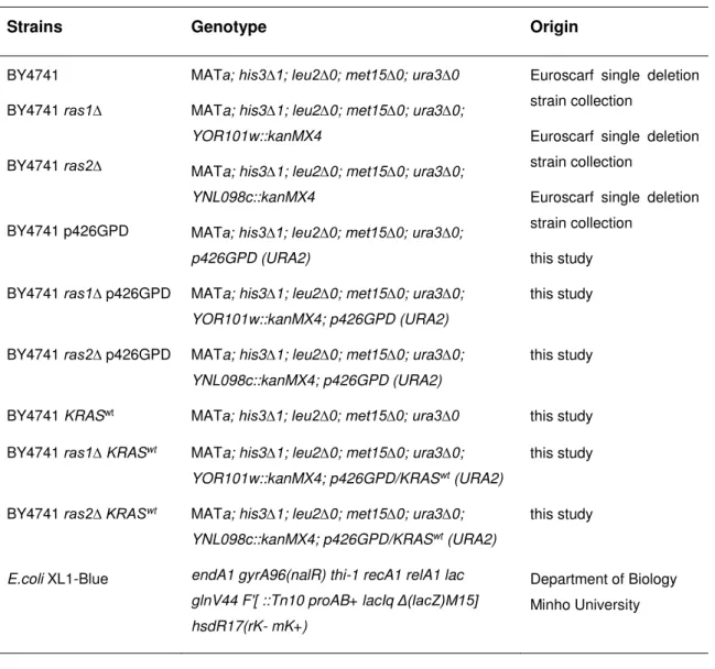
Table 1. The S. cerevisiae BY4741 and E.coli strains used in present work. ... 25
Table 2. Primers used for amplification of human KRASwt fragment. ... 28
Table 3. Growth conditions tested using YNB as base medium. ... 32
Table 4: Compared genetic backgrounds of BY4741 and the different strains used in the literature to describe Ras-related phenotypes. ……….. 51
Table 5. Specific growth rates (µg) of S. cerevisiae BY4741 wt, ras1∆ and ras2∆ untransformed, harbouring p426ø, and expressing KRASwt strains, grown in YNB medium with 2% glucose. Each value is the mean of three independent experiments. ………... 53
Chapter I
3
1. Introduction
1.1 Saccharomyces cerevisiae as a model system in biological
research
The Saccharomyces cerevisiae genome was the first eukaryotic genome to be fully sequenced in April 1996 [1], and over the years the data thus generated fuelled whole genome scale screening methods, including microarrays, two-hybrid analysis and the application of deletion and overexpression libraries [2]. The whole yeast nuclear genome contains a haploid set of 16 well-characterized chromosomes, ranging in size from 200 to 2,200 kb. In contrast to the genomes of multicellular organisms, the yeast genome is highly compact, with genes representing 72% of the total sequence (<2% in the human genome). S. cerevisiae has therefore a genome with a small size and low degree of complexity as compared with higher Eukaryotes. Furthermore, as all other eukaryotic organisms, the yeast S. cerevisiae also contains an additional extranuclear mitochondrial genome [3].
A considerable amount of knowledge about basic cellular processes originated from model organisms that include the simpler eukaryote S. cerevisiae [4]. The increasing amount and complexity of work performed in this organism, which occurred during the last decades, solidified yeast position at the forefront of eukaryotic cellular and molecular biology, facilitating the establishment of new fields of study like systems biology. This goes beyond the functions of individual genes and proteins, focusing on how these interact and work together to determine major properties underlying life and major biological properties and behaviours [5]. The reasons underlying include the many technical advantages that yeasts present over other systems for using in research and biotechnology, namely the short generation time and the high amenability to genetic modifications, as well as the ability to control its growth and division by adjusting environmental conditions [6]. Importantly, it accrues the fact of yeast being a GRAS organism, inexpensive to maintain and propagate, and easy to cultivate and store [3, 7, 8].
Yeast has been recognised the status of model organism for research of complex processes, as a simple eukaryotic organism whose genome can be easily manipulated allowing suitable analysis and efficient assessment of gene products from other Eukaryotes, including man [6, 9]. Although important aspects of human diseases lie
4
complexity, yeast is still used for unveiling molecular processes involved in some diseases based on their high degree of conservation [2, 7]. These are mostly basic cellular processes such as cell cycle control, DNA replication, recombination, repair, protein folding, trafficking, programmed cell death, and metabolic and regulatory mechanisms [10-12].
Moreover, many human genes that are mutated or that have their expression changed in tumour cells have yeast orthologues (approximately 30% of genes according to Françoise Foury) [13]. The fact that a gene codifying for a protein implied in a disease is conserved in yeast, opens the possibility to directly study its function by expressing it in yeast, or by integrating it into the yeast genome [5], namely to complement the correspondent yeast gene deletion [9, 14]. Yeast was used to analyse molecular mechanisms of several human diseases, sometimes of rather unexpected nature like neurological diseases [15-18], or cancer related signalling pathways [7, 8]. As said above, this was achieved by directly studying an endogenous protein orthologue of a human involved in the disease [15-18] or through the heterologous expression of human disease associated proteins [16, 19]. The use of yeast, expressing human genes or not, is therefore also of great value for high-throughput phenotyping and pharmacological assaying [6].
1.1.1 The yeast life cycle
The yeast life cycle (Fig. 1) comprehends two possible reproductive cycles, a mitotic asexual cycle in which cells reproduce by budding, and a sexual-like meiotic cycle in which cells sporulate, germinate, mate and then reproduce by budding. Mating cells can be genetically different from each other - heterothallic yeast – or identical – homothallic yeast. S. cerevisiae is heterothallic and produces two haploid mating types,
MATa and MATα. After conjugation, diploid cells can reproduce asexually in identical
fashion to haploid cells (by budding), or can suffer sporulation entering meiosis and consequently producing four haploid spores, which in turn can also germinate and reproduce asexually (by budding), or can mate returning the culture to diploid state [20].
5
Figure 1. Scheme of yeast life cycle showing haploid and diploid states. Withdrawn from Herskowitz I
(1988) [20].
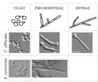
Inversely to humans, S. cerevisiae lives mostly as a unicellular organism, yet as all other microbes, it can form multicellular aggregates, such as colonies or biofilms that are complex, organized, multicellular structures [20, 21]. Additionally, in certain stress conditions such as carbon or nitrogen starvation or oxygen limitation, S. cerevisiae cells can suffer a morphological switch characterized by changes in cell polarity and shape with consequences in cell adhesion and invasive behaviour [22, 23]. Cells become elongated, exhibit a polar budding and remain attached after cytokinesis, switching from growth as independent yeast-form cells on the surface of medium to branching chains of cells, that spread over and into the agar to forage for nutrients, known as filamentous growth [22-24]. In diploid strains this dimorphic switch is characterized by pseudohyphae formation (Fig. 2), while the haploid strains have an more subtle switching displaying only limited changes in cell morphology but an increased cell-cell adhesion, known as haploid invasive growth phenotype (Fig. 2) [23]. In addition, the pseudohyphal growth is a behaviour characteristic of diploids starved for nitrogen, whereas the similar haploid invasive growth is induced by fermentable carbon limitation [25]. Accordingly, stress-induced signalling has an important role in filamentous growth [23, 26].
6
Like all eukaryotic cells, yeasts reproduce while they are young, age and die when they are old. There are two ways of describing aging in yeasts. Replicative aging quantifies aging by the number of daughter cells produced by each mother cell before senescence, and chronological aging, quantifies aging by the length of time that a yeast cell can survive in a non-dividing state [27]. Finally, death can happen via necrose or apoptose, which can also happen upon different stimuli regardless to the yeast age [28].
Figure 2. Morphological forms possible in S. cerevisiae as compared with the human commensalist
and opportunistic pathogen Candida albicans. Adapted from Gancedo J (2001) [24], Cullen et al. (2000) [25], William et al. (2012) [29], Wightman et al. (2004) [30] and www.uni-goettingen.de/en/425121.html.
S. cerevisiae asexual cycle, called mitotic cell cycle initiates with an unbudded cell
(mother cell) in the G1 checkpoint (Fig. 3). The end of G1 checkpoint is marked by the occurrence of the spindle plaque duplication and the beginning of DNA synthesis and bud emergence (daughter cell). At this stage, the unbudded cell starts polarized growth, predominantly at its apex, ensuring correct assembly of a new bud and septum formation before cell division [31]. Cells then enter S phase - DNA synthesis - and plaque separation occurs. The plaques separate until the complete spindle is formed and the bud size continues to grow along the remaining cycle. The finish of G2 checkpoint is marked by the nucleus migration to the cell neck where the spindle elongation occurs and the nuclear division begins, which in turn finishes during M phase followed by cytokinesis. The final event is the cell wall separation with the consequent formation of two unbudded cells, which does not occurs during filamentous growth [32]. This polarized cell division is characterized by two genetically programmed different spatial patterns, axial for haploid cells, and bipolar for diploid cells [33].
7
The major cell cycle control point is in the regulatory G1 time lapse called START. At start, yeast cell integrates many intra and extracellular signals, being then committed to continue proliferation, being switched to differentiation pathways, or entre stationary phase [34]. At START point the yeast cell needs to achieve a critical size in order to bud and progress into S phase, being the G1 phase delayed until this critical mass is reached [35]. The critical cell mass required for budding relates to the nutritional conditions of the cells, which also modulate the degree of asymmetry of cell division. For example, in poor medium usually the parent cells are large and the daughters are very small, whereas in rich medium the asymmetry between parent and daughter cells
is reduced [35, 36].The switch between yeast and pseudohyphae forms also involves
important changes in the pattern of progression through the cell cycle. In pseudohyphal growth, there is an extended G2/M period and the daughter cell reaches the same size of the mother cell. So that both cells can start a new cycle synchronously, while in the yeast form, G1 phase presents itself as the extended cell cycle period, and the bud separates from the mother before reaching considerable size, starting both cells a new cell cycle independently [24].
Figure 3. Phases of S. cerevisiae mitotic cell cycle. Adapted from Hartwell L.H (1974) [32].
1.1.2 The signalling pathways
1.1.2.1
Mitogen activated protein kinases (MAPK) pathways
MAPK pathways are highly conserved signalling operating units, required in all the eukaryotic cells to properly activate specific or general responses, which allow cells to
8
cope with different external stimuli, like all types of stress, nutrients availability, growth factors and cytokines, ensuring cell survival [37]. In S. cerevisiae MAPKs cascades participates in transmitting several extracellular signals controlling mating, morphogenesis, proliferation and cellular integrity in response to nutrients limitation, or osmotic, oxidative, temperature and pH stresses [38]. S. cerevisiae has several MAP kinases, allocated to six distinct MAPK pathways [39, 40] (Fig. 4):
Fus3 – from the mating pheromone response pathway,
Kss1 – common to the filamentous development and the cell wall integrity
pathways,
Hog1 – from the high osmolarity glycerol (HOG) pathway,
Slt2/Mpk1 – cell wall integrity (CWI)/ protein kinase C (PKC) pathway, and
Smk1 – from the spore wall assembly pathway.
Figure 4. Brief outline of the yeast MAPK pathways. Withdrawn from Hohmann S (2002) [40].
These are upstream controlled by MAPKKs and MAPKKKs that may ensure some degree of crosstalk and a common answer to different stimuli. An example is the stimulation of the synthesis and subsequent function of the general stress response
9
transcription factors Msn2 and Msn4 which fine tuning allows the cell to respond to a series of different environmental constraints, including osmotic, heat and oxidative stresses [41].
1.1.1.1
Nutrient signalling pathways
In yeast, a major signalling pathway activated by glucose is the cAMP/PKA (cyclic adenosine monophosphate/ protein kinase A) pathway, which regulates many aspects of cellular physiology, according to nutrients availability [36]. cAMP is synthetized from ATP from adenylate cyclase, which activity is controlled by two distinct G-protein systems, the Ras1/Ras2 and the Gpr1/Gpa2 proteins. Ras1/Ras2-dependent adenylate cyclase activity is further controlled by the exchange factors Cdc25 as well as the GTPase-activating proteins Ira1 and Ira2 (Fig. 5).
Figure 5. RAS/cAMP/PKA signalling pathway in S. cerevisiae. Adapted from Tamanoi F (2011) [42].
The activation of cAMP/PKA pathway can be triggered by transferring yeasts from a poor to a rich carbon source environment [43], which favours rapid growth and cell proliferation by stimulating the glycolytic flux, and repressing the stress response and the expression of genes required for respiratory metabolism [36, 44]. Alternatively, intracellular acidification can have an identical effect [43]. Gpr1/Gpa2 G-protein-coupled receptor system is required for glucose activation of cAMP synthesis, while Ras1/Ras2, in particular Ras2, may stimulate cAMP synthesis in response to either glucose or intracellular acidification [36, 43].
10
In addition to cAMP/ PKA pathway, the yeast also has other two nutrient signalling pathways, TOR (target of rapamicyn) and the less well-known Sch9, partially conserved in higher Eukaryotes [39, 45]. When glucose or other nutrients are present, the three pathways are active conveying signals that promote cell growth and division. In contrast, if the nutrients are scarce, the reduction of the Sch9, TOR, and cAMP/PKA signalling activity, causes cell division arrest and the activation of mechanisms responsible for cellular protection [46]. TOR and Sch9 act cooperatively with PKA to regulate multiple downstream processes, including autophagy, protein synthesis, mitochondrial function and stress resistance, in response to nutrient availability but also to other environmental signals [47]. Studies of chronological aging in S. cerevisiae led to the discovery that Sch9, TOR, and cAMP/PKA pathways are pro-aging pathways, negatively influencing the life span [48]. In particular, the nutrient-responsive Sch9 kinase, adenylate cyclase (Cyr1) and Tor1 are negative regulators of chronological life span (CLS). Accordingly, the deletion of SCH9 and TOR1 increased CLS [49]. This extension involves multiple stress-responsive transcription factors, namely the above mentioned Msn2 and Msn4, which once up-regulated lead to an increase of the levels of superoxide dismutase and catalase, thereby minimizing oxidative stress and cellular
damage [50]. This demonstrates the association between longevity extension and
ability to respond to stress, which has beenobserved in several organisms [27, 51].
1.1.1.2
Signalling crosstalk in particular cell responses
1.1.1.2.1 MAPK cascade and RAS/cAMP/PKA pathway in filamentous
growth response
Evolutionarily conserved signalling pathways encompassing crosstalk and feedback mechanisms regulate the filamentous growth in S. cerevisiae. In particular, the morphogenetic switch requires the cooperation of two different signalling pathways, the filamentous MAPK cascade and the RAS/cAMP/PKA pathway [24]. PKA stimulates
the expression of the MAPK cascade–controlled genes - TEC1 [52] and FLO11 [53]
possessing a Filamentation Response Element (FRE) in their promoters. FLO11 encodes a cell-surface flocculin required for filamentous growth in haploid and diploid strains [53]. As above referred, filamentous growth involves changes in the budding and elongation patterns, and invasive ability. Although these cellular processes are interconnect, they are not always controlled by the same elements. For instance,
11
Ste12p of MAPK cascade is required for cell elongation, while Tpk2p of cAMP/PKA pathway controls the pattern of budding, namely the budding switch, and both are involved in the invasive process [24].
1.1.1.2.2 CWI/PKC, TOR and RAS/cAMP/PKA pathways in oxidative stress
response
Oxidative stress has been implicated in a large variety of biological processes including aging and apoptosis. Reactive oxygen species (ROS), including hydrogen peroxide (H2O2), are generated endogenously in many cells as a consequence of
metabolic processes, mostly at mitochondrial level [54]. By contributing to mitochondrial damage, ROS are also important in redox signalling from the organelle to the rest of the cell. As a result, aerobic organisms sense redox perturbations and develop several different adaptive mechanisms in order to acquire survival capacity [55]. S. cerevisiae was used to study the signal transduction pathways involved in the response to oxidative stress. In this yeast, TOR, CWI/PKC and RAS/cAMP/PKA, are the best characterized routes crosstalking in transducing the oxidative signal [38, 56]. They further react to glucose starvation in a similar manner [57] (Fig. 6). Mtl1 is a cell surface protein required for survival under oxidative stress [58]. Mtl1 transmits the
oxidative signal to the CWI/PKC pathway’s Rom2, which then activates the Rho1, this
way inducing the activation of Slt2 [59]. Furthermore, in conditions of nutrient depletion and oxidative stress Mtl1 negatively influences CWI/PKC through the inhibition of Rom2, which is also inhibited indirectly via Tor1 and Ras2 inhibition through the inhibition by the same stimuli (Fig. 7). This repression eventually has several outputs, such as the decrease of cAMP, the activation of a wide subset of genes potentially regulated by the general stress response dual transcription factor Msn2/Msn4, and the ribosomal gene repression [38, 56]. This extensive crosstalk between culminates in the further interaction with the RAS/cAMP/PKA pathway in processes that ultimately lead to aging (Fig. 8).
12
Figure 6: Schematic representation of the integrative yeast cellular response to glucose depletion and
oxidative stress. Withdrawn from Weinberger et al. (2010) [57].
Figure 7: Schematic representation of the influence of TOR over WCI/PKC pathway via Rom2/Rho1
that also react to the effect of cell surface sensors of cell wall integrity, namely Mtl1. A) Adapted from Torres et al. (2002) [60]; B) adapted from Heinisch (2014) https://www.biologie.uni-osnabrueck.de/forschung/sfb_944/sfb_944_home/projects.html.
13
Figure 8: Schematic representation of the balance between different pathways that contributes to
opposite effects depending on external stimuli, namely TOR, RAS/ cAMP/PKA. Withdrawn from Ruckenstuhl et al. (2010) [61].
1.2 Mammalian RAS pathway
Ras proteins are the founders of a large superfamily of GTPases, very conserved throughout all eukaryotic species, including 150 human members distributed into 5 distinct families, RAS, RHO, RAB, ARF and RAN [62, 63]. Each family is responsible for the control of certain cellular processes. For example, the RAS family controls cell growth, having thus a potential role in the cancer development [64]. Ras proteins are guanine nucleotide-binding proteins that function as GDP/GTP molecular switches. They are activate in response to the activation of a growth factor receptor by the Guanine Nucleotide Exchange Factors (GEFs). These stimulate the intrinsic GDP/GTP exchange activity of Ras proteins promoting the formation of the active RAS-GTP form. Otherwise, GTPase-Activating Proteins (GAPs) stimulate the intrinsic GTP hydrolysis activity of Ras proteins promoting the formation of the inactive RAS–GDP form. Consecutively, the active Ras protein will work as an adaptor protein recruiting
14
effectors from cytosol to the plasma membrane, where they are activated through the interaction with proteins and lipids, producing intracellular signals to modulate cellular behaviour [62, 65, 66].
In mammalian cells, there are at least three functional RAS genes, KRAS, HRAS
and NRAS, encoding 21 kD proteins with 189 (KRas4B) and 188 (KRas4A, HRas and
NRas) amino acids. KRas4A and 4B are alternative splice variants of KRAS at the level of C-terminal, which is important for post-translational modification [67]. These human RAS isoforms are particularly homologous between each other, sharing nearly 85% sequence homology in the first 165 amino acids (the N-terminal) and having a significant variation in 25 remaining amino acids (the terminal), except for the C-terminal CAAX box hypervariable region [67, 68]. This region is useful to distinguish between different Ras proteins [69] and does not implicate significant structural differences, since RAS family members, although functionally different, are closely related at structurally level [70].
Ras proteins regulate cell fates by coupling receptor activation to downstream effector pathways that control several cellular responses including proliferation, differentiation, survival and apoptosis [71]. To regulate signal transmission from cell surface receptors to intracellular signalling cascades, Ras proteins were firstly considered to necessarily localize in the plasma membrane [64, 66]. Nevertheless, Ras proteins localized in different cellular membranes could also signal, recruiting distinct upstream and downstream partner proteins and activating different signalling pathways [62, 65, 70]. Exception is the role of Ras proteins in oncogenesis-related processes, which apparently require their association with the inner face of plasma membrane [72].
The above-mentioned “hypervariable” region of RAS promotes the traffic of Ras proteins to the plasma membrane and their specific localization. Thus, the interactions of Ras proteins with the plasma membrane can differ from one RAS isoform to the
other due to their different structure of the hypervariable domain [69].All Ras proteins
undergo a series of posttranslational modifications at their C-terminus namely in their CAAX motif. These modifications, which comprise farnesylation, cleavage, methyl esterification and palmitoylation, are essential for protein localization and biological activity. In particular, palmitoylation is essential for the tight association of Ras proteins with the plasma membrane, since after the first three modification steps (farnesylation, proteolysis, and methylation), Ras proteins remains largely cytosolic. For Kras, which is not palmitoylated, a stretch of basic residues (lysine) located just upstream of the
15
CAAX motif is required for plasma membrane localization [73]. RAS-membrane interactions can also depend on the activation state of specific RAS isoforms [72].
In mammalian cells, activated Ras proteins stimulate various pathways, being the
Raf-mitogen-activated kinase (MAPK) and PI3K the best characterize Ras-controlled effectors (Fig. 9) [71]. The RAS/Raf/MEK/ERK signalling pathway has been shown to play key role in the transmission of proliferative signals originating from membrane-bound receptors [74]. Raf stimulates cell proliferation and differentiation by the MAPK pathway, while PI3K pathway is involved in the regulation of cell survival, cell cycle progression, cell size and cellular proliferation through numerous downstream effectors including Akt [65, 75, 76]. Furthermore, the survival signals induced by some receptors are mostly mediated by PI3K/Akt and it is known that mutations in this pathway are frequent in many cancers, namely, activating mutations of the PI3K catalytic subunit, and loss-of-function mutations of the PTEN tumour suppressor [75, 76].
Figure 9. A general view of RAS signalling pathway in human cells. Adapted from Hezel A.F (2006) [63].
The HRas, KRas and NRas proteins are widely expressed, being KRas expressed in almost all types of eukaryotic cells [64]. KRAS is essential for normal cellular development while neither N- nor HRAS are, therefore having a well recognize role in tumorigenesis [77, 78]. The most prevalent oncogenic RAS-associated mutations in KRAS are point mutations causing amino acids substitution in codons 12, 13 and 61 [65, 67, 79]. These mutations hamper the capacity of GEFs to interact with KRas protein, producing constitutively activated KRas, and consequently, an improper activation of the pathway, ultimately generating a malignant phenotype [65, 71]. Concurrently, the duration and the intensity of RAS signalling regulate the
16
developmental programs in specific cellular types [71]. Furthermore, KRAS locus in humans encodes two splice variants, for which mutations in KRAS have the potential to interfere in splicing events [69].
1.3 RAS/cAMP/PKA pathway in Saccharomyces cerevisiae
1.3.1 RAS genes
S. cerevisiae has two isoforms of RAS genes, expressing extremely homologous
small GTPases moderately redundant at phenotype level [26, 80]. Moreover, RAS1 and RAS2 are structurally and functionally homologs of the mammalian RAS proto-oncogenes [81]. RAS1 and RAS2 genes constitute an essential yeast gene family, since the deletion of both is lethal to the yeast cells [82]. RAS1 is located on
chromosome XV, 7 cM from ADE2 and 63 cM from HIS3, and encodes a protein of 309 amino acid residues. On the other hand, RAS2 is located on chromosome XIV, 2 cM from MET4, encoding proteins of 322 amino acid residues [42, 83]. Ras1 and Ras2 are nearly 90% homologous in the region corresponding to the N-terminal first 180 amino acids, but diverge in the remaining amino acid sequence. The N-terminal domain is also the region of greatest similarity with the mammalian Ras proteins, with nearly 90% homology between positions 10 - 90 and 3 - 83 of, respectively, the yeast and the mammalian Ras proteins. On the other hand, another major difference between these proteins lies in the sequence and size of their C-terminal, particularly in the hypervariable region including CAAX box, which in the case of Ras1 and Ras2 is much more extended [73, 84].
As mammalian Ras proteins, also yeast Ras proteins undergo a series of post-translational modifications of their C-termini, including farnesylation and palmitoylation that are required for targeting the proteins to the cytoplasmic face of the plasma membrane and for biological activity of the protein. Ras proteins biological activity requires farnesylation, while palmitoylation is required for their localization to the plasma membrane. The essential role of the farnesyl moiety on yeast Ras1/2 proteins is to enable the efficient activation of adenylyl cyclase and response to membrane-associated GEFs, i.e., nucleotide exchange activity mediated by membrane bound full-length Cdc25 and Sdc25 exchange factors [73, 84]. In the N-terminal domain positions 12, 13, 59, 61, or 63, amino acid substitutions can activate the transforming potential of
17
the mammalian Ras proteins. At the equivalent positions, the yeast RAS genes encode the same amino acids. This homology could reflect both a highly conserved biochemical function and a mechanism for regulating that function [83, 85].
As mentioned above, Ras1 and Ras2 proteins are biologically equivalent, although transcriptional and translational controls determine under what physiological conditions either Ras protein will be expressed [86]. As a consequence of a fluid transcriptional and translational regulation, no clear-cut phenotype was ever apparent for ras1∆
mutant strains [86]. The following examples allow a deeper understanding of the resulting phenotyping conundrum:
a) Ras2 is the major regulator of adenylate cyclase, while Ras1 is only a minor one.
Accordingly, ras2∆ mutants present low levels of intracellular cAMP [87];
b) ras2∆ mutants are defective for growth on non-fermentable carbon sources [80],
however this growth defect can be suppressed by overproduction of the RAS1 gene product [82];
c) ras1∆ mutants with a further temperature-sensitive RAS2 mutation, accumulate
unbudded cells at non-permissive temperatures because they arrest at G1 phase [42];
d) ras2∆ras2∆ diploids an increase in storage carbohydrates, sporulation and an high
heat shock resistance was observed, even in rich medium. On the contrary, an
activating mutation of RAS2 such as RAS2Val19, causes a reduction of the glycogen
storage level, and an increase in sensitivity to nutrient starvation and to heat shock [42, 51, 88];
e) ras2∆ have increased longevity and resistance to oxidative stress [89];
f) strains carrying hypo-active RAS2 alleles exhibit a delay in recovery from glucose starvation [90].
1.3.2 RAS/cAMP/PKA signalling pathway
Similar to the mammalian cells, in the yeast S. cerevisiae RAS pathway is activated by growth signals, importantly glucose [91]. In S. cerevisiae, Ras proteins connect nutrient availability to cell growth through regulation of PKA activity, being thus an element of cAMP/PKA pathway [92] as above mentioned. RAS/cAMP/PKA is involved in cell adaptation to environmental changes, responding to nutrient status and various
18
types of stress, such as, oxidative, osmotic and heat shock stresses [28]. RAS/cAMP/PKA pathway performs a key role in the modulation of growth, metabolism, aging, stress resistance, morphogenesis (stimulates filamentous and invasive growth), and cell cycle progression, to ensure that growth occurs only when overall conditions are favourable [36, 42, 92]. This pathway negatively regulates cellular physiology characteristic of stationary phase/nutrient starvation. Consistently, cells with constitutive RAS/cAMP/PKA signalling fail to adapt their growth program in response to nutrient starvation and rapidly lose viability [93].
Ras1 and Ras2 are two small monomeric GTP-binding proteins capable to switch between an active GTP-bound state and an inactive GDP-bound form. In yeast as in mammals, the guanine adenosine phosphate (GAPs) encoded by IRA1 and IRA2 in yeast, down-regulate Ras1 and Ras2 through GTP hydrolysis, resulting in the accumulation of the inactive Ras protein form. Otherwise, the Cdc25 and Sdc25 GEFs promote GDP re-charging to GTP. When Ras1 and Ras2 are in their active conformation (phosphorylated), their hyper-variable domain binds directly to the adenylate cyclase (Cyr1p), which is the only RAS effector protein identified in S.
cerevisiae and the main component of this signalling transduction pathway (Fig. 5-8)
[92]. Cyr1 catalyses the synthesis of cAMP, that binds to the regulatory subunit of the PKA (Bcy1p) triggering the PKA activity by inducing its dissociation from the PKA catalytic subunits (encoded by TPK1, TPK2, and TPK3) [94]. In turn, PKA signals to the nucleus to carry out many functions of which the essential one is G1 cell cycle progression [95]. Ras proteins are required to maintain basal adenylate cyclase activity, being thus essential for cell viability [36, 73].
The constitutive activation of the RAS/cAMP/PKA pathway prevents several rapamycin-induced TOR responses, such as the nuclear translocation of the transcription factor Msn2 and the consequent induction of stress genes, the accumulation of storage carbohydrates, the induction of autophagy, and the down-regulation of ribosome biogenesis [93]. The constitutive activation of the RAS pathway also can suppress a TOR deficiency [93]. Moreover, many of TOR-mediated responses are signalled through the RAS/cAMP/PKA pathway, independently of TOR effectors, and acting upstream of Ras proteins to regulate the PKA activity [36] (Fig. 7, 8). This relationship between these two pathways could go as far as suggesting RAS pathway as a novel TOR effector branch [93]. This is emphasised by the fact that enhanced sensitivity to rapamycin is often a result of the hyperactivation of RAS/cAMP/PKA signalling [96].
19
1.3.2.1
RAS/cAMP/PKA pathway in response to different stresses
Many pathways, including the RAS/cAMP/PKA pathway, converge on the related transcription factors Msn2 and Msn4, which induce stress genes in response to a wide variety of environmental conditions including nutritional, osmotic, acidic, heat shock and oxidative stresses, as well as diauxic transition [31, 38].
The RAS/cAMP/PKA pathway is a negative regulator of the these stress response pathway genes, once in optimal growth conditions, RAS/cAMP/PKA pathway is activated and repress the function of the general stress transcription factor Msn2/Msn4 [97], also negatively regulating the Msn2/Msn4 nuclear localization, which is mandatory under stress condition [38, 40]. On the contrary, the nutrient starvation, oxidative stress, heat shock and entry into stationary phase demand RAS/cAMP/PKA repression [56]. In S. cerevisiae, the RAS/cAMP/PKA pathway must be shut down to allow cell cycle exit, and a full stress response [98]. Concurrently, mutants with high PKA activity display a low tolerance to stress usually associated to optimal growth conditions, while mutants with low PKA activity have high stress tolerance, which in turn is related to suboptimal growth conditions [40].
As above mentioned, in response to oxidative stress, CWI/PKC, TOR and RAS/cAMP/PKA pathways crosstalk through Rom2/Rho1 (Fig. 7). Two possible models were proposed to this crosstalk: (a) Rho1p signals simultaneously but independently of Tor1 and Ras2 inactivation; and (b) Rho1 first inactivates Tor1 protein and then this transmits the signal to inhibit Ras2. Moreover, crosstalk also occurs in a reverse flow, from TOR and RAS to the CWI/PKC pathway. In this case the signal flows from Ras2 and Tor1 inactivation to induce the phosphorylation of Slt2, activating the CWI/PKC pathway in the absence of the Mtl1 protein, assuring the proper adaptive response to oxidative and glucose deprivation [38, 56]. In addition, the hyperactivation of RAS/cAMP/PKA pathway decreases the cell tolerance to various stress conditions, including wall-damaging high-temperature [99-102].
1.3.2.2
RAS/cAMP/PKA pathway in filamentous growth
Ras proteins are central regulators, activators of the two pathways involved in the filamentous growth response [23, 26]. However, Ras1 and Ras2 have distinct importance in the regulation of invasive growth:
20
b) RAS2 is needed to induce invasive growth, since RAS1 expression in a ras2∆
mutant allows growth but not invasive growth;
c) The expression of specific RAS2 alleles can uncouple the two effectors pathways; d) The loss of RAS2 prevents invasion, while the hyperactivation of RAS pathway by
integration of the RAS2V19 allele, causes hyperfilamentation.
The activation of Ras2-dependent pathway is therefore required for filamentous growth [22, 23, 26]. Ras2 activates invasive growth using either of two downstream signalling pathways, the filamentation MAPK (Cdc42p/Ste20p/MAPK) cascade or the cAMP dependent protein kinase (Cyr1p/cAMP/PKA) pathway, which were above mentioned [22, 23, 26]. Surprisingly, although Ras2 has been placed upstream of pathways responsible for the morphogenetic switch, there is at present no information on the mechanisms by which Ras2 might act. Ras2 activity also decreases the activity of the stress responsive transcription factors Msn2p and Msn4p by an overactive Ras2/cAMP/PKA cascade, which is essential for invasive growth phenotype [91]. In addition, Ras2 is also involved in the yeast life span [26].
1.3.2.3
RAS/cAMP/PKA pathway in cellular aging
In fact, Ras2 downstream pathway has been argued to be a key regulator of aging in yeast, and to share similarities with the insulin/IGF1-(like) longevity pathway of mammals [27]. RAS constitutes a pro-aging pathway, negatively regulating the longevity. Mutations that decrease the activity of the RAS/Cyr1/PKA pathway extend longevity and increase stress resistance by activating the above-mentioned transcription factors Msn2/Msn4 (Fig. 8) [103, 104]. It is also remarkable that mutation or overexpression of several other components of the PKA cascade like RAS1,
CDC25, CYR1 affect lifespan, suggesting that in fact, this pathway modulates the life
span [105].
RAS1 and RAS2 show opposite roles in replicative aging. The deletion of RAS1
extends, whereas the deletion of RAS2 shortens replicative longevity, in turn the induced expression of the RAS2 extends, while the overexpression of RAS1 does not affect replicative longevity [87] [51]. Moreover, RAS genes also play opposite roles in the regulation of CLS. The deletion of RAS2 causes CLS extension, while the
constitutive activation of RAS signalling pathway (e.g. RAS2val19) causes a decrease of
CLS [27]. These opposite effects show that the two aging models are somehow related, but that at the same time each has particular characteristics [44].
21
1.3.2.4
The expression of human Ras isoforms in S. cerevisiae
S. cerevisiae has been use as model system to study mammalian RAS genes via
expression of their correspondent cDNAs in strains deleted for either yeast RAS gene. The mammalian HRAS cDNA was expressed for the first time in yeast using a clever construction strategy that enabled the deletion of both yeast RAS genes without the yeast losing viability [106]. Reversely, a modified RAS1 transformed NIH-3T3 cells [107]. The functional interchangeability between yeast and mammalian RAS genes [42] indicates a profound conservation not only of the amino acid sequence, but also of detailed biological function.
1.4 Rationale and aims of the present work
This master thesis was develop within the scope of Glycopharm - Marie Curie Initial Training Network, which scientific objectives are: the development and testing of selective blocking compounds and the development and testing of galectin-mimetic peptides with respective target selectivity. This project impacts enormously on a wide variety of diseases in need for novel therapeutic solutions, namely through the aim of developing a rapid and easy-to-use tool for primary pharmacological testing. This thesis is enclose in the wide objectives of the network and aims at building and validating a high throughput-screening platform of yeast strains displaying phenotypes that can enable the survey of putative galectin-related ligands/inhibitors. This platform was designed to consist of two types of strains, the ones expressing tout court human galectins (in particular Gal3 and Gal1), and the ones expressing these human proteins
together with the human KRASwt cDNA. An additional set of yeast strains expressing
KRASwt without galectin will also be use, in order to study the effect of KRASwt alone
and compare it with the effect resulting from the co-expression of KRASwt and
galectins. The rationale behind this relates with the putative dialogue between galectins and RAS signalling pathway in mammals.
RAS1 and RAS2 genes encode low molecular weight, GTP-binding/hydrolysing
proteins that are highly conserved throughout all eukaryotic species [108]. In human cells, the RAS proto-oncogenes encode proteins that are involved in the control of cell growth, differentiation and survival, having thus a potential role in cancer development [109]. In humans as in S. cerevisiae, Ras proteins are signal switch molecules that regulate cell fates by coupling receptor activation to downstream effector pathways that control diverse cellular responses including proliferation and survival [42, 110, 111].
22
Beyond the protein structural similarities, also the genes from both organisms are functionally homologous and carry out similar functions [112]. Among all human RAS genes, KRAS is expressed in most cellular types, and is the one most frequently mutated in diverse types of cancer [110].
S. cerevisiae harbours one functional RAS pathway, mediated by RAS1 and RAS2
genes [113]. RAS signalling in yeast controls similar biological processes as in higher eukaryotes in response to environmental changes, namely cell proliferation, growth, metabolism, aging, morphogenesis, and cell cycle progression [42]. This functional conservation allows the use of yeast as a model organism to study mammalian proteins and molecular processes [5], including RAS signalling [108]. Moreover, in the last decades, the yeast S. cerevisiae has been identified as a powerful tool to study the relationship between genotype and phenotype in eukaryotic cells, due to an easy experimental tractability, allowing inference of individual gene functions or of network structures through various kinds of experiments [5].
The principal aim of this thesis was the construction of KRASwt expressing strains
of S. cerevisiae, and the subsequent phenotype screening, as part of the development of a yeast-based phenotypic platform for future pharmacological testing. Human
KRASwt cDNA will be express in S. cerevisiae BY4741 genetic background deleted for
RAS1 and RAS2. This genetic background was chosen according with the genetic
marks availability, and for phenotyping control identical strains from the W303-1A background (also haploid and from the same mating type - a) will be used (Brito A.S., unpublished work).
The subsequent phenotypic characterization of the KRASwt expressing strains will
focus on assaying growth-influencing conditions, like carbon source, temperature, pH and different types of environmental stress, as well as proliferation, chronological aging, cell cycle progression and haploid invasive growth. The data to be obtain are expect to contribute to gain insight into the heterologous expression of KRAS in yeasts to allow the future utilization of the above-mentioned platform. The results of this thesis will be complemented with identically obtained phenotypes using BY4741 harbouring
the chromosomal insertion of KRASwt (Cazzanelli J., unpublished work), and using the
control strains from S. cerevisiae W303-1A background, (Brito A.S., unpublished work). Altogether, this data will serve for the development of the yeast-based high throughput phenotype platform. This knowledge is expect to be subsequently validated in human derived cell lines, paving the way for the utilization of the platform in the development of new diagnostic/prognostic tests and new bioactive ligands/inhibitors.
Chapter II
25
2. Material and Methods
2.1 Strains and growth conditions
The Saccharomyces cerevisiae and Escherichia coli strains used in this work are listed on Table 1. E. coli XL1-Blue strain was used for plasmid propagation.
Table 1. The S. cerevisiae BY4741 and E.coli strains used in present work.
Strains Genotype Origin
BY4741 BY4741 ras1∆
BY4741 ras2∆
BY4741 p426GPD
MATa; his3∆1; leu2∆0; met15∆0; ura3∆0
MATa; his3∆1; leu2∆0; met15∆0; ura3∆0; YOR101w::kanMX4
MATa; his3∆1; leu2∆0; met15∆0; ura3∆0; YNL098c::kanMX4
MATa; his3∆1; leu2∆0; met15∆0; ura3∆0; p426GPD (URA2)
Euroscarf single deletion strain collection
Euroscarf single deletion strain collection
Euroscarf single deletion strain collection
this study BY4741 ras1∆ p426GPD MATa; his3∆1; leu2∆0; met15∆0; ura3∆0;
YOR101w::kanMX4; p426GPD (URA2)
this study
BY4741 ras2∆ p426GPD MATa; his3∆1; leu2∆0; met15∆0; ura3∆0; YNL098c::kanMX4; p426GPD (URA2)
this study
BY4741 KRASwt MATa; his3∆1; leu2∆0; met15∆0; ura3∆0 this study
BY4741 ras1∆ KRASwt MATa; his3∆1; leu2∆0; met15∆0; ura3∆0;
YOR101w::kanMX4; p426GPD/KRASwt (URA2)
this study
BY4741 ras2∆ KRASwt
E.coli XL1-Blue
MATa; his3∆1; leu2∆0; met15∆0; ura3∆0; YNL098c::kanMX4; p426GPD/KRASwt (URA2)
endA1 gyrA96(nalR) thi-1 recA1 relA1 lac glnV44 F'[ ::Tn10 proAB+ lacIq Δ(lacZ)M15] hsdR17(rK- mK+)
this study
Department of Biology Minho University
Yeast strains were grown and maintained in rich medium (YPD - yeast extract
(1% w/v), peptone (2% w/v), glucose (2%, w/v)) or in minimal medium (YNB – yeast
nitrogen base without amino acids and ammonium sulphate (0.17% w/v) [Difco], glucose (2% w/v), ammonium sulphate (0.5% w/v)). When appropriate, YNB was supplemented with adequate quantities of amino acids for auxotrophic
26
complementation (0.1 g/l leucine, 0.02 g/l histidine, 0.02 g/l methionine and 0.02 g/l uracil). All strains were batch-grown aerobically in liquid medium, at 30 ºC with orbital shaking at 200 rpm and air/liquid ratio of 3/1. The strains maintenance was done in solid medium (YNB or YPD supplemented with 2% agar), grown at 30 ºC until colonies were observed, and then kept at 4 ºC.
Bacterial cells were grown in liquid Luria-Bertani (LB) medium (yeast extract (0.5%, w/v); tryptone (1%, w/v); NaCl (1%, w/v)), overnight at 37 ºC with shaking at 200 rpm in an orbital shaker. Strains were maintained in LB medium supplemented with 2% agar, previously grown overnight at 37 ºC and kept/storage at 4 ºC. For the selection of transformants, LB was supplemented with 100 µg/ml ampicillin.
2.2 Plasmids
Two plasmids, p416GPD and p426GPD [114], were used in this work to clone human KRASwt (Fig. 10). These are episomal shuttle plasmids, containing the strong
GPD promoter and selective marks for yeast (uracil) and bacteria (ampicillin). p416 is centromeric, while p426 harbours the 2µ element and produces 10-30 copies/cell. Both plasmids have their multicloning site (MCS) adjacent to a strong promoter (originating from GPD1), and have an identical terminator (originating from CYC1). The pUC19 vector was used as a positive control of E. coli transformation, serving to quantify the
competence of E. coli cells. The cDNA from human KRASwt was received in a
pLenti/KRASwt plasmid, provided by IPO-Porto.
![Figure 1. Scheme of yeast life cycle showing haploid and diploid states. Withdrawn from Herskowitz I (1988) [20]](https://thumb-eu.123doks.com/thumbv2/123dok_br/17678359.826233/19.892.350.549.102.472/figure-scheme-showing-haploid-diploid-states-withdrawn-herskowitz.webp)
![Figure 3. Phases of S. cerevisiae mitotic cell cycle. Adapted from Hartwell L.H (1974) [32]](https://thumb-eu.123doks.com/thumbv2/123dok_br/17678359.826233/21.892.308.569.593.864/figure-phases-cerevisiae-mitotic-cell-cycle-adapted-hartwell.webp)
![Figure 4. Brief outline of the yeast MAPK pathways. Withdrawn from Hohmann S (2002) [40].](https://thumb-eu.123doks.com/thumbv2/123dok_br/17678359.826233/22.892.138.771.505.955/figure-brief-outline-yeast-mapk-pathways-withdrawn-hohmann.webp)
![Figure 5. RAS/cAMP/PKA signalling pathway in S. cerevisiae. Adapted from Tamanoi F (2011) [42]](https://thumb-eu.123doks.com/thumbv2/123dok_br/17678359.826233/23.892.333.560.533.844/figure-ras-camp-signalling-pathway-cerevisiae-adapted-tamanoi.webp)


![Figure 9. A general view of RAS signalling pathway in human cells. Adapted from Hezel A.F (2006) [63].](https://thumb-eu.123doks.com/thumbv2/123dok_br/17678359.826233/29.892.291.628.525.799/figure-general-signalling-pathway-human-cells-adapted-hezel.webp)