RESUMO.- [Indução de resposta imune em frangos de corte imunizados com FliC recombinante e desafiados por Salmonella Typhimurium.] Este estudo investigou a resposta imunitária de frangos de corte após a
imuniza-ção oral com flagelina recombinante (rFliC) de Salmonella Typhimurium conjugada com micropartículas de alginato de sódio, e como intensificador de resposta imune foi as -sociada a proteína subunidade B da toxina colérica (rCTB) e pool de Lactobacillus spp. (PL). As respostas imunes fo -ram avaliadas por dosagem de IgY sérica e IgA do fluído intestinal e imunomarcação de linfócitos T CD8+ presentes
no ceco. Os animais imunizados foram desafiados aos 21 dias após tratamento com Salmonella Typhimurium (ST). Foi observado em todos os grupos imunizados um aumento significativo (p<0,05) nos níveis de IgA (mg/mL) principal -mente três semanas após as imunizações. Os níveis de IgY sérica (mg/mL) foram pouco influenciados pelos tratamen -tos, apenas na segunda semana após imunização obser
-Induction of immune response in broiler chickens immunized with
recombinant FliC and challenged by
Salmonella
Typhimurium
1Ana Angelita S. Baptista2*, Tais C. Donato2, Edmárcia E. Souza3, Guilherme A.M. Gonçalves 2, Keila C.O.D. Garcia2, João C.Z. Rodrigues2, Júlio L. Sequeira2
and Raphael L. Andreatti Filho2
ABSTRACT.- Baptista A.A.S., Donato T.C., Souza, E.E., Gonçalves G.A.M., Garcia, K.C.O.D., Ro -drigues J.C.Z., Sequeira J.L. & Andreatti Filho R.L. 2013. Induction of immune response in broiler chickens immunized with recombinant FliC and challenged by Salmonella Typhimurium.Pesquisa Veterinária Brasileira 33(10):1215-1221. Departamento de Clínica Veterinária, Faculdade de Medicina Veterinária e Zootecnia, Universidade Estadual Pau -lista Júlio de Mesquita Filho, Unesp-Botucatu, Distrito de Rubião Júnior s/n, Botucatu, SP 18608-970, Brazil. E-mail: [email protected]
The study examined (1) the immune response in broiler chickens after oral immu -nization with recombinant flagellin (rFliC) from Salmonella Typhimurium conjugated with sodium alginate microparticles, and the immune response enhancement in as-sociation with recombinant cholera toxin B subunit protein (rCTB) and pool of Lacto-bacillus spp. (PL). The immune responses were evaluated by dosage of IgY serum and IgA from intestinal fluid and immunostaining of CD8+ T lymphocytes in the cecum. The
immunized animals were challenged with Salmonella Typhimurium (ST) 21 days after treatment. In all immunized groups, a significant increase (p<0.05) was observed in IgA levels (mg/mL), especially three weeks after immunization. The serum IgY levels (mg/mL) were little affected by the treatments and differed significantly among groups only in the second post-immunization week (p<0.05). After the challenge, the number of CD8+ T cells differed significantly between the treatments and negative control. Re
-trieval of Salmonella Typhimurium was not detected at 48 hours after the challenge in T2 (rFliC+rCTb), T3 (rFliC+PL) and T4 (rFliC+rCTB PL). The rFliC administered orally with or without rCTB and Lactobacillus spp. produces significant induction of humoral immune response, and the immunized chickens were more effective in eliminating Sal-monella after challenge.
INDEX TERMS: Salmonella Typhimurium, FliC, flagellin, immune response, broiler chickens.
1 Received on July 12, 2013.
Accepted for publication on September 10, 2013.
2 Departamento de Clínica Veterinária, Faculdade de Medicina Veteriná
-ria e Zootecnia (FMVZ), Universidade Estadual Paulista (Unesp), Distrito de Rubião Júnior s/n, Botucatu, SP 18608-970, Brazil. *Corresponding au -thor: [email protected]
3 Laboratório Nacional de Biociências (LNBio), Centro Nacional de Pes
vou-se diferenças significativas (p<0,05) entre os grupos. Observou-se que o número de linfócitos T CD8+ apresentou
diferença significativa entre os tratamentos e o controle ne -gativo após o desafio. Quanto a recuperação de Salmonella Typhimurium, observou-se que 48 horas após o desafio já não havia detecção do agente nos grupos T2 (rFliC+rCTb), T3 (rFliC+PL) e T4 (rFliC+rCTB+PL). Concluí-se que rFliC administrada, via oral, associada ou não a Lactobacillus spp e rCTB, demonstrou induzir significativamente a resposta imune humoral e que as aves imunizadas foram mais efi -cientes na eliminação de Salmonella após desafio.
TERMOS DE INDEXAÇÃO: Salmonella Typhimurium, FliC, flageli -na, resposta imune, frangos de corte.
INTRODUCTION
Salmonellosis is a relevant foodborne disease occurring throughout the world. Salmonella Enteritidis and S. Typhi-murium represent the principle serovars associated with foodborne infection in humans (Berndt & Methner 2001, Calenge et al. 2010).
Salmonella infects humans and other animals by the oral route through contaminated food or water (López et al. 2012). A large variety of food products, especially eggs and broiler meat, represent the main infection sources of Sal-monella for humans (Orji et al. 2005, Bernadt et al. 2007). Therefore, the ultimate objective of developing measures to prevent and control salmonellas in chickens is to prevent these microorganisms from entering the food chain (Ber -nadt et al. 2007).
The implementation of biosecurity and the use of pre -biotics, pro-biotics, bacteriophages and vaccines (Chambers & Gong 2011) are among the measures adopted by the avi -culture industry to prevent and control salmonellosis.
The protection against colonization by enteric patho -gens is highly dependent on secretory IgA and on cellular immune response mediated by cytotoxic T cells (Vandeplas et al. 2010).
Studies show that flagellin (FliC), the main structural component of flagella, is capable of interacting with the innate immune system of the host via Toll-like receptor 5 (TLR5) triggering immune responses (Cunningham et al. 2004, Alexan et al. 2009). Recently, this protein has been described as a candidate for a subunit vaccine against Sal-monella (Toyota-Hanatani et al. 2008, 2009, Okamura et al. 2012).
The recognition of FliC via TLR5, expressed in epithelial cells and monocytes, for example, induces the secretion of pro-inflammatory mediators, such as IL-6, TNF-a, IL-1b, as well as the anti-inflammatory cytokine IL-10 (Bedoui et al. 2012). The oral administration of flagellin induces a type Th2 immune response causes an increase mucosal and sys-temic IgA levels (Chaung et al. 2012).
In light of the immunostimulatory characteristics of FliC, the current study evaluated the immune response of com-mercial chickens immunized with recombinant FliC in asso -ciation with intensifiers of immune response, the B subunit of cholera toxin (rCTB) and strains of Lactobacillus spp.
MATERIALS AND METHODS Bacterial samples and culturing conditions
The strains of Lactobacillus spp. used in this study were iso-lated from chicken and selected according to their adhesion ca -pacity and effect as described by Rocha et al. (2012). The strains were cultured separately in 15 mL of Man Rogosa Sharp (MRS) liquid medium, incubated at 37° C, for 24h in an anaerobic system. On the following day, the cultures were mixed in equal parts and administered to the birds.
The challenge was accomplished through a culture of
Salmo-nella Typhimurium, a mutant resistant to nalidixic acid (Nalr) and
rifampicin (Rifr). For this, S. Typhimurium was cultured in 20 mL
of brain heart infusion (BHI) liquid medium, containing Nal and
Rif (100mg/mL) and incubated at 40°C, for 18-24 h. Subsequently, the culture was diluted 100 times in BHI broth and orally admi -nistered to the birds with the aid of a sterile plastic pipette. The number of colony-forming units (CFU) was determined through decimal dilutions in PBS (pH 7.2) and plated in brilliant green agar (BGA).
Recombinant proteins
rCTB. The expression of the recombinant CTB protein (rCTB) was carried out in a strain of Escherichia coli BL21(SI), which was
transformed with the plasmid pAE/ctb (generously donated by Dr. Paulo Lee Ho). Next, recombinant colonies were selected and incubated at 30°C in LB broth added with ampicillin (100 mg/ mL), until reaching the optical density (OD600) of 0.6. Thereafter,
300mM of NaCl was added to induce expression, and the product was incubated again at 30°C under shaking (180rpm). The rCTB expression was verified by electrophoresis in 12% polyacryla -mide gel (SDS-PAGE). After confirmation of recombinant protein expression, the culturing and large-scale induction were accom -plished.
The rCTB protein was expressed as inclusion corpuscles; the -refore, the rCTB was purified using the refolding protocol as des-cribed by Arêas et al. (2002).
To confirm the expression of the rCTB protein in pentame -ric (native) and monome-ric form, the western blotting assay was performed using the purified and renatured (refolding) protein.
For this, the rCTB protein was submitted to SDS-PAGE (12%) and subsequently transferred to a nitrocellulose membrane (Sigma, 0.2um). The result was blocked for one hour in a blocking buffer (PBS+0.1% Tween 20+5% skim powdered milk) and incubated for one more hour with the primary antibody rabbit anti-Cholera
toxin (Sigma) at a 1:10.000 dilution, which was submitted to five
washings (5 min) with PBS solution added with 0.05% Tween 20, and again incubated (1h) with secondary antibody stained with peroxidase (anti-rabbit ), 1:2.500 dilution. After incubation,
five washings of 5 min were performed as previously described and the system was revealed with DAB (DAKO), according to the manufacturer’s recommendations.
After amplification, the PCR product was visualized in 1% aga -rose gel and purified with a Qiaquick PCR purification kit (Qia -gen), following the manufacturer’s protocol. The PCR product was cloned in pGEM T-easy (Promega) and sub-cloned in the vector pET28b (Novagem) using the sites XhoI and NdeI.
Expression and purification. To induce the expression, E.coli
BL21(DE) pLySs was transformed with the plasmid pET28b/fliC. The recombinant colonies were selected and cultured in LB broth added with kanamycin (30mg/mL) and chloranphenicol (50mg/ ml) at 37°C until reaching the optical density (OD600) of 0.6. For induction, 1mM of IPTG was added and the culture was incubated at 37°C under shaking (180 rpm). Aliquots were withdrawn hour -ly to select the best induction time. The rFliC expression was veri -fied in SDS-PAGE (15%). After the expression of recombinant pro -tein was confirmed, the culture was carried out on a large scale.
The rFliC protein was expressed in a soluble form and puri -fied under native conditions by Ni-NTA immobilized-metal affi -nity chromatography (QIAGEN - Integrated Solutions for the Life Sciences), according to the manufacturer’s protocol.
Protein expression was confirmed by the western blotting as -say, similarly to the description for rCTB; however, employing the primary antibody anti-FliC (Flagellin, BioLegend) at 1:5.000 dilu -tion ratio and secondary antibody stained with peroxidase (anti --mouse ) at 1:2.500 dilution ratio.
After protein purification, the protocol described by Aida et al. (1990) was adopted to remove the endotoxins.
Microencapsulation of the recombinant proteins
The purified proteins were microencapsulated to avoid pos -sible degradation of the initial portion from the digestive tract. To this end, the protein solution was emulsified in soybean oil containing 5% sorbitan monooleate. Next, the emulsion was re --emulsified in a sodium alginate solution (1%) and subsequently the double emulsion was dripped into a calcium chloride solution (1%), and maintained at -20°C until the moment of use. A pilot assay was performed with the microcapsules to verify integrity along the length of the digestive tract.
Experimental design
The procedures performed with the animals were approved by Ethics Committee on the use of animals from College of Vete -rinary Medicine and Animal Science, UNESP (nº 71/2009-CEUA). The one-day-old commercial chickens (210) of Ross lineage were housed in experimental cages in the Ornithopathology In -fectorium at College of Veterinary Medicine and Animal Science, Unesp, where they received water and non-medicated ration ad
libitum and necessary heating according to age. The birds were
divided into seven groups composed of 30 animals each. The tre -atments are described in Table 1.
On days two, three and four, the animals in treatment T4 recei-ved 500mL (109 CFU/mL) of pool of Lactobacillus spp. by oral route,
with the aid of a gavage needle. Between the fifth and seventh day of life, the animals in treatments T1, T2, T3 and T6 received the encapsulated recombinant proteins orally, with the aid of a plas -tic spatula. Next, they were supplied 500 mL of carbonated buffer (0.3M NaHCO3; pH 8.0) by means of a gavage needle. One reinfor -cement dose of protein, according to the group, was administered after 15 days. The animals remained fasting 2h before and 2h after the treatments.
The challenge was applied 21 days after the treatments, with an inoculum of 2x10⁵ CFU/ mL of Salmonella Typhimurium (Nalr
and Rifr).
Serological monitoring
On days zero, seven, 14, 21, 22, 23, 26 after the treatments, blood was collected from birds, by means of venous or jugular puncture using sterile needles and syringes. The blood samples were centrifuged at 8000 x g for 5 min. The serum obtained was
maintained at -20° C until analysis to determine IgY.
Collection of intestinal fluid
Soon after blood collection, five birds from each group were euthanized by cervical dislocation. The intestines were collected and, with the aid of a sterile disposable syringe, 2mL of washing solution (PBS pH 7.0 added with BSA (1%), thymerosal (0.01%), 5mM of EDTA and 1 mM of phenylmethyl sulfonyl fluoride – PMSF) were injected into the proximal portion of the duodenum to ensure that the entire length of the intestine would be washed. The fluid collected was centrifuged at 1200xg for 7 min. The resultant su -pernatant was retrieved and stored at -20oC for subsequent quan -tification of secretory IgA.
Collections of ceca for re-isolation ofSalmonella Typhimurium For the bacterial determinations of the cecal content, the ceca were collected from all birds and placed individually in sterile plas -tic collection bags. Next, they were macerated and diluted in PBS (pH 7.2) at 1:10 ratio. Subsequently, dilutions were performed in series, and plated in BGA added with 100mg/mL of Nal and Rif. The reading of the plates was carried out 18-24 hours after incubation.
All the cecal samples were cultured in tetrathionate broth (100mg/mL of Nal and Rif) at 1:10 ratio, incubated at 40°C for 18-24 hours. Subsequently, they were plated in BGA medium (100mg/ mL of Nal and Rif).
Cecal collections for immunohistochemistry assays
One-centimeter portions were collected from ceca. The frag-ments were opened with a longitudinal cut, deposited in his-tological cassettes and fixed in buffered formol (10%) for 24h. Subsequently, the material was washed in distilled water and con -ditioned in ethanol (70%) until they were blocked in paraffin and the slides were formulated.
Enzyme Linked Immunosorbent Assay - ELISA
Secretory IgA intestinal and serum IgY.The measurement of antibodies was accomplished by Kits Chicken IgA ELISA quantifi -cation and Chicken IgY ELISA quantifi-cation (Bethyl Laboratories, Montgomery, TX) according to the manufacturer’s recommenda -tions. The serum samples were diluted at 1:6.400 and intestinal fluid samples were diluted at 1:3.200 and tested in quadruplicate.
Immunohistochemistry
An immunohistochemistry assay was conducted to determine in influx of CD8+ T lymphocytes in the cecum of chickens after im -munizations. Slides were assembled with cecal segments of four animals from each group, euthanized on days 21 and 28 after the Table 1. Description of treatments and immunization
scheme of chickens with recombinant proteins (FliC, CTB) and Lactobacillus spp. Pool (PL)
Treatments Dose of proteins/ Age of animals PL Duration [3d] 1st dose/booster**
T1 - rFliC 50 mg 5-7/21 T2 - rFliC + rCTB 50 mg 5-7/21 T3 - rFliC + PL* 500 ml/ 50 mg 5-7/21
T4 - rFliC + rCTB + PL 500 ml (109) 2-4
treatments. The tissue cuts (3mm) were submitted to deparaffi -nization at 56°C for 24h. On the following day, the samples were submitted to antigen retrieval, according to the protocol descri -bed by Kajiya et al., (2009), with modifications , where the sam -ples were incubated in citrate-phosphate-borate buffer(33mM of Na3C6H5O7; 33mM of NaH2PO4, 57mM of Na2BO4O7.10H2O, pH 3.0) and incubated in a water bath at 96° C for 20 min. Subsequently, we adopted the protocol recommended by the manufacturer of the detection kit Envision-HRP (Dako, USA). The primary antibo -dy used was CD8+ anti-chicken (CT-8; NBP1-28270, Novus Biologi -cals, USA), diluted 1:50, whereas the revelation was accomplished with 3.3-diaminobenzidine (DAB, Dako, USA). The tissues were photographed randomly in 10 histological fields (229mm2), at 40X objective in an optical microscope (Zeiss).
Statistical analysis
The experimental design was completely randomized using a factorial scheme of independent groups. The data obtained were submitted to the analysis of variance, followed by the Tukey test at 5% significance.
The IgA concentrations and the number of CD8+ T lymphocytes were transformed into log10 and the results are presented as ge-ometric means.
RESULTS Measurement of IgA and IgY
The results of IgA concentration (mg/mL) are presented in Table 2.
We observed that the IgA concentrations increased in all the groups until 21 days after the treatments (dat). Afterwards, the treated groups presented values higher (p<0.05) than the control groups and the highest concen -tration of 943 mg/mL was observed for group T1 (rFliC).
On the days (22 and 23) following the challenge with S. Typhimurium, we observed a significant drop in IgA concentrations in groups T1, T2 (rFliC+rCTB) and T3 (rFliC+PL), while group T4 (rFliC+ rCTB+ PL) showed a significant decline starting from day 23. However, we found that on day 26 dat, five days after the challenge and the IgA values of all the groups had resumed a significant increase and reached levels similar to those observed prior to the challenge (21 dat).
On day 26 dat, groups T4 and T5 (positive control) pre -sented the highest IgA concentrations, approximately 1511 and 1546 mg/mL, respectively, differing (p<0.05) from ne -gative controls T6 and T7.
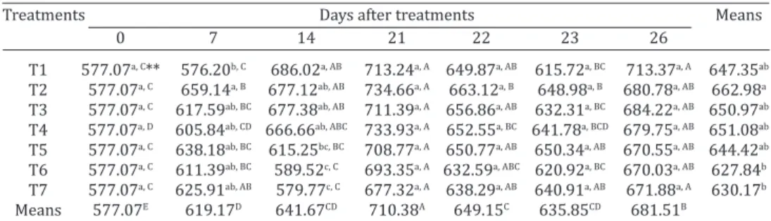
Table 3 details the serum IgY concentrations. At 14 dat, we observed a significant difference (p<0.05) between the groups (T1 - T4) as compared to the controls (T6 and T7) and group T1 (rFliC) had the highest concentration, 686mg/mL. Nevertheless, from 21 dat until 26 dat, we did not observe significant differences in the IgY values among the different groups.
Analysis of the IgY means throughout the experiment
Table 2. Concentration of IgA (mg/mL) in intestinal fluid of commercial chickens at
zero, seven, 14, 21, 22, 23 and 26 days after treatment with: T1 - rFliC, T2 - rFliC + rCTB, T3 - rFliC + Lactobacillus spp., T4 - rFliC + rCTB + Lactobacillus spp., T5 - Salmonella
Typhimurium, T6 - microcapsules or T7 - negative control
Treatments Days after treatment Means*
0 7 14 21 22 23 26
T1 22.01a, D** 55.76bc, C 260.27a, B 943.45a, A 396.67b, B 245.17cd, B 54.67ab, A 223.64b
T2 22.01a, D 86.76b, D 157.23bc, C 927.39a, A 381.46b, B 275.87bcd, B 938.12ab, A 223.07b
T3 22.01a, E 89.92b, D 181.27ab, C 916.64a, A 520.96ab, B 366.55abc, B 592.77b, AB 232.95b
T4 22.01a, A 154.64a, D 193.87ab, D 832.73a, B 701.09a, BC 470.94a, C 1511.05a, A 309.84a
T5 22.01a, E 73.66b, D 172.41ab, C 223.44c, C 657.38a, B 406.11ab, B 1546.69a, A 221.05b
T6 22.01a, E 33.47c, E 99.69c, D 165.00c, C 529.21ab, A 306.29abcd, B 809.60b, A 148.47c
T7 22.01a, E 37.59cd, D 173.92ab, C 399.80b, B 470.16ab, B 212.22d, C 785.61b, A 172.31c
Means* 22.01F 67.27E 171.13E 516.78B 510.53B 315.07C 966.74A
* Geometric mean. ** Means followed by the same upper-case letter on the line or lower-case letter in the column do not differ statistically by the test of Tukey (p<0.05).
Table 3. Concentration of IgY (mg/mL) in serum of commercial chickens at zero, seven, 14, 21, 22, 23 and 26 days after treatment with: T1 - rFliC, T2 - rFliC + rCTB, T3 - rFliC +
Lactobacillus spp., T4 0 rFliC + rCTB + Lactobacillus spp., T5 - Salmonella Typhimurium, T6 - microcapsules or T7 - negative control
Treatments Days after treatments Means
0 7 14 21 22 23 26
T1 577.07a, C** 576.20b, C 686.02a, AB 713.24a, A 649.87a, AB 615.72a, BC 713.37a, A 647.35ab
T2 577.07a, C 659.14a, B 677.12ab, AB 734.66a, A 663.12a, B 648.98a, B 680.78a, AB 662.98a
T3 577.07a, C 617.59ab, BC 677.38ab, AB 711.39a, A 656.86a, AB 632.31a, BC 684.22a, AB 650.97ab
T4 577.07a, D 605.84ab, CD 666.66ab, ABC 733.93a, A 652.55a, BC 641.78a, BCD 679.75a, AB 651.08ab
T5 577.07a, C 638.18ab, BC 615.25bc, BC 708.77a, A 650.77a, AB 650.34a, AB 670.55a, AB 644.42ab
T6 577.07a, C 611.39ab, BC 589.52c, C 693.35a, A 632.59a, ABC 620.92a, BC 670.03a, AB 627.84b
T7 577.07a, C 625.91ab, AB 579.77c, C 677.32a, A 638.29a, AB 640.91a, AB 671.88a, A 630.17b
Means 577.07E 619.17D 641.67CD 710.38A 649.15C 635.85CD 681.51B
revealed that the values were most pronounced at 21. The general mean on 21 dat was 710mg/mL.
Retrieval of Salmonella Typhimurium(ST)
As shown in Table 4, all the treated groups had a signifi -cant reduction (p<0.05) in ST retrieval after the challenge. At 24h after the challenge, group T3 (rFliC+PL) showed a significant difference (p<0.05) in relation to the positive control (T5) and the other groups.
Yet, starting from 48 h post-challenge (23 dat), there was no ST retrieval in groups T2, T3 or T4. Furthermore, all the treated groups differed significantly (p<0.05) from the positive control.
The presence of ST in the cecum of challenged chickens was not detected 120 h (26 dat) after the challenge in the different groups.
The results from culturing samples of cecal content (data not shown) confirm the findings of the counting of CFU/mL.
T2 (rFliC+rCTB) and T5 (positive control) as compared to group T6, which was not submitted to the challenge (Table 5).
On day 23, all groups showed a significant increase (p<0.05) in the number of CD8+ T lymphocytes compared
to T6. Furthermore, on the same day, we found no differen -ce (p<0.05) between the treated groups and T5. This beha -vior was also observed on 26 dat (Table 5).
DISCUSSION
The FliC protein, the largest structural component of bac -terial flagella, had highly conserved domains that are re -cognized by TLR5 receptors (Mizel & Bates 2010). Studies show adjuvant activity of FliC when it is associated with other antigens (McSorley et al. 2000, Skountzou et al. 2009, Bedoui et al. 2012) and its capacity to induce humoral and cellular immune response (Bobat et al. 2011)
Alexan et al. (2009) evaluated the immunomodulatory activity of FliC in mice and reported that high titers of an -tibodies at six weeks after immunization have the capacity to protect against the challenge of virulent strains of Salmo-nella spp. Although recent studies show the immunostimu -latory capacity of FliC in chickens (Toyota-Hanatani 2008, 2009, Chaung et al. 2012, Okamura et al. 2012), there is lit -tle information in the literature on this subject.
In our study, we have investigated the capacity of the FliC recombinant to induce immune response in commer -cial chickens, when associated with Lactobacillus spp. and CTB and immune response enhancers in the mucosa.
We detected a significant increase of intestinal IgA le -vels in all the immunized groups, with the most pronoun-ced response observed three weeks after immunization. Strindilius et al. (2004) found similar results and reported high IgA levels in mice immunized with flagellin. Chaung et al. (2012) studied the association de FliC with an inacti -vated influenza virus (H5N2) and observed that the birds immunized with this combination of antigens showed high IgA levels against H5N2, and concluded that FliC acts as an efficient mucosal adjuvant in birds.
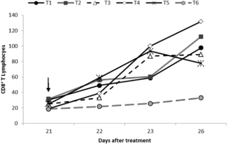
Table 5. Mean of CD8+ T lymphocytes in ceca of commercial chickens at 21, 22, 23 and 26 days after treatment with: T1 - rFliC, T2 - rFliC + rCTB, T3 - rFliC + Lactobacillus spp.,
T4 - rFliC + rCTB + Lactobacillus spp., T5 - Salmonella
Typhimurium or T6 - microcapsules
Treatments Days after treatment Means*
21 22 23 26
T1 30.20a, C** 48.98a, BC 58.88a, AB 97.72a, A 53.95a
T2 31.62a, B 56.23a, B 60.25a, AB 112.20a, A 58.88a
T3 25.12a, B 33.11ab, B 87.09a, A 89.12a, A 50.12a
T4 19.05a, C 38.90ab, B 100.00a, A 131.82a, A 56.23a
T5 25.12a, B 58.88a, A 93.32a, A 77.62a, A 57.54a
T6 18.62a, A 21.88b, A 25.70b, A 33.11b, A 24.43a
Means* 24.55C 40.74B 64.56A 83.17A
* Geometric means. ** Means followed by the same upper-case letter on the line or lower-case letter in the column do not differ statistically by the test of Tukey (p<0.05).
Number of CD8+ T lymphocytes
We observed no significant differences (p<0.05) in the number of CD8+ T lymphocytes in cecum of chickens at 21
dat among the different groups in relation to the negative control (T6) (Table 5, Fig.1).
Soon after the challenge (24h), corresponding to 22 dat, we observed a significant increase (p<0.05) in the CD8+ T lymphocytepopulation in the groups T1 (rFliC),
Fig.1. Geometric mean of CD8+ T lymphocytes in cecum of chi -ckens at 21, 22, 23 and 26 days after treatment with: T1 - rFliC, T2 - rFliC + rCTB, T3 - rFliC + Lactobacillus spp., T4 - rFliC +
rCTB + Lactobacillus spp., T5 - Salmonella Typhimurium; T6
- microcapsules. () indicates the day of Salmonella Typhimu-rium challenge.
Table 4. Quantity of Salmonella Typhimurium in cecal content of chickens after oral administration of 2x10⁵ CFU/mL
Hours after Bacterial count (log10 CFU/mL) Means
infection Treatments
T1 T2 T3 T4 T5 T6 24 2.03a, AB** 2.22a, AB 1.20a, BC 1.29a, ABC 2.55a, A 0.00a,C 1.55a
48 0.20b, B 0.00b, B 0.00b, B 0.00b, B 1.90a, A 0.00a, B 0.35b
120 0.00b, A 0.00b, A 0.00b, A 0.00b, A 0.00b, A 0.00a, A 0.00b
Means 0.74AB 0.74AB 0.40B 0.43B 1.48A 0.00B
The IgA levels of intestinal fluid observed in our study, at two days after the challenge, showed a significant decre -ase. Although this behavior is not very clear, we attribute it to a possible local immunological reaction where the presence of opsonized or secretory-IgA-linked microorga -nisms may have interfered with the method used to detect immunoglobulin.
Nevertheless, on five days after the challenge, IgA levels significantly increased in the challenged groups. These re -sults corroborate Marcq et al. (2011) who demonstrated that after challenge with Salmonella Typhimurium, the bir -ds showed higher titers of secretory IgA.
The systemic humoral immune response was not gre-atly influenced by the treatments regarding mucosal im -munity. We observed that two weeks after immunization, the treated groups differed significantly from the control groups; however, this difference disappeared from the third week onwards, when the IgY levels continued highly similar among all groups, and no further significant inter --group differences were observed until the final collection day. These results show that flagellin has little capacity to induce serum IgY and corroborate Sbrogio-Almeida et al. (2001), who verified that oral immunization of mice with flagellin does not provide higher IgG titers. Similar to the results found by Revolledo et al. (2009), in the current stu -dy, there were no significant changes in serum IgY levels after the challenge.
The fact that the experimental conditions in our study did not produce significant pre-challenge differences in the population of CD8+ T lymphocytes among the different
groups allows to infer that rFliC, even when associated with Lactobacillus and rCTB, does not cause a significant increase in the number of CD8+ T lymphocytes in the cecal
content of treated chicken meat. However, we observed a significant increase in lymphocytes after the challenge with Salmonella spp.
The effect of the different treatments on intestinal co-lonization with Salmonella Typhimurium showed early elimination of microorganisms by the immunized animals (Table 4). We observed that 48 h after the challenge, ST was not detectable in groups T2 (rFliC+rCTb), T3 (rFliC+PL) and T4 (rFliC+rCTB+PL). Although these results did not show a significant difference to group T1 (rFliC), we can infer that the association of rFliC with Lactobacillus and/or CTB enabled greater efficacy in the Salmonella elimination. Thus, we can conclude that the different rFliC treat -ments were capable of inducing mucosal immunity in the animals, conferring high levels of secretory IgA, which may have contributed to the elimination of Salmonella Typhi-murium soon after the challenge. There was a slight sti-mulation of the systemic immune response while the po-pulation of CD8+ T lymphocyteswas not influenced by the
treatments before the challenge.
Acknowledgements.- The authors thank the Sao Paulo Research Foun -dation (FAPESP) for granting the academic scholarship (Grant number 2009/52980-3) and financial support for the project (Grant number 2009/53570-3).
REFERENCES
Aida Y. & Pabst M.J. 1990. Removal of endotoxin from protein solutions by phase separation using Triton X-114. J. Immunol. Methods132:191-195. Alexan A.F., Mohamed S.H. & Ibrahim A.M. 2009. Immune response elic
-ited in mice after immunization with flagellin from Salmonella enterica
Serovar Enteritidis. Global Vet. 3:465-471.
Arêas A.P.M., Oliveira M.L.S., Ramos C.R.R., Sbrogio-Almeida M.E., Raw I. & Ho P.L. 2002. Synthesis of cholera toxin B subunit gene: cloning and ex -pression of a functional 6XHis-tagged protein in Escherichia coli. Protein
Exp. Purif. 25:481-487.
Bedoui S., Kupz A., Wijburg O.L., Walduck A.K., Rescigno M. & Strugnell R.A. 2012 Different bacterial pathogens, different strategies, yet the aim is the same: evasion of intestinal Dendritic cell recognition. J. Immunol. 184:2237-2242.
Berndt A. & Methner U. 2001. Gamma/delta T cell response of chickens after oral administration of attenuated and non-attenuated Salmonella
Typhimurium strains. Vet. Immunol. Immunopathol. 78:143-161. Berndt A., Wilhelm A., Jugert C., Pieper J., Sachse K. & Methner U. 2007.
Chicken cecum immune response to Salmonella enterica Serovars of dif -ferent levels of invasiveness. Infect. Immun. 75:5993-6007.
Bobat S., Langarica A.F. & Hitchcock J. 2001. Soluble flagellin, FliC, induces an Ag-specific Th2 response, yet promotes T-bet-regulated Th1 clear -ance of Salmonella Typhimurium infection. Eur. J. Microbiol. Immunol.
41:1606-1618.
Calenge F., Kaiser P., Vignal A. & Beaumont C. 2010. Genetic control of re -sistance to salmonellosis and to Salmonella carrier-state in fowl: a re-view. Genetics Selection Evolution42:2-11.
Chambers J.R. & Gong J. 2011. The intestinal microbiota and its modula -tion for Salmonella control in chickens. Food Res. Int. 44:3149-3159.
Chaung H.C., Cheng L.T., Hung L.H., Tsai P.C., Skountzou I., Wangd B., Com -pans R.W. & Lien Y.Y. 2012. Salmonella flagellin enhances mucosal im -munity of avian influenza vaccine in chickens. Vet. Microbiol. 157:69-77. Cunningham A.F., Khan M., Ball J., Toellner K.M., Serre K., Mohr E. & Ma -clennan I.C.M. 2004. Responses to the soluble flagellar protein FliC areTh2, while those to FliC on Salmonella are Th1. Eur. J. Microbiol. Im -munol. 34:2986-2995.
Kajiya H.,·Takekoshi S., Takei M., Egashira N., Miyakoshi T., Serizawa A., Teramoto A. & Osamura R.Y. 2009. Selection of buffer pH by the isoelec -tric point of the antigen for the effcient heat-induced epitope retrieval: re-appraisal for nuclear protein pathobiology. Histochem. Cell Biol. 132:659-667.
López F.E., Pescaretti M.M., Morero R. & Delgado M.A. 2012. Salmonella Ty-phimurium general virulence factors: a battle of David against Goliath? Food Res. Int. 45:842-851.
McSorley S.J., Cookson T. & Jenkins M.K. 2000. Characterization of CD4+ T
Cell responses during natural infections with Salmonella Typhimurium. J. Immunol. 164:968-993.
Marcq C., Cox E., Szalo I.M., Théwis A. & Beckers Y. 2011. Salmonella Typhi-murium oral challenge model in mature broilers: bacteriological, immu -nological, and growth performance aspects. Poult. Sci. 90:59-67. Mizel S.B. & Bates J.T. 2010. Flagellin as an adjuvant: cellular mechanisms
and potential. J. Immun. 185:5677-5682.
Okamura M., Matsumoto W., Seike Y., Teratani C., Tozuka M., Kashimoto T., Takehara K., Nakamura M. & Yoshikawa Y. 2012. Efficacy of Soluble Re -combinant FliC Protein from Salmonella enterica Serovar Enteritidis as a
Potential Vaccine Candidate Against Homologous Challenge in Chickens. Avian Dis. 56:354-358.
Orji M.U., Onuigbo H.C. & Mbata T.I. 2005. Isolation of Salmonella from poultry droppings and other environmental sources in Awka, Nigeria. Int. J. Infect. Dis. 9:86-89.
Revolledo L., Ferreira C.S.A. & Ferreira A.J.P. 2009. Prevention of Salmo-nella Typhimurium colonization and organ invasion by combination
treatment in broiler chicks. Poult. Sci. 88:734-743.
Sbrogio-Almeida M.E. & Ferreira L.C.S. 2001 Flagellin expressed by live
Salmonella vaccine strains induces distinct antibody responses follow -ing delivery via systemic or mucosal immunization routes. FEMS Immu -nol. Med. Mic. 30:203-208.
Skountzou I., Martin M.P., Wang B., Ye L., Koutsonanos D., Weldon W., Jacob J. & Compans R.W.2010 Salmonella flagellins are potent adjuvants for
intranasally administered whole inactivated influenza vaccine. Vaccine 28:4103-4112
Strindelius L., Filler M. & Sjöholm I. 2004. Mucosal immunization with pu -rified flagellin from Salmonella induces systemic and mucosal immune responses in C3H/HeJ mice. Vaccine 22:3797-3808.
Toyota-Hanatani Y., Inoue M., Ekawa T., Ohta H., Igimi S. & Baba E. 2008. Importance of the major Fli C antigenic site of Salmonella enteritidis as a subunit vaccine antigen. Vaccine 26:4135-4137.
Toyota-Hanatani Y., Kyoumoto Y., Baba E., Ekawa T., Ohta H., Tani H. & Sa -sai K. 2009. Importance of subunit vaccine antigen of major FliC an -tigenic site of Salmonella Enteritidis II: a challenge trial. Vaccine 27:
1680-1684.