Article
Salvia elegans
,
Salvia greggii
and
Salvia officinalis
Decoctions: Antioxidant Activities and Inhibition of
Carbohydrate and Lipid Metabolic Enzymes
Olívia R. Pereira1 , Marcelo D. Catarino2 , Andrea F. Afonso1,2,3, Artur M. S. Silva2 and Susana M. Cardoso2,*
1 Centro de Investigação de Montanha (CIMO), Instituto Politécnico de Bragança, Campus de Santa Apolónia, 5300-253 Bragança, Portugal; oliviapereira@ipb.pt (O.R.P.); andrea@ipb.pt (A.F.A.)
2 QOPNA & LAQV-REQUIMTE, Department of Chemistry, University of Aveiro, 3810-193 Aveiro, Portugal;
mcatarino@ua.pt (M.D.C.); artur.silva@ua.pt (A.M.S.S.)
3 Public Health Laboratory of Bragança, Local Health Unit, Rua Eng. Adelino Amaro da Costa,
5300-146 Bragança, Portugal
* Correspondence: susanacardoso@ua.pt; Tel.: +351-234-370-360; Fax: +351-234-370-084
Received: 30 October 2018; Accepted: 28 November 2018; Published: 1 December 2018
Abstract: Salvia elegans Vahl., Salvia greggii A. Gray, and Salvia officinalis L. decoctions were
investigated for their health-benefit properties, in particular with respect to antioxidant activity and inhibitory ability towards key enzymes with impact in diabetes and obesity (α-glucosidase, α-amylase and pancreatic lipase). Additionally, the phenolic profiles of the three decoctions were
determined and correlated with the beneficial properties. TheS. elegansdecoction was the most promising in regard to the antioxidant effects, namely in the scavenging capacity of the free radicals DPPH•
, NO•
and O2•–, and the ability to reduce Fe3+, as well as the most effective inhibitor of
α-glucosidase (EC50 = 36.0± 2.7µg/mL vs. EC50 = 345.3± 6.4µg/mL and 71.2 ± 5.0µg/mL
forS. greggiiandS. officinalis,respectively). This superior activity of theS. elegansdecoction over those ofS. greggiiandS. officinaliswas, overall, highly correlated with its richness in caffeic acid and derivatives. In turn, theS. officinalisdecoction exhibited good inhibitory capacity against xanthine
oxidase activity, a fact that could be associated with its high content of flavones, in particular the glycosidic forms of apigenin, scutellarein and luteolin.
Keywords: sage; phenolic compounds; antioxidant; α-glucosidase; pancreatic lipase; α-amylase;
LC-MS analysis
1. Introduction
Salviagenus (Salviaspp.), belonging to the Lamiaceae family, comprises more than 900 species that are used for distinct purposes, including the culinary and cosmetic industries or in traditional medicines due to their claimed health benefits [1,2]. Among them,Salvia officinalisL., i.e., “common
sage” or “Dalmatian sage”, is widely cultivated. These plants usually grow 30–70 cm tall, with a woody stem, whitish beneath and grayish-green above, and with purple-blue flowers up to 3 cm long appearing from early summer to early autumn [1]. Due to its worldwide spread,S. officinalis
has been the most monitored species in relation to the biological potential of the whole plant as well as of its essential oils and polar extracts. For example, promising results were obtained in clinical studies with aqueous or ethanolic extracts of thisSalviaspecies when focused on memory and cognitive functions, pain, and the biochemical profile of glucose and lipids [3]. In addition, in vivo assays in an ear edema induced by croton oil model pointed out the good anti-inflammatory activity of hydroethanolic extracts [4]. Indeed, in vitro experiments demonstrated that its ability to
inhibit 5-lipoxygenase activity [5] and to reduce the levels of interleukin 8 (IL-8) [6] might be based on these anti-inflammatory properties. Moreover, several authors also reported the benefits ofS. officinalispolar extracts towards cell protection in distinct cell-based studies such as in HepG2, HeLa and Caco-2 cell lines, evidencing their possible usage as DNA-protective agents [7,8]. Notably, polar extracts fromS. officinalis(aqueous, methanolic, ethanolic, and hydroalcoholic) have also been proven
to have protective effects against oxidative events [9,10] or oxidative stress-related processes [11,12], as demonstrated inin chemico, cell-based or in vivo models [5,10–17].
In addition toS. officinalis, other widely distributed sage species such asSalvia miltiorrhizaBge.
andSalvia hispanicaL. were highlighted by their richness in bioactive compounds and their potential
health-promoting properties [1,2,18–20]. Still, many less-distributed species, includingSalvia greggii
A. Gray andSalvia elegansVahl., remain poorly studied regardless of their broad use for culinary and medicinal purposes.S. greggii, also known as “autumn sage”, is originated from Mexico and Texas,
although it is currently spread in southwestern United States and Arizona and cultivated in some parts of the world. It grows as a soft, evergreen shrub taller than about 1.2 m, and, similarly toS. officinalis, its leaves are green and smooth [21]. Its flowers, which appear between spring to autumn, can be of different colors (red, pink, purple, white or orange) and are characterized by an intense aroma and abundant nectar. In turn,Salvia elegansis a species native of Mexico and is currently grown in the
United States, Canada, and other regions of the world [22]. It grows as a sub-bush of 1–1.5 m high with a pineapple aroma and flavor, opposite leaves and oval, hairy, elliptic, pale green, and ruby red flowers [23].
S. greggiiandS. elegans are widely used in traditional medicine, particularly in the form of
infusions or decoctions, to treat digestive and oral problems (S. greggii) [1] or to lower blood pressure and combat central nervous system disorders for anxiety and insomnia (S. elegans) [23–25]. However,
as far as we know, the phytochemical composition and beneficial effects of polar extracts, in particular those related to traditional usage (aqueous), remain unexplored. Regardless of this,S. greggiihas been screened for terpenic compounds [26,27], and the antigerminative activity of its essential oils [28], while aspects related to polar extracts have not yet been studied. In turn, polar extracts fromS. elegans
have been the focus of some attention, and, in particular, hydroalcoholic extracts have been shown to exhibit antihypertensive, antidepressant, and anxiolytic effects [23–25] in in vitro models. Yet, to our knowledge, bioactive constituents ofS. eleganspolar extracts and their ability to counteract oxidative-stress-related events have not been previously elucidated.
Hence, the present study aimed to elucidate the phenolic composition and biological effects ofS. elegansandS. greggiidecoctions (mainly focusing on their potential antioxidant activity and inhibitory capacities towards key metabolic enzymes with impact in diabetes and obesity), while comparing the findings to those of the well-knownS. officinalisspecies.
2. Results and Discussion
2.1. Phytochemical Composition
The decoction yields of the threeSalviaspecies were approximately 20%, with slightly higher
levels observed forS. elegansandS. greggiiin comparison toS. officinalis(22.1±2.2% and 22.2±
1.5% vs. 19.3±2.3%, respectively). Consistent with their prevalence inSalviaplants [1,2,29], caffeic
acid derivatives (particularly rosmarinic acid) were dominant compounds inS. officinalis,S. elegans, andS. greggiidecoctions, accounting for about one third of the global identified phenolic species
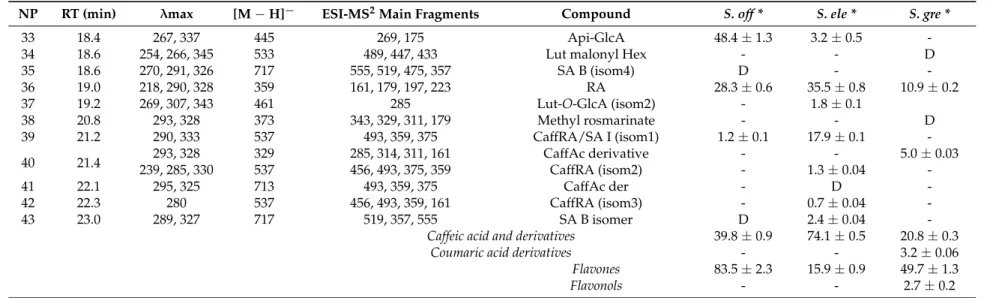
(Table1, Figure1, Figure S1). Nevertheless, significant differences could be found between extracts.
S. eleganswas distinguished by its richness in caffeic acid derivatives, namely rosmarinic acid (peak 36, [M−H]−atm/z359→161, 179), caffeoylrosmarinic acid (peak 39, [M−H]−atm/z537→493, 359)
and salvianolic acid B (peak 27, [M−H]−atm/z717→519), which overall represented approximately
70% of the total quantified phenolics, while the aqueous extract ofS. greggiiwas characterized by high
(peaks 24 and 20, [M−H]−atm/z447 and 431, respectively), representing 33% and 20%, respectively,
of total quantified phenolic compounds. Moderate amounts of luteolin-C-hexoside (peak 15, [M−
H]−
atm/z447→327, 357), quercetin-O-hexoside (peak 22, [M− H]− atm/z463→301) and two
coumaric acid derivatives (peaks 6 and 8, [M−H]−atm/z295 and 265, respectively) have also been
detected in this extract. Interestingly, none of these compounds were detected in the decoctions of the two other species. Hence, globally, the extracts obtained fromS. elegansandS. greggiispecies were clearly distinguishable from that of theS. officinalis,which was dominated by theO-hexuronic form of apigenin ([M−H]−atm/z445→269, 48.4±1.3 mg/g extract) and scutellarein ([M−H]−atm/z
461→285, 13.4±0.6 mg/g extract), in addition to rosmarinic acid (28.3±0.6 mg/g extract). Note that
the predominance of apigenin-O-glucuronide and rosmarinic acid in this decoction is coherent with the abundance of these two constituents previously reported for polar extracts of this species (e.g., ethanol, methanol, aqueous, hydroalcoholic) [2,30]; however, it is worth noting that this is the first time that scutellarein-O-glucuronide was detected inS. officinalisextracts, while the previous studies
Table 1.Phytochemical composition ofS. officinalis,S. elegans, andS. greggiidecoctions determined by UHPLC-DAD-ESI-MSn.
NP RT (min) λmax [M−H]− ESI-MS2Main Fragments Compound S. off * S. ele * S. gre *
1 1.5 275 149 103, 87, 131, 59 2,4-DimethylBA 4.1±0.2 5.1±0.1 7.3±0.2
2 1.7 205 191 111, 173 Quinic acid 0.6±0.1 0.6±0.1 0.5±0.01
3 3.6 280 197 179, 73, 153 Danshensu D D D
4 5.0 290, 324 353 191, 179, 135, 173 cis3-O-CQA - - 3.1±0.03
5 5.7 220, 278 137 109, 93, 119 HydroxyBA D 2.0±0.1
-6 8.3 313 295 163 p-Coum Ac Pent - - 0.3±0.02
7 8.8 290, 325 353 191, 179 trans-5-O-CQA - - 1.9±0.1
8 9.4 313ND 265325 177, 149, 119163, 119 Coumaric Ac DerCaff Hex D- -- 2.9±-0.05
9 9.7 290, 323 179 135 CaffAc 1.8±0.04 1.5±0.02
-10 9.8 314 325 265, 235, 163 Coum Hex D -
-11 9.9 255, 265, 350 625 463, 301 Querc diHex - - D
12 12.1 271, 336 593 473, 503, 353 Api-6-C-Glc-7-O-Glc 4.3±0.1 -
-13 13.1 291, 311 637 351, 285, 193 Ferulic Ac Der - D
-14 13.5 274 571 527, 483, 439, 373 YA E (isom1) - 1.9±0.1
-15 13.9 256, 267, 345 447 327, 357 Lut-C-Hex - - 4.6±0.09
16 13.9 281, 345 477 301, 373, 343, 397 Hydroxy-Lut-GlcA D 1.9±0.2
-17 14.1 276 571 527, 439, 553, 483 YA E (isom2) D -
-18 14.4 269, 304 473 311, 293, 179, 135 Cichoric acid - 1.6±0.07
-19 14.8 267, 345 621 351, 269 Api-diGlcA 4.6±0.3 -
-20 15.2 268, 336 431 311, 341, 269 Api-C-Hex - - 15.7±0.3
21 15.4 274 555571 527, 553, 509, 329313, 357 YA E (isom3)SA K 1.6±D0.2 -- -
-22 15.8 255, 350 463 301 Querc-O-Hex - - 2.7±0.2
23 15.9 280, 333 461 285 Scut-O-GlcA 13.4±0.6 3.9±0.1
-24 16.0 255, 265, 348 447 285 Lut-7-O-Glc - - 26.1±0.9
25 16.1 255, 266, 345 461 285 Lut-7-O-GlcA (isom1) 8.4±0.3 5.1±0.3
-26 16.9 271, 306 521 359, 197, 179, 135 Salviaflaside - D
-27 17.2 278 717 519, 475, 537, 339 SA B (isom1) - 7.8±0.4
-28 17.3 279 571 527, 553, 329 YA E (isom4) 0.9±0.1 -
-29 17.7 279 717 537, 519, 339, 295 SA B (isom2) - 1.7±0.6
-30 17.9 268, 334283 577719 359, 539, 521, 341269 Sagerinic acidApi-rut 4.56.0±±0.30.1 DD -
-31 18.1 271, 304 717 519, 607, 339, 537 SA B (isom3) - 1.7±0.1
Table 1.Cont.
NP RT (min) λmax [M−H]− ESI-MS2Main Fragments Compound S. off * S. ele * S. gre *
33 18.4 267, 337 445 269, 175 Api-GlcA 48.4±1.3 3.2±0.5
-34 18.6 254, 266, 345 533 489, 447, 433 Lut malonyl Hex - - D
35 18.6 270, 291, 326 717 555, 519, 475, 357 SA B (isom4) D -
-36 19.0 218, 290, 328 359 161, 179, 197, 223 RA 28.3±0.6 35.5±0.8 10.9±0.2
37 19.2 269, 307, 343 461 285 Lut-O-GlcA (isom2) - 1.8±0.1
38 20.8 293, 328 373 343, 329, 311, 179 Methyl rosmarinate - - D
39 21.2 290, 333 537 493, 359, 375 CaffRA/SA I (isom1) 1.2±0.1 17.9±0.1
-40 21.4 239, 285, 330293, 328 329537 456, 493, 375, 359285, 314, 311, 161 CaffAc derivativeCaffRA (isom2) -- 1.3±-0.04 5.0±-0.03
41 22.1 295, 325 713 493, 359, 375 CaffAc der - D
-42 22.3 280 537 456, 493, 359, 161 CaffRA (isom3) - 0.7±0.04
-43 23.0 289, 327 717 519, 357, 555 SA B isomer D 2.4±0.04
-Caffeic acid and derivatives 39.8±0.9 74.1±0.5 20.8±0.3
Coumaric acid derivatives - - 3.2±0.06
Flavones 83.5±2.3 15.9±0.9 49.7±1.3
Flavonols - - 2.7±0.2
NP—Number of peak represented in Figure1; D—Detected; Ac—acid; Api- Apigenin; BA—Benzoic acid; CaffAc—Caffeic acid; Caff—Caffeoyl; CQA—Caffeoylquinic acid;
Coum—Coumaroyl; Der—Derivative; Glc—Glucoside; GlcA—Glucuronide; Hex—Hexoside; Lut—Luteolin; Pent—Pentoside; Querc—Quercetin; Rut—Rutinoside; RA—Rosmarinic acid;
0 25 50 75 100
0 5 10 15 20 25
R e la ti ve A b sor b a n ce ( %) Time (min) 0 25 50 75 100
0 5 10 15 20 25
R e la ti ve A b sor b a n ce ( %) Time (min) 21 0 25 50 75 100
0 5 10 15 20 25
R e la ti ve A b sor b a n ce ( %) Time (min) 34
1 12
8 10 9
5 3
2 16
18 16 17 13 9 5 3 2 (a) 24 11 20 15 7 4 3 2 8 6 (b) (c) 1 14 19 25 21 22 23 23 25 26 27 28 29 31 30 33 32 36 32 36 35 30 37 38 39 39 40 40 43 41 42 43 33 36 1
2.2. Biological Activities
2.2.1. Antioxidant Activity
The antioxidant ability of the aqueous extracts obtained from theSalviaplants were evaluated
in regard to their ability to scavenge free radicals, namely DPPH•
(2,2-diphenyl-1-picrylhydrazyl), superoxide (O2•–), nitric oxide (NO•), and peroxyl (RO2•), and their capacity to reduce Fe3+to Fe2+.
Furthermore, all the extracts were screened for their potency in inhibiting xanthine oxidase.
Globally, theS. elegansdecoction was more promising thanS. greggiiin regard to its ability to
scavenge free radicals and to reduce Fe3+ (Table2). It presented EC50 values about 1.8–2.5 lower
than the latter in DPPH•
, NO•
, O2•–, and reducing power tests, and a tendentially higher ability to
capture RO2•. Notably, theS. elegansextract also presented tendentially better antioxidant potential
thanS. officinalis, with tendentially reduced EC50values being registered for NO•, O2•–, and reducing
power tests, and even three times lower for the DPPH•
assay. In addition, one must highlight that the potency ofS. elegansdecoction to counteract DPPH•and NO•was 0.6- and 2.3-fold that of the ascorbic
acid, respectively. The only exception was observed for the oxygen radical absorbance capacity (ORAC) assay for which the result observed forS. officinaliswas better than that forS. elegans, although it was
not statistically significant.
Table 2.Antioxidant properties ofS. officinalis, S. elegans, andS. greggiidecoctions.
S. officinalis S. elegans S. greggii Standard
DPPH•
(EC50µg/mL)(1) 34.8±3.3a 10.7±2.1b 21.1±2.5c 6.69±0.7b
Reducing Power (EC50µg/mL)(2) 40.0±11.2a 31.3±5.0a,c 77.9±5.6b 16.30±1.5c
NO•
(EC50µg/mL)(1) 118.2±16.4a 91.5±14.5a 167.8±23.9b 212.1±9.7c
O2•–(EC50µg/mL)(3) 32.8±0.6a 30.6±1.3a 61.7±3.4b 7.8±0.5c
ORAC (µM TE/mg ext)(4) 404.4±1.80a 373.1±28.1a 335.6±69.6a
-Xanthine oxidase (EC50µg/mL)(5) 55.1±10.6a 71.8±3.8b 70.1±4.0a,b 0.09±0.01c
(1)Ascorbic acid was used as the reference compound. (2)Amount of extract able to provide 0.5 of absorbance
by reducing 3.5µM Fe3+to Fe2+. Butylated hydroxyanisole (BHA) was used as a reference compound.(3)Gallic
acid was used as the reference compound.(4)TE—Trolox Equivalent. (5)Allopurinol was used as the reference
compound. Mean values±SD; statistical analysis was performed by one-way ANOVA followed by Tukey’s test.
In each line, different letters mean significant differences (p< 0.05).
Table3 summarizes the correlation coefficients between the amounts of classes of phenolic components found in theSalviadecoctions (caffeic acid and derivatives, coumaric acid derivatives, flavones and flavonols) and the data from the distinct biological experiments. According to these results, it is possible to suggest that the superior antioxidant activity of theS. elegansdecoction is
strongly associated with its richness in caffeic acid and derivatives, since correlation factors in DPPH•
, reducing power, NO•
, and O2•–assays were 0.801, 0.948, 0.986, and 0.844, respectively.
The comparison of the herein gathered data with that previously reported for other solvent-extracts or other Salviaspecies is not an easy task, since methodologic adaptations (e.g., radical
precursor concentrations and their producing conditions) cause inevitable changes in EC50values. This
difficulty can be partly overcome by the comparison of the extract’s potencies with that of reference compounds. Unfortunately, this approach is often not addressed by the authors. Moreover, there is no universal reference compound for a specific antioxidant assay, and variations in the selected standards are frequent within literature. Regardless of that, one must note thatS. officinalispolar extracts have been commonly used as a reference for the assessment of antioxidant properties of other less-investigated plants [14,15], showing EC50values in the range of 2.0 to 233.0µg/mL for the
DPPH•
assay [5,7,13–15,31]. Other ethanolic, methanolic, or aqueous extracts ofSalviaorigin, including those obtained fromSalvia amplexicaulis[32],Salvia ringens[33],Salvia verbenaca,S. sclarea[34],Salvia argentea[15], andSalvia nemorosa[35], have been claimed to be good DPPH•scavengers as well, with
butylated hydroxytoluene—BHT, or butylated hydroxyanisole—BHA). Hence, one might conclude that, in agreement with other studies reported for polar extracts of severalSalviaspecies,S. officinalis, S. elegans, andS. greggiidecoctions have a high ability to scavenge DPPH•
, withS. elegansshowing the most promising activity, followed byS. greggiiandS. officinalis.
Table 3. Correlation coefficients between the amounts of phenolic components found in theSalvia decoctions (caffeic acid and derivatives, coumaric acid derivatives, flavones and flavonols) and the data from the distinct biological experiments.
DPPH RP ORAC NO O2 XO AG L
Flavones −0.971 −0.357 0.454 −0.498 −0.123 0.901 −0.551 −0.367
Flavonols −0.239 −0.934 −0.891 −0.868 −0.992 −0.434 −0.835 0.930
CafAcD 0.801 0.948 0.400 0.986 0.844 −0.237 0.995 −0.485
CouAcD −0.239 −0.934 −0.891 −0.868 −0.992 −0.434 −0.835 0.930
DPPH 0.570 −0.228 0.690 0.356 −0.771 0.734 0.134
RP 0.670 0.988 0.971 0.084 0.976 −0.738
ORAC 0.547 0.829 0.796 0.493 −0.995
NO 0.922 −0.071 0.998 * −0.624
O2 0.321 0.996 −0.878
XO −0.134 −0.735
AG −0.574
Values expressed as Pearson correlation coefficientR; AG—α-glycosidase inhibitory activity; CafAcD—caffeic
acid and derivatives; CouAcD—coumaric acid derivatives; DPPH—DPPH radical scavenging activity; L—lipase inhibitory activity; NO—nitric oxide radical scavenging capacity; ORAC—oxygen radical absorbance capacity; O2—superoxide anion scavenging activity; RP—reducing power potential; XO—xanthine oxidase inhibitory activity;
*p< 0.05.
Polar extracts obtained fromSalviaplants have also been previously screened for antioxidant abilities through other assays, although not as frequent as for DPPH•
. In this context, Hamrouni-Sellami et al. [36] reported that the Fe3+reducing ability ofS. officinalismethanolic extracts was 6.5-fold
less that of ascorbic acid, being in agreement with our results which also pointed to good effectiveness for decoctions of the same species. In general, our results also indicate that the three sage species herein studied possess promising NO•
scavenging capacities, as all the extracts had a lower EC50compared
to ascorbic acid. Moreover, their activity seems to be superior to that described by Chen and Kang [37] for the methanolic extracts ofSalvia plebeia(EC50= 216±2.9µg/mL), albeit that the absence of a
reference compound in that study hampered solid conclusions. Furthermore, in our study,S. elegans
andS. officinalisdecoctions showed good O2•–scavenging capacity, also suggesting that these extracts
might be more active than the methanolic extracts ofSalvia splendens(EC50= 527µg/mL) [38]. Likewise,
decoctions ofS. officinalis,S. elegans, andS. greggiishowed high capacity to scavenge RO2•(336–404µM
TE/mg), which was significantly superior to those previously reported for the aqueous and ethanolic extracts ofS. officinalis(1143 and 2535µM TE/g) and that otherSalviaspecies (279–4735µM TE/g).
Phenolic compounds have been previously reported to counteract the activity of xanthine oxidase (XO) [39,40], i.e., the enzyme that catalyzes the oxidation of hypoxanthine to xanthine, and further catalyzes xanthine to uric acid with a concomitant production of O2•–, thus contributing to increment of
oxidative stress events in cells. As can be observed in Table2, the decoctions of the threeSalviaspecies
could effectively inhibit the activity of XO, albeit being less potent than the commercial drug allopurinol (EC50= 55.1–71.8µg/mL forSalviaextracts vs. 0.09±0.01µg/mL for allopurinol, respectively). Among
the extracts, the most powerful was that fromS. officinalis, a fact that could be related to its richness
in apigenin glucuronide or in other flavones (i.e., scutellarein and luteolin glycosides), since these compounds have been described as strong inhibitors of this enzyme [41–44]. Although the individual effect of the compounds has not been tested by us, the correlation coefficients between the antioxidant assays and the main compounds of each aqueous extract are in good agreement (Table3). In XO inhibitory assay, the flavones content of theS. officinalisdecoction was highly correlated (0.901) with its
2.2.2. Metabolic Enzyme Activity
α-Glucosidase, α-amylase and pancreatic lipase are key digestive enzymes involved in the
metabolism of carbohydrates and lipids, which make them important targets for therapeutic control of diabetes and obesity.α-Amylase andα-glucosidase catalyze the hydrolysis of carbohydrates into
simple sugars, thus their inhibition retards the digestion of starch and oligosaccharides contributing to the reduction of postprandial increase in plasma glucose levels. In turn, lipase inhibition decreases the digestion of dietary triglycerides, hence reducing the levels of free fatty acids and monoacylglycerols in the intestinal lumen [40,45,46]. In this study, the ability ofS. officinalis, S. elegans,
andS. greggiidecoctions to inhibit the activity of these three digestive enzymes were assessed through in chemicomodels.
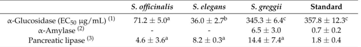
Notably, the inhibitory activities of the threeSalvia extracts againstα-glucosidase were very
promising, especially forS. elegansandS. officinalis(EC50= 36.0±2.7µg/mL and 71.2±5.0µg/mL,
respectively), demonstrating activities of 9- and 4-times that of the antidiabetic pharmaceutical drug, acarbose, respectively (Table4). Moreover, despite being less effective thanS. elegansandS. officinalis,
S. greggiidecoction was as effective as acarbose. Hence, our results suggest that the decoctions of
S. elegans,S. officinalis, andS. greggiicould serve as natural antidiabetic and anti-obesity agents to help
in the control of glucose levels through the control ofα-glucosidase activity. This hypothesis is also
consistent with previous studies that reported identical results for polar extracts ofSalviaagainst this enzyme, e.g., hydroethanolic extracts of S.officinalis(EC50value of 69.7µg/mL) [47] and methanolic
extracts ofSalvia acetabulosa, S. nemorosa, andSalvia chloroleuca(EC50= 76.9µg/mL, EC50= 19µg/mL
and EC50= 13.3µg/mL, respectively) [35,48,49]. In vivo experiments have even demonstrated that
the administration of a daily dose ofS. officinalismethanolic extracts (500 mg/kg body weight) to alloxan-induced diabetic rats caused the inhibition ofα-glucosidase activity comparable to that of the
administration of acarbose (20 mg/kg bw) [45].
Table 4.Enzyme inhibitory properties ofS. officinalis, S. elegans, andS. greggiidecoctions.
S. officinalis S. elegans S. greggii Standard α-Glucosidase (EC50µg/mL)(1) 71.2±5.0a 36.0±2.7b 345.3±6.4c 357.8±12.3c
α-Amylase(2) - - 6.5±3.0 0.7±0.2
Pancreatic lipase(3) 4.6±3.6a 8.2±0.3a 14.4±7.4a 1.8±0.4
(1)Acarbose was used as standard.(2)Results are expressed as percentage (%) inhibition at the concentration of
0.5 mg/mL (Salviadecoctions) or as EC50(µg/mL), for the reference compound acarbose.(3)Results are expressed
as percentage (%) inhibition at the concentration of 0.2 mg/mL (Salviadecoctions) or as EC50(ng/mL), for the
reference compound orlistat. In each line, different letters mean significant differences (p< 0.05).
The inhibitory capacity of polar extracts ofSalviaspecies towardsα-glucosidase have been mostly
correlated with their phenolic constituents. In fact, Chen and Kang [37] reported that the inhibition of this enzyme byS. plebeiamethanolic extracts increased proportionally to their total phenolic content. Moreover, Kocak et al. [50] reported that aqueous and methanolic extracts ofS. cadmica, both rich
in rosmarinic acid, luteolin, and apigenin, had high inhibitory effects towardsα-glucosidase and α-amylase. Moreover, several phenolic compounds isolated fromS. miltiorrhiza, namely, tanshinone IIA, rosmarinic acid, rosmarinic acid methyl ester and salvianolic acid C methyl ester, were reported to be stronger inhibitors ofα-glucosidase than acarbose (EC50= 0.042–0.23µM and EC50= 5.8µM,
respectively) [51]. The flavonoid compounds luteolin-7-O-glucoside, luteolin-7-O-glucuronide, and
diosmetin-7-O-glucuronide, isolated from the aerial parts of S. chloroleuca, also showed potent
α-glucosidase inhibitory effects with EC50values of 18.3, 14.7, and 17.1µM, respectively, exhibiting
an inhibitory effect close to that of acarbose (EC50 = 16.1µM) [49]. Note that rosmarinic acid and
caffeoyl rosmarinic acid are two major phenolic components inS. elegansdecoctions and, based on the
inhibitory activities onα-glucosidase and the content in caffeic acid and derivatives of the extracts
(0.995). Interestingly, high correlation coefficients were also observed between theα-glucosidase
and the antioxidant assays (0.976, 0.998, and 0.996 for reducing power, nitric oxide scavenging, and superoxide anion scavenging, respectively; Table3), suggesting that metabolic and antioxidant effects might possibly be related.
However, regardless the greatα-glucosidase inhibitory capacity and the fact that some authors
have previously found potential inhibitory capacities in polar extracts ofSalvia, namely for aqueous and methanolic extracts ofS. cadmica, as well as for some individual phenolic compounds fromSalvia
origin [49], our results showed no substantial inhibition towardsα-amylase up to the concentration of
0.5 mg/mL. Moreover, at 0.2 mg/mL, onlyS. greggiishowed an anti-lipase activity higher than 10%. This could possibly be owed to its main phenolic component, i.e., luteolin-7-O-glucoside, since its aglycone has been reported to be a good lipase inhibitor [52,53], a hypothesis also supported by the high correlation found between the content of this flavone and the anti-lipase activity (0.930, data not shown). Interestingly, the anti-lipase activity of polar extracts ofSalviaspecies has been previously described, namely for the methanolic extract of the leaves ofS. officinalis(EC50 = 94µg/mL) [54],
the methanol extract ofSalvia spinosa(EC50= 156.2µg/mL) [55], and methanol extracts ofSalvia triloba
(EC50= 100.8µg/mL) [56]. Hence, despite data from literature that seems to suggest that at least
some polar extracts fromSalviaspecies might be promising with respect to their abilities to control the activity ofα-amylase and pancreatic lipase, this was not the observed forS. officinalis, S. elegansandS. greggiidecoctions herein studied.
3. Materials and Methods
3.1. Chemicals
Ethanol, potassium di-hydrogen phosphate, and gallic acid were purchased from Panreac. Dimethylsulfoxide (DMSO), sodium chloride, potato starch, sodium and potassium tartrate, sodium hydroxide, and tris-HCl were purchased from Fisher (Pittsburgh, PA, USA). Fluorescein, 2,2′
-azobis(2-amidinopropane)di- hydrochloride (AAPH), sodium nitroprusside, sulfanilamide, and 3,5-dinitrosalicylic acid (DNS) were purchased from Acros Organics (Hampton, NH, USA). Trolox, xanthine oxidase from bovine milk, allopurinol,α-glucosidase fromSaccharomyces cerevisiae,
4-nitrophenylα-D-glucopyranoside (pNPG), lipase from porcine pancreas and 4-nitrophenyl butyrate, α-amylase from porcine pancreas, β-nicotinamide adenine dinucleotide (β-NADH), phenazine
methosulphate (PMS), nitrotetrazolium blue chloride (NBT), BHA (butylated hydroxyanisole), DPPH radical (2,2-diphenyl-2-picrylhydrazyl), ascorbic acid, and BHT (2,6-di-tert-butyl-4-methylphenol)
were obtained from Sigma (St. Louis, MO, USA). Calcium chloride and sodium di-hydrogen phosphate were purchased from ChemLab (Eernegem, Belgium). Orlistat was purchased from TCI (Tokyo, Japan), acarbose from Fluka (Bucharest, Romania), xanthine from AlfaAesar (Ward Hill, MA, USA), and N-(1-naphthyl)ethylenediamine dihydrochloride from VWR (Radnor, PA, USA). Standard phenolics used for quantitative analysis were obtained from Extrasynthese. Folin-Ciocalteu reagent, Na2CO3, formic acid, and ethanol were purchased from Panreac (Barcelona, Spain).n-Hexane,
methanol, and acetonitrile with high performance chromatography (HPLC) purity were purchased from Lab-Scan (Lisbon, Portugal). Water was treated in a Direct-Q® water purification system (Merck Life Science, Darmstadt, Germany). All reagents were of analytical grade or of the highest available purity.
3.2. Plant Sampling and Preparation of Extracts
S. officinallis,S. elegans¸andS. greggiiwere purchased from Ervital (Viseu, Portugal) as a mixture
of flowers and leaves, and stems where were cultivated under an organic regime. After collection, the aerial parts were dried in a ventilated incubator at 20 to 35◦
C for 3 to 5 days.
plant material (0.5 mm mesh powder) and the mixture was heated and then boiled for 15 min and filtered under reduced pressure through a G3 sintered plate filters. The resulting filtrated solution was concentrated in a rotary evaporator at 37◦
C, followed by defatting withn-hexane (1:1v/v). The resulting fraction was frozen, freeze-dried, and kept under vacuum in a desiccator in the dark for subsequent use [58].
3.3. Identification and Quantification of Phenolic Compounds
UHPLC-DAD-ESI/MSnanalyses of phenolic profiles fromS. officinalis,S. elegans, andS. greggii
decoctions (5 mg/mL) were carried out on an Ultimate 3000 (Dionex Co., San Jose, CA, USA) apparatus equipped with an ultimate 3000 Diode Array Detector (Dionex Co., San Jose, CA„ USA) and coupled to a Thermo LTQ XL mass spectrometer (Thermo Scientific, San Jose, CA, USA), an ion trap MS equipped with an electrospray ionization (ESI) source, following a method previous described [58]. Control and data acquisition were carried out with the Thermo Xcalibur Qual Browser data system (Thermo Scientific, San Jose, CA, USA). Nitrogen above 99% purity was used, and the gas pressure was 520 kPa (75 psi). The instrument was operated in negative-ion mode with the ESI needle voltage set at 5.00 kV and an ESI capillary temperature of 275◦C. The full scan covered the mass range from
m/z100 to 2000.
CID–MS/MS and MSnexperiments were simultaneously acquired for precursor ions using helium as
the collision gas with a collision energy of 25–35 arbitrary units.
Gradient elution was carried out with a mixture of two solvents. Solvent A consisted of 0.1% (v/v) of formic acid in water, and solvent B consisted of acetonitrile, which was degassed and filtrated,
using a 0.2µm Nylon filter (Whatman International, Ltd., Maidstone, England) before use. The solvent
gradient used consisted of a series of linear gradients starting from 5% of solvent B and increasing to 23% at 14.8 min, to 35% at 18 min, and to 100% at 21 min over three minutes, followed by a return to the initial conditions.
For quantitative determinations, the parameters of calibration curves, obtained by injection of known concentrations of the exact or structurally-related standard compounds, allowed the calculation of the limits of detection (LOD) and quantification (LOQ) [58].
3.4. Antioxidant Activities
3.4.1. DPPH•
Scavenging Assay
Extracts capacity for scavenging DPPH•
were evaluated following the procedure previously described by Catarino et al. [59]. Ascorbic acid was used as positive control. The concentration of the extract/standard able to scavenge 50% of DPPH•
(EC50) was determined using linear regression by
plotting the percentage of inhibition against the concentration of the extracts.
3.4.2. Ferric Reducing Antioxidant Power (FRAP) Assay
For the reducing power assay, five different concentrations of each extract were prepared (0.05–0.25 mg/mL), and the assay was carried out according to a procedure described previously [59]. BHA was used as the positive control. A linear regression analysis was carried out by plotting the mean absorbance against the concentrations, and the EC50 value was determined considering the
extract/standard concentration that provided 0.5 of absorbance.
3.4.3. Oxygen Radical Absorbance Capacity (ORAC) Assay
The ORAC assay was performed according to the method previously described by Catarino et al. [60]. In a 96-well plate, 150µL of fluorescein (10 nM), diluted from the stock solution of 250µM, with
75 mM phosphate buffer (pH 7.4) were placed together with 25µL of different trolox concentrations
(3.13–25µM). The same process was repeated for the extracts with final concentrations ranging between
0.4–6.3µg/mL. For blanks, 25µL of phosphate buffer was added instead of antioxidant solutions.
After 10 min incubation at 37◦C, 25
solution was added to each well to reach a final reaction volume of 200µL. The plate was immediately
placed in the plate reader (Biotek, Austria), and fluorescence was monitored every minute over 60 min. The measurement was carried out at 37◦
C with automatic agitation for 5 s prior to each reading. Excitation was conducted at 485 nm with a 20 nm bandpass, and emission was measured at 528 nm with a 20 nm bandpass. Six concentration-dependent kinetic curves were obtained for each sample and for trolox as well. The area under the curve (AUC) of the fluorescence decay and Net AUC were calculated according to the following equations (1–3):
AUC= 1+
ti=60 min
∑
t0=60 min
Ri
R0, (1)
Net AUC =AUCsample−AUCblank, (2)
where R0is the fluorescence reading at the initiation of the reaction and Riis the fluorescence reading at
the time i. Antioxidant activities (ORAC values) of the extracts were calculated by using the following ratio:
ORAC value= me
mT, (3)
whereme is the slope of the curve of Net AUC vs. extract concentrations, andmT is the slope of
the curve of Net AUC vs. trolox concentrations. The final results were expressed inµM of trolox
equivalents (µM TE) perµg of sample extract.
3.4.4. NO•
Scavenging Assay The NO•
scavenging method was adapted from Catarino et al. [60]. Briefly, 100µL of six different
extract concentrations (0–1 mg/mL) were mixed with 100µL of sodium nitroprusside (3.33 mM in
100 mM sodium phosphate buffer pH 7.4) and incubated for 15 min under a fluorescent lamp (Tryun 26 W). Afterwards, 100µL of Griess reagent (0.5% sulphanilamide and 0.05% naphthyletylenediamine
dihydrochloride in 2.5% H3PO4) were added to the mixture, which was allowed to react for another
10 min in the dark. The absorbance was then measured at 562 nm, and the percentage of NO•
scavenging was calculated using the equation described by Yen and Der Duh [61] as follows:
% NO•
scavenging= Ac− Ae
Ac
×100, (4)
where Acis the absorbance of the control (without extract addition) and Aeis the absorbance of the
extract. Ascorbic acid was used as the reference compound. The concentration of the extract/standard able to scavenge 50% of NO• (EC
50) was then calculated by plotting the percentage of inhibition
against the extract concentrations.
3.4.5. Superoxide Anion (O2•–) Scavenging Assay
The O2•– scavenging method was carried out according to the method described by
Catarino et al. [60]. Briefly, in a 96-well plate, 75 µL of six different sample concentrations
(0.0–250µg/mL) were mixed with 100µL ofβ-NADH (300µM), 75 µL of NBT (200µM), and 50 µL of PMS (15µM). After 5 min, the absorbances at 560 nm were recorded and the scavenging activity
of superoxide radicals was calculated according to Equation (4). Gallic acid was used as the reference compound. The concentration of the extract/standard able to scavenge 50% of O2•–(EC50) was
determined using linear regression by plotting the percentage of inhibition against the concentration of the extracts.
3.4.6. Inhibition of Xanthine Oxidase Activity
(0–2 mg/mL) were mixed with 45 µL of sodium dihydrogen phosphate buffer (100 mM, pH 7.5)
and 40µL of enzyme (5 mU/mL). After 5 min incubation at 25◦C, the reaction was started with
the addition of 125µL of xanthine (0.1 mM dissolved in buffer) and the absorbance at 295 nm was
measured every 45 s over 10 min at 25◦
C. The inhibitory effects towards xanthine oxidase activity was calculated as follows:
% inhibiton= mc− me
mc
×100, (5)
wheremcis the slope of the straight-line portion of the curve generated by the control (no inhibitor)
andmeis the slope of the straight-line portion of the curve generated by each extract. Allopurinol was
used as a positive control of inhibition. The concentration of the extract/standard able to inhibit 50% (EC50) of the activity of the enzyme was determined using linear regression by plotting the percentage
of inhibition against the concentration of the extracts.
3.5. Inhibition of Enzymatic Activities
3.5.1. Inhibition ofα-Glucosidase Activity
Inhibition ofα-glucosidase activity was measured following the method described by Neto et al. [63],
with slight modifications. In short, 50µL of different extract concentrations (0–2 mg/mL, in 50 mM
phosphate buffer pH 6.8) were mixed with 50µL of 6 mM 4-nitrophenylα-D-glucopyranoside (pNPG),
dissolved in deionized water. The reaction was started with the addition of 100µL ofα-glucosidase
solution, and the absorbance was monitored at 405 nm every 60 s for 20 min at 37◦
C. The inhibitory effects towardsα-glucosidase activity was calculated as in Equation (5). Acarbose was used as a
positive control of inhibition. The concentration of the extract/standard able to inhibit 50% (EC50)
of the activity of the enzyme was determined using linear regression by plotting the percentage of inhibition against the concentration of the extracts.
3.5.2. Inhibition ofα-Amylase Activity
Inhibition ofα-amylase activity was measured according to Wickramaratne et al. [64], with slight
modifications. Briefly, 200µL of extract six different extract concentrations (0–2 mg/mL) dissolved in
20 mM phosphate buffer (pH 6.9, containing 6 mM of NaCl) were added to 400µL of a 0.8% (w/v)
starch solution in the same phosphate buffer, and the mixture was incubated for 5 min at 37◦
C. The reaction was then started with the addition of 200µL ofα-amylase solution, and after 5 min of
incubation, 200µL of the reaction mixture was collected and immediately mixed with 600µL of DNS
reagent (10 g/L of 3,5-dinitrosalicylic acid, 300 g/L of potassium and sodium tartrate tetrahydrate, and 0.4 M NaOH) to stop the reaction. A second aliquot of 200µL was further collected 15 min
later and mixed with DNS reagent as well. Samples were then boiled for 10 min, and, once they had cooled, 250µL were transferred to each well in a 96-well microplate for absorbance reading at 450 nm.
Blank readings (no enzyme) were then subtracted from each well and the inhibitory effects towards
α-amylase activity was calculated as follows:
%inhibiton= ∆Absc
− ∆Abse
∆Absc ×100, (6)
where∆Abscis the variation in the absorbance of the negative control and∆Abseis the variation in
the absorbance of the extract. Acarbose was used as a positive control of inhibition.
3.5.3. Inhibition of Pancreatic Lipase Activity
The lipase activity was measured according to the procedure described by Neto et al. [63], with slight modifications. The reaction mixture was prepared in a microtube by mixing 55 µL of five
different concentrations of extract (0–2 mg/mL) dissolved in tris buffer 100 mM (pH 7.0) with 467.5µL
started by adding 11µL of 20 mM 4-nitrophenyl butyrate diluted in DMSO. Final DMSO concentration
in the reaction mixture did not exceed 2%. The reaction mixture was then quickly transferred to a 96-well plate and incubated for 35 min at 37◦
C while the absorbance was being measured every 60 s at 410 nm. The inhibitory effects towards pancreatic lipase activity was calculated as in Equation (5). Orlistat was used as a positive control of inhibition.
3.6. Statistical Analysis
All data are presented as mean±standard deviations from three independent assays performed
at least in duplicate. One-way analysis of variance (ANOVA) followed by Tukey´s test was used to detect any significant differences among different means. Correlation analyses were performed using a two-tailed Pearson’s correlation test. Ap-value less than 0.05 was assumed as significant. The results
were analyzed using GraphPad Prism 6 (GraphPad Software, La Jolla, CA, USA) and SPSS v 23.0 (Statistical Package for the Social Sciences).
4. Conclusions
This work clarifies the antioxidant properties ofS. elegans, S. greggii, andS. officinalisdecoctions as well as their inhibition towards the activity of carbohydrate and lipid metabolic enzymes, highlighting possible correlations with their phenolic components. It was shown that among the three plants,
S. elegansdecoctions were the most promising regarding antioxidant activity and inhibitory potential
againstα-glucosidase, a fact that might be related to its richness in caffeic acid and its derivatives.
In turn, despite all the three decoctions ofSalviaspecies could effectively inhibit the activity of xanthine
oxidase, one should highlight the superior inhibitory capacity of S. officinalis, which is possibly
associated with the presence of flavones. In conclusion, similarly to the well-knownS. officinalis
species,S. elegansandS. greggiiare a valuable source of natural metabolites and could be used for commercial applications in novel functional foods or pharmaceutical ingredients targeting diabetes and obesity prevention.
Supplementary Materials:Supplementary Materials can be found in a separate file: Figure S1: UV spectra of the main peaks identified inS. elegans, S. greggii, andS. officinalisdecoctions.
Author Contributions:M.D.C. performed the experimental work and co-wrote the manuscript; O.R.P. and A.F.A.
performed the analysis and interpretation of data and wrote the manuscript; A.M.S.S. co-wrote the manuscript; S.M.C. coordinated the work and co-wrote the manuscript.
Funding:The Science and Technology Foundation/Ministry of Education and Science (FCT/MEC) funded the Organic Chemistry, Natural Products and Food Stuffs Research Unit (QOPNA) research unit (FCT UID/QUI/ 00062/2013) through national funds and, where applicable, was co-financed by the European Regional Development Fund (FEDER), within the Portugal 2020. Project AgroForWealth (CENTRO-01-0145-FEDER-000001), funded by Centro2020, through FEDER and PT2020, financed the research contract of Susana M. Cardoso. The Science and Technology Foundation financed Marcelo D. Catarino (fellowship PD/BD/114577/2016).
Conflicts of Interest:The authors declare no conflict of interest.
References
1. Kintzios, S.E.Sage The Genus Salvia; Harwood Academic Publisher: Amsterdam, The Netherlands, 2000; ISBN 0203303660.
2. Lu, Y.; Foo, L. Polyphenolics ofSalvia—A review.Phytochemistry2002,59, 117–140. [CrossRef]
3. Ghorbani, A.; Esmaeilizadeh, M. Pharmacological properties ofSalvia officinalisand its components.J. Tradit. Complement. Med.2017,7, 433–440. [CrossRef] [PubMed]
4. De Melo, G.A.N. Anti-inflammatory activity ofSalvia officinalisL.J. Med. Plants Res. 2012,6, 4934–4939. [CrossRef]
5. Albano, S.M.; Miguel, M.G. Biological activities of extracts of plants grown in Portugal.Ind. Crops Prod.2011,
6. Chohan, M.; Naughton, D.P.; Jones, L.; Opara, E.I. An investigation of the relationship between the anti-inflammatory activity, polyphenolic content, and antioxidant activities of cooked and in vitro digested culinary herbs.Oxidative Med. Cell. Longev.2012,2012, 627843. [CrossRef] [PubMed]
7. Kozics, K.; Klusová, V.; Sranˇcíková, A.; Muˇcaji, P.; Slame ˇnová, D.; Hunáková, L’.; Kusznierewicz, B.; Horváthová, E. Effects ofSalvia officinalisandThymus vulgarison oxidant-induced DNA damage and antioxidant status in HepG2 cells.Food Chem.2013,141, 2198–2206. [CrossRef] [PubMed]
8. Ramos, A.A.; Azqueta, A.; Pereira-Wilson, C.; Collins, A.R. Polyphenolic compounds fromSalviaspecies protect cellular DNA from oxidation and stimulate DNA repair in cultured human cells.J. Agric. Food Chem.
2010,58, 7465–7471. [CrossRef]
9. Walch, S.G.; Tinzoh, L.N.; Zimmermann, B.F.; Stühlinger, W.; Lachenmeier, D.W. Antioxidant capacity and polyphenolic composition as quality indicators for aqueous infusions ofSalvia officinalisL. (sage tea). Front. Pharmacol.2011,2, 79. [CrossRef]
10. Bettaieb, I.; Hamrouni-Sellami, I.; Bourgou, S.; Limam, F.; Marzouk, B. Drought effects on polyphenol composition and antioxidant activities in aerial parts ofSalvia officinalisL.Acta Physiol. Plant. 2011,33,
1103–1111. [CrossRef]
11. Ozkan, G.; Kamiloglu, S.; Ozdal, T.; Boyacioglu, D.; Capanoglu, E. Potential use of Turkish medicinal plants in the treatment of various diseases.Molecules2016,21, 257. [CrossRef]
12. Li, A.N.; Li, S.; Zhang, Y.J.; Xu, X.R.; Chen, Y.M.; Li, H.B. Resources and biological activities of natural polyphenols.Nutrients2014,6, 6020–6047. [CrossRef]
13. Martins, N.; Barros, L.; Santos-Buelga, C.; Henriques, M.; Silva, S.; Ferreira, I.C.F.R. Evaluation of bioactive properties and phenolic compounds in different extracts prepared fromSalvia officinalisL.Food Chem.2014, 170, 378–385. [CrossRef] [PubMed]
14. Jeshvaghani, Z.A.; Rahimmalek, M.; Talebi, M.; Goli, S.A.H. Comparison of total phenolic content and antioxidant activity in differentSalviaspecies using three model systems.Ind. Crops Prod.2015,77, 409–414. [CrossRef]
15. Farhat, M.B.; Landoulsi, A.; Chaouch-Hamada, R.; Sotomayor, J.A.; Jordán, M.J. Characterization and quantification of phenolic compounds and antioxidant properties ofSalviaspecies growing in different habitats.Ind. Crops Prod.2013,49, 904–914. [CrossRef]
16. Šulni ¯ut˙e, V.; Ragažinskien˙e, O.; Venskutonis, P.R. Comprehensive Evaluation of Antioxidant Potential of 10 SalviaSpecies Using High Pressure Methods for the Isolation of Lipophilic and Hydrophilic Plant Fractions. Plant Foods Hum. Nutr.2016,71, 64–71. [CrossRef]
17. Paun, G.; Neagu, E.; Moroeanu, V.; Ungureanu, O.; Cretu, R.; Ionescu, E.; Tebrencu, C.E. Phytochemical analysis and in vitro biological activity ofBetonica officinalisandSalvia officinalisextracts.Rom. Biotechnol. Lett.2017,22, 12751–12761.
18. Ullah, R.; Nadeem, M.; Khalique, A.; Imran, M.; Mehmood, S.; Javid, A.; Hussain, J. Nutritional and therapeutic perspectives of Chia (Salvia hispanicaL.): A review.J. Food Sci. Technol.2016,53, 1750–1758, Epub
2015 Oct 1. [CrossRef]
19. Min-hui, L.; Jian-min, C.; Yong, P.; Pei-gen, X. Distribution of Phenolic Acids in ChineseSalviaPlants. World Sci. Technol.2008,10, 46–52. [CrossRef]
20. Zettel, V.; Hitzmann, B. Applications of chia (Salvia hispanicaL.) in food products.Trends Food Sci. Technol.
2018,80, 43–50. [CrossRef]
21. Frett, J.F. Influence of nutrient salts, auxins and cytokinins on the in vitro growth ofSalvia greggii.Plant Cell Tissue Organ Cult.1987,9, 89–93. [CrossRef]
22. Starr, G. New WorldSalviasfor Cultivation in Southern Arizona.Desert Plants1985,7, 184.
23. Lim, T.K.Edible Medicinal and Non Medicinal Plants; Springer: Basel, Switzerland, 2014; ISBN 978-94-017-7276-1.
24. Herrera-Ruiz, M.; García-Beltrán, Y.; Mora, S.; Díaz-Véliz, G.; Viana, G.S.B.; Tortoriello, J.; Ramírez, G. Antidepressant and anxiolytic effects of hydroalcoholic extract fromSalvia elegans.J. Ethnopharmacol.2006, 107, 53–58. [CrossRef] [PubMed]
25. Jiménez-Ferrer, E.; Badillo, F.H.; González-Cortazar, M.; Tortoriello, J.; Herrera-Ruiz, M. Antihypertensive activity ofSalvia elegansVahl. (Lamiaceae): ACE inhibition and angiotensin II antagonism.J. Ethnopharmacol.
26. Kawahara, N.; Tamura, T.; Inoue, M.; Hosoe, T.; Kawai, K.I.; Sekita, S.; Satake, M.; Goda, Y. Diterpenoid glucosides fromSalvia greggii.Phytochemistry2004,65, 2577–2581. [CrossRef] [PubMed]
27. Kawahara, N.; Inoue, M.; Kawai, K.I.; Sekita, S.; Satake, M.; Goda, Y. Diterpenoid fromSalvia greggii. Phytochemistry2003,63, 859–862. [CrossRef]
28. De Martino, L.; Roscigno, G.; Mancini, E.; De Falco, E.; De Feo, V. Chemical composition and antigerminative activity of the essential oils from five salvia species.Molecules2010,15, 735–746. [CrossRef] [PubMed]
29. Pereira, O.R.; Afonso, A.F.; Cardoso, S.M. Advances in Analysis of Phenolic Compounds ofSalvia, Thymus andLavandulaPlants. InRecent Progress in Medicinal Plants Series; Govil, J.N., Pathak, M., Eds.; Studium Press LLC: New Delhi, India, 2016; pp. 1–33.
30. Cvetkovikj, I.; Stefkov, G.; Acevska, J.; Stanoeva, J.P.; Karapandzova, M.; Stefova, M.; Dimitrovska, A.; Kulevanova, S. Polyphenolic characterization and chromatographic methods for fast assessment of culinary Salviaspecies from South East Europe.J. Chromatogr. A2013,1282, 38–45. [CrossRef]
31. Brahmi, N.; Scognamiglio, M.; Pacifico, S.; Mekhoukhe, A.; Madani, K.; Fiorentino, A.; Monaco, P. 1H NMR based metabolic profiling of eleven Algerian aromatic plants and evaluation of their antioxidant and cytotoxic properties.Food Res. Int.2015,76, 334–341. [CrossRef]
32. Alimpi´c, A.; Kneževi´c, A.; Milutinovi´c, M.; Stevi´c, T.; Šavikin, K.; Staji´c, M.; Markovi´c, S.; Marin, P.D.; Matevski, V.; Duleti´c-Lauševi´c, S. Biological activities and chemical composition ofSalvia amplexicaulisLam. extracts.Ind. Crops Prod.2017,105, 1–9. [CrossRef]
33. Alimpi´c, A.; Pljevljakuši´c, D.; Šavikin, K.; Kneževi´c, A.; ´Curˇci´c, M.; Veliˇckovi´c, D.; Stevi´c, T.; Petrovi´c, G.; Matevski, V.; Vukojevi´c, J.; et al. Composition and biological effects ofSalvia ringens(Lamiaceae) essential oil and extracts.Ind. Crops Prod.2015,76, 702–709. [CrossRef]
34. Kostic, M.; Petrovic, M.B.; Jevtovic, T.; Jovic, M.; Petrovic, A.; Slavoljub, Ž. Anti-inflammatory effect of the Salvia sclareaL. ethanolic extract on lipopolysaccharide-induced periodontitis in rats.J. Ethnopharmacol.2017, 199, 52–59. [CrossRef] [PubMed]
35. Bahadori, M.B.; Asghari, B.; Dinparast, L.; Zengin, G.; Sarikurkcu, C.; Abbas-Mohammadi, M.; Bahadori, S. Salvia nemorosaL.: A novel source of bioactive agents with functional connections.Food Sci. Thecnol.2017,75, 42–50. [CrossRef]
36. Wang, B.Thousand Formulas and Thousand Herbs of Traditional Chinese Medicine; Heilongjiang Education Press: Harbin, China, 1993; Volume 2, ISBN 7531618990.
37. Chen, L.; Kang, Y.H. Antioxidant and Enzyme Inhibitory Activities of Plebeian Herba (Salvia plebeiaR. Br.) under Different Cultivation Conditions.J. Agric. Food Chem.2014,62, 2190–2197. [CrossRef] [PubMed] 38. Mahesh, K.P.; Sasmal, D.; Papiya, M.M. In vitro antioxidant activity of methanolic extract of aerial parts of
Salvia splendens(Scarlet sage).Pharmacogn. J.2010,2, 7–12.
39. Liu, X.; Chen, R.; Shang, Y.; Jiao, B.; Huang, C. Superoxide radicals scavenging and xanthine oxidase inhibitory activity of magnesium lithospermate B fromSalvia miltiorrhiza.J. Enzyme Inhib. Med. Chem.2009,
24, 663–668. [CrossRef]
40. Lin, D.; Xiao, M.; Zhao, J.; Li, Z.; Xing, B.; Li, X.; Kong, M.; Li, L.; Zhang, Q.; Liu, Y.; et al. An overview of plant phenolic compounds and their importance in human nutrition and management of type 2 diabetes. Molecules2016,21, 1374. [CrossRef] [PubMed]
41. Kim, J.K.; Kim, W.J.; Hyun, J.M.; Lee, J.S.; Kwon, J.G.; Seo, C.; Song, M.-J.; Choi, C.; Hong, S.S.; Park, K.; et al. Salvia plebeiaExtract Inhibits Xanthine Oxidase Activity In Vitro and Reduces Serum Uric Acid in an Animal Model of Hyperuricemia.Planta Med.2017,83, 1335–1341. [CrossRef]
42. Lin, C.-M.; Chen, C.-S.; Chen, C.-T.; Liang, Y.-C.; Lin, J.-K. Molecular modeling of flavonoids that inhibits XO.Biochem. Biophys. Res. Commun.2002,294, 167–172. [CrossRef]
43. Jiao, R.H.; Ge, H.M.; Shi, D.H.; Tan, R.X. An apigenin-derived xanthine oxidase inhibitor fromPalhinhaea cernua.J. Nat. Prod.2006,69, 1089–1091. [CrossRef]
44. Vitaminol, J.N.S. Chemical Evidence for Potent Xanthine Oxidase Inhibitory Activity ofGlechoma hederacea var. grandis leaves.J. Nutr. Sci. Vitaminol.2013,59, 570–575. [CrossRef]
45. Moradabadi, L.; Kouhsari, S.M.; Sani, M.F. Hypoglycemic effects of three medicinal plants in experimental diabetes: Inhibition of rat intestinalα-glucosidase and enhanced pancreatic Insulin and cardiac Glut-4 mRNAs expression.Iran. J. Pharm. Res.2013,12, 385–397.
47. Hamza, A.A.; Ksiksi, T.S.; Shamsi, O.A.A.; Balfaqh, S.A.α-Glucosidase Inhibitory Activity of Common Traditional Medicinal Plants Used for Diabetes Mellitus.J. Dev. Drugs2015,4, 2. [CrossRef]
48. Loizzo, M.R.; Saab, A.M.; Tundis, R.; Menichini, F.; Bonesi, M.; Piccolo, V.; Statti, G.A.; de Cindio, B.; Houghton, P.J.; Menichini, F. In vitro inhibitory activities of plants used in Lebanon traditional medicine against angiotensin converting enzyme (ACE) and digestive enzymes related to diabetes.J. Ethnopharmacol.
2008,119, 109–116. [CrossRef] [PubMed]
49. Asghari, B.; Salehi, P.; Sonboli, A.; Ebrahimi, S.N. Flavonoids fromSalvia chloroleucawith alpha-Amylsae and alpha-Glucosidase Inhibitory Effect.Iran J. Pharm. Res.2015,14, 609–615. [PubMed]
50. Kocak, M.S.; Sarikurkcu, C.; Cengiz, M.; Kocak, S.; Uren, M.C.; Tepe, B.Salvia cadmica: Phenolic composition and biological activity.Ind. Crops Prod.2016,85, 204–212. [CrossRef]
51. Ma, H.Y.; Gao, H.Y.; Sun, L.; Huang, J.; Xu, X.M.; Wu, L.J. Constituents withα-glucosidase and advanced
glycation end-product formation inhibitory activities fromSalvia miltiorrhizaBge.J. Nat. Med.2011,65, 37–42. [CrossRef] [PubMed]
52. Ramirez, G.; Zamilpa, A.; Zavala, M.; Perez, J.; Morales, D.; Tortoriello, J. Chrysoeriol and other polyphenols from Tecoma stans with lipase inhibitory activity. J. Ethnopharmacol. 2016,5, 1–8, Epub 2016 Mar 10. [CrossRef] [PubMed]
53. Watcharachaisoponsiri, T.; Sornchan, P.; Charoenkiatkul, S.; Suttisansanee, U. Theα-glucosidase and α-amylase inhibitory activity from different chili pepper extracts.Int. Food Res. J.2016,23, 1439–1445. 54. Ninomiya, K.; Matsuda, H.; Shimoda, H.; Nishida, N.; Kasajima, N.; Yoshino, T.; Morikawa, T.; Yoshikawa, M.
Carnosic acid, a new class of lipid absorption inhibitor from sage.Bioorg. Med. Chem. Lett.2004,14, 1943–1946. [CrossRef]
55. Saad, B.; Zaid, H.; Shanak, S.; Kadan, S.Anti-Diabetes and Anti-Obesity Medicinal Plants and Phytochemicals; Springer: Basel, Switzerland, 2017; ISBN 978-3-319-54102-0.
56. Arabiyat, S.; Al-Rabi’ee, A.; Zalloum, H.; Hudaib, M.; Mohammad, M.; Bustanji, Y. Antilipolytic and hypotriglyceridemic effects of dietarySalvia trilobaLf (Lamiaceae) in experimental rats.Trop. J. Pharm. Res.
2016,15, 723–728. [CrossRef]
57. Ferreira, F.M.; Dinis, L.T.; Azedo, P.; Galhano, C.I.C.; Simões, A.; Cardoso, S.M.; Domingues, M.R.M.; Pereira, O.R.; Palmeira, C.M.; Peixoto, F.P. Antioxidant capacity and toxicological evaluation ofPterospartum tridentatumflower extracts.CYTA J. Food2012,10, 92–102. [CrossRef]
58. Afonso, A.F.; Pereira, O.R.; Neto, R.T.; Silva, A.M.S.; Cardoso, S.M. Health-promoting effects ofThymus herba-barona,Thymus pseudolanuginosus, andThymus caespititiusdecoctions.Int. J. Mol. Sci.2017,18, 1879. [CrossRef] [PubMed]
59. Catarino, M.D.; Silva, A.M.S.; Saraiva, S.C.; Sobral, A.J.F.N.; Cardoso, S.M. Characterization of phenolic constituents and evaluation of antioxidant properties of leaves and stems of Eriocephalus africanus. Arab. J. Chem.2018,11, 62–69. [CrossRef]
60. Catarino, M.D.; Silva, A.M.S.; Cruz, M.T.; Cardoso, S.M. Antioxidant and anti-inflammatory activities of Geranium robertianumL. decoctions.Food Funct.2017,8, 3355–3365. [CrossRef] [PubMed]
61. Yen, G.C.; Duh, P. Der. Scavenging Effect of Methanolic Extracts of Peanut Hulls on Free-Radical and Active-Oxygen Species.J. Agric. Food Chem.1994,42, 629–632. [CrossRef]
62. Filha, Z.S.F.; Vitolo, I.F.; Fietto, L.G.; Lombardi, J.A.; Saúde-Guimarães, D.A. Xanthine oxidase inhibitory activity ofLychnophoraspecies from Brazil (“Arnica”).J. Ethnopharmacol.2006,107, 79–82. [CrossRef]
63. Neto, R.T.; Marçal, C.; Queirós, A.S.; Abreu, H.; Silva, A.M.S.; Cardoso, S.M. Screening ofUlva rigida, Gracilariasp.,Fucus vesiculosusandSaccharina latissimaas Functional Ingredients.Int. J. Mol. Sci.2018,19, 2987. [CrossRef]
64. Wickramaratne, M.N.; Punchihewa, J.C.; Wickramaratne, D.B.M. In-vitro alpha amylase inhibitory activity of the leaf extracts ofAdenanthera pavonina.BMC Complement. Altern. Med.2016,16, 466. [CrossRef]
Sample Availability:Samples of extracts/compounds are available from the authors.