Development and validation of a whole-cell ELISA for
serologically diagnosing
Helicobacter pylori
infection in
Brazilian children and adults: a diagnostic accuracy study
Silvio Kazuo Ogata
I, Margarita Camorlinga-Ponce
II, Celso Francisco Hernandes Granato
III, Maria Rachel da Silveira Rohr
IV,
Ricardo Artigiani Neto
V, Elisabete Kawakami
VIEscola Paulista de Medicina, Universidade Federal de São Paulo (EPM-UNIFESP), São Paulo (SP), Brazil
INTRODUCTION
Helicobacter pylori (H. pylori) colonizes the stomach of more than half of the world’s population, mainly in developing countries.1,2 In fact, the burden of H. pylori infection is mostly borne by
developing countries and specific high-risk groups in poor communities.3
This infection is associated with chronic gastritis, peptic ulcer disease and mucosa-associated lymphoid tissue (MALT) lymphoma in children and adults, and with development of gastric can-cer, which occurs typically in adulthood.4 Although the incidence and mortality rates for gastric
cancer have been slowly decreasing in many countries over the last five decades, it is still the fifth most common cause of cancer-related deaths, and more than 70% of gastric cancer cases occur in developing countries.5 Thus, gastric cancer will still remain an important healthcare problem
over the coming decades, particularly in poor communities.
Recently, we demonstrated that H. pylori eradication plays a role in the treatment of chronic immune thrombocytopenic purpura (ITP) in children.6 Moreover, a systematic review study has
shown that, among adults, detection and eradication of H. pylori infection should be considered in the management of patients with seemingly typical ITP.7
Different diagnostic methods are used to assess H. pylori infection. To perform the rapid ure-ase test, culturing, polymerure-ase chain reaction (PCR) and histological analysis, the invasive pro-cedures of esophagogastroduodenoscopy and gastric biopsy are required. Among the methods that are usually considered “noninvasive” for Helicobacter infection(serological tests, C13-urea
IMD, PhD. Clinical Instructor, Discipline of
Pediatric Gastroenterology, Department of Pediatrics, Escola Paulista de Medicina, Universidade Federal de São Paulo (EPM‑UNIFESP), São Paulo (SP), Brazil.
orcid.org/0000‑0003‑1953‑8700
IIMBD. Senior Researcher, Infectious Disease
Research Unit, UMAE Hospital de Pediatria, Instituto Mexicano del Seguro Social, Mexico City (DF), Mexico.
orcid.org/0000‑0003‑0075‑3910
IIIMD, PhD. Associate Professor, Discipline
of Parasitic and Infectious Diseases, Department of Medicine, Escola Paulista de Medicina, Universidade Federal de São Paulo (EPM‑UNIFESP), São Paulo (SP), Brazil.
orcid.org/0000‑0003‑2820‑3005
IVMD, PhD. Clinical Instructor, Discipline of
Gastroenterology, Department of Medicine, Escola Paulista de Medicina, Universidade Federal de São Paulo (EPM‑UNIFESP), São Paulo (SP), Brazil.
orcid.org/0000‑0001‑6934‑8740
VMD, PhD. Full Professor, Discipline of Clinical
Pathology, Department of Pathology, Escola Paulista de Medicina, Universidade Federal de São Paulo (EPM‑UNIFESP), São Paulo (SP), Brazil.
orcid.org/0000‑0002‑4378‑076X
VIMD, PhD. Full Professor, Discipline of Pediatric
Gastroenterology, Department of Pediatrics, Escola Paulista de Medicina, Universidade Federal de São Paulo (EPM‑UNIFESP), São Paulo (SP), Brazil.
orcid.org/0000‑0002‑4322‑4667
KEY WORDS:
Helicobacter pylori. Serology.
Enzyme‑linked immunosorbent assay. Child.
Adult.
ABSTRACT
BACKGROUND: Serological tests are practical, with low cost, but no noninvasive tests are available for
diagnosing Helicobacter pylori (H. pylori) infection in Brazil. The aim here was to develop and validate en‑
zyme‑linked immunosorbent assay (ELISA) serological tests to detect anti‑H. pylori immunoglobulin G an‑
tibodies, based on cultured strains from Brazilian patients.
DESIGN AND SETTING: Cross‑sectional, diagnostic accuracy study comparing a locally developed and validated ELISA and invasive tests among dyspeptic patients at two public hospitals in São Paulo, Brazil.
METHODS: An ELISA test was prepared using whole‑cell antigen from 56 strains. After genotypic charac‑ terization, it was standardized and optical density (OD) cutoffs were determined based on the serum an‑
tibody response of 100 H. pylori‑negative samples, compared with 82 H. pylori‑positive samples. Validation
was performed on 174 symptomatic patients.
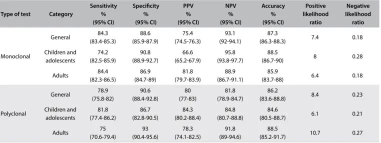
RESULTS: The optimal OD cutoffs established (for monoclonal and polyclonal tests, respectively) were 0.167 and 0.164; overall ELISA sensitivity: 84.3%, 78.9%; specificity: 88.6%, 90.6%; positive predictive value (PPV): 75.4%, 80%; negative predictive value (NPV): 93.1%, 81.8%; accuracy: 87.3%, 86.2%; child and ad‑ olescent ELISA sensitivity: 74.2%, 81.8%; specificity: 90.8%, 86.7%; PPV: 66.6%, 84.3%; NPV: 95.8%, 84.8%; accuracy: 88.5%, 84.6; adult ELISA sensitivity: 84.4%, 75%; specificity: 86.9%, 93%; PPV: 81.8%, 78.3%; NPV: 88.9%, 91.8%; accuracy: 85.9%, 88.5%.
CONCLUSION: The polyclonal serological test developed using local strains presented better diagnos‑ tic performance among children and adolescents, while the monoclonal test was better among adults.
The results from both tests suggest that these in‑house serological tests could be used to detect anti‑H.
breath test and stool antigen test), detection of serum-specific anti-H. pylori antibodies is the only one available in Brazil, and this is limited to a few laboratories in big cities.
Anti-H. pylori immunoglobulin G (IgG) antibody detection by means of the enzyme-linked immunosorbent assay (ELISA) is the third-best method for noninvasive screening for H. pylori
infection.8 It might be suggested as the first method, in
high-prev-alence areas, if the urea breath test and stool antigen test are not available.9 Furthermore, serological tests are considered to be the
most efficient diagnostic method under certain clinical situations in which local changes in the stomach may affect the results from other tests (i.e. in cases of gastrointestinal bleeding, atrophic gas-tritis, gastric MALT lymphoma and gastric carcinoma), or when antibiotics or proton pump inhibitors are used.8 ELISA tests
devel-oped using H. pylori antigens from local samples usually present better accuracy than that of commercial tests based on non-local
H. pylori antigen. One of the main criticisms of commercial tests is that they are widely used in any population (including in devel-oping countries). Therefore, development of a local test has been encouraged.10-17 Hence, an easily performed, low-cost, locally
devel-oped and validated noninvasive test, for widespread use, might be recommendable for screening for H. pylori infection. In particu-lar, use of such tests might potentially reduce the need to perform invasive tests, in a test-and-treat strategy.
OBJECTIVE
The aims of this study were to develop and validate an ELISA-based serological test using whole-cell antigen of cultured
H. pylori from gastric biopsies on Brazilian patients, in order to detect anti-H. pylori IgG antibodies in children and adults.
METHODS
Design and setting
This was a cross-sectional diagnostic accuracy study, in which a serological test to diagnose Helicobacter pylori infection was developed. The locally developed ELISA serological test was standardized and the optical density (OD) cutoff was determined based on the serum antibody responses of 100 H. pylori-negative uninfected patients. A cross-sectional study comparing this in-house ELISA serological test with invasive tests was conducted among 174 consecutive dyspeptic patients at two public hospitals in São Paulo, Brazil.
Gastric biopsy samples and strain isolation
We examined a series of preserved frozen cultures (-80 °C) that had been isolated from gastric biopsy samples from 29 chil-dren and adolescents and 27 adult patients who were attended at the endoscopy units of two public hospitals: Hospital São
Paulo, a university hospital, and Hospital Infantil Cândido Fon-toura, a secondary-level children’s hospital, which are both in the city of São Paulo, Brazil. Antral gastric biopsies were pro-cessed and cultured in brain-heart infusion (BHI) agar (BHI Agar, BD Difco, Becton-Dickinson, NJ, USA), on plates that were incubated for 10-14 days under microaerophilic conditions, as previously described.18
Virulence gene detection by means of conventional PCR
Samples of bacteria were harvested from a three-day-old cul-ture and were suspended in 1 ml of distilled water for deoxyri-bonucleic acid (DNA) extraction (Wizard genomic DNA puri-fication kit, Promega), in accordance with the manufacturer’s instructions. For H. pylori DNA confirmation, a PCR specific for the H. pylori UreA and B genes was used.19 PCR amplification of
the VacA signal sequence and midregion, and of the gene CagA, was performed as previously described.20,21 Negative and positive
controls were included in all reactions.
The gene CagA was identified in 27/56 (48.2%). The gene VacA s1 was observed in 41/56 (73.2%) and VacA s2 in 15/56 (26.8%). The allele VacA m1 was identified in 35/56 (60.7%) and m2 in 21/56 (39.3%): s1m1 occurred in 31/56 (55.3%), s1m2 in 10/56 (17.8%), s2m1 in 3/56 (5.3%) and s2m2 in 12/56 (21.4%).
Antigen preparation
Whole-cell antigen from a sonicated pool (Ultrasonic Disrup-tor QR500W, Ultronique, Brazil) of 56 strains was obtained as pre-viously described.22 Antigen preparation was adjusted to a protein
concentration of 1 mg/ml,23 and aliquots of 100 µl were stored at
-20 °C until used.
ELISA standardization
Detection of anti-H. pylori IgG antibodies was, firstly, tested on 10 H. pylori-positive patients (five adults and five children) and 10 H. pylori-negative patients (five adults and five children), who formed positive and negative serum controls, respectively. Their diagnoses were based on invasive methods: the gold stan-dard for positive diagnoses was a positive culture and/or positive histological test and a positive rapid urease test; and, for negative diagnoses, all three tests needed to be negative.
Standardization was performed as previously described by Camorlinga-Ponce et al.23 For each assay, the optimal
anti-gen concentration and working serum dilution were determined based on checkerboard titrations. ELISA plates were prepared as previously described.22
even for detecting proteins that are present in low quantities in a sample. In contrast, monoclonal antibodies comprise antibod-ies from a single antibody-producing B cell. Thus, they bind with a single epitope. This is highly specific, with only a small risk of cross-reactivity, and it can provide better results in assays requir-ing quantification of the protein levels.
For the polyclonal test (peroxidase-conjugated polyclonal anti-human IgG antibodies; Sigma), color was developed using 100 µl of 0.4 mg/ml ortho-phenylenediamine dihydrochloride diluted in citrate-phosphate buffer and 0.03% sodium perbo-rate (Sigma) as the substperbo-rate. The plates were incubated under dark conditions at 37 °C and, after 20 minutes, 50 µl of stop solu-tion (2M H2SO4) was added to each well. Absorbance was read at 492 nm (Multiskan FC microplate photometer). For the mono-clonal test (alkaline phosphatase-conjugated monomono-clonal anti-hu-man IgG antibodies; Sigma), color was developed using 100 µl of 1 mg/ ml p-nitrophenylphosphate (Sigma) and absorbance was read at 405 nm (Multiskan FC microplate photometer). All reac-tions were performed in triplicate, and the mean of three optical density (OD) measurements was used.
Analysis on the checkerboard titration for polyclonal antibodies showed that the following were the optimal concentrations: for anti-gen preparation, 1:500 (2 µg/ml); for working serum dilution, 1:400; and for peroxidase-conjugated anti-human antibodies, 1:60,000. For monoclonal antibodies, the following were the optimal con-centrations: for antigen preparation, 1:300 (3.3 µg/ ml); for work-ing serum dilution, 1:400; and for alkaline phosphatase-conjugated anti-human antibodies, 1:25,000. The best reading time for mono-clonal antibodies was 25 minutes. ELISA tests were run for four consecutive days to evaluate interoperability and reproducibility.
Antigen specificity
The cross-reactivity of the in-house ELISA serological tests for
H. pylori antigens was evaluated against whole-cell antigens or membrane proteins of 14 heterologous bacterial species: Cam-pylobacter jejuni, Escherichia coli, Pseudomonas aeruginosa
(mucoid), Pseudomonas aeruginosa (nonmucoid), Burkholderia cepacia, Klebsiella pneumoniae, Klebsiella sp., Proteus mirabilis, Proteus vulgaris, Shigella flexneri, Shigella sp., Salmonella typhi, Salmonella enterica and Salmonella sp. These reactions were per-formed through competitive inhibition assays, as previously described.24 No positive reactions were observed.
Determination of cutoff
The optical density (OD) cutoffs for the monoclonal and poly-clonal tests were determined based on the serum antibody responses of 100 samples (50 adults and 50 children and adoles-cents) from H. pylori-negative uninfected patients, based on the gold standard. Thus, a pool of these 100 serum samples was used
as the negative serum control. The threshold for positivity was defined as the mean value plus three standard deviations of the optical density, as previously described.23
These values were compared with the values from a collection of 82 serum samples (33 adults and 49 children and adolescents) from H. pylori-positive infected patients, based on the gold stan-dard, which presented OD values above the corresponding cutoff, without showing any overlapping values. A pool of these 82 sam-ples was used as the positive serum control.
During the testing of the unknown samples, a positive serum pool was included in quadruplicate in every plate and the mean of the four OD values was used to calculate the threshold for that plate. The results from each serum sample were defined as the ratio of the OD value of the sample to the threshold value and were expressed in ELISA units (EU). Serum samples with EU > 1.0 were considered seropositive.
Validation of in-house ELISA serological assay
Peripheral blood samples (10 ml) were collected by means of venous puncture from 174 patients on whom esophagogastro-duodenoscopy had been performed. The serum was stored at -20 °C. The following were taken to be exclusion criteria: recent use of antibiotics, H2 receptor antagonists, proton pump inhib-itors and/or bismuth salts, and presence of digestive or extra-digestive chronic diseases. Six gastric biopsies were taken for the rapid urease test and for histological evaluation (one from the gastric body and one from the antrum, for each of these), and for culturing (two from the antrum). The procedures were per-formed as previously described.18,25
Statistical analysis
Qualitative variables were described in terms of their propor-tions, and quantitative variables were described in terms of their means and standard deviations. Analyses on continuous vari-ables were based on the negative gold standard (negative rapid urease test, histological evaluation and culturing). The thresh-old for positivity was defined as the mean value plus three stan-dard deviations of the optical density. The positive serum control was standardized using the positive gold standard (positive cul-ture and/or positive histological evaluation and rapid urease test) and presence of a positive serological test, i.e. OD over the estab-lished cutoff value. The OD for each serum sample was deter-mined in triplicate. The optimal OD cutoff was deterdeter-mined based on receiver operating characteristic (ROC) curves using differ-ent sensitivity and specificity values, and the area under the ROC curve (AUC) was estimated taking the significance level to be an accuracy value of 0.5.
around 30% in children and 50% in adults in Brazil. The desirable AUC was considered to be 0.90, with a significance level of 0.05 and absolute precision of 5%. Thus, for this study, the sample size was estimated to be 78 adults and 95 children.
The sensitivity, specificity, accuracy and positive and negative predictive values of the in-house ELISA serological tests were eval-uated by comparing OD values, based on cutoffs that were deter-mined using the ROC curve, against the gold standard (rapid urease test, histological evaluation and culturing). Negative and positive likelihood ratios were evaluated.
Ethical considerations
This study was approved by the Institutional Review Board of the Federal University of São Paulo (Universidade Federal de São Paulo), under registration number 180.606, on Decem-ber 21, 2012, and under Brazil Platform registration numDecem-ber (CAAE) 10235612.0.0000.5505. All patients and/or their guard-ians were informed about the purposes of this study and signed an informed consent form.
RESULTS
Patients
Between November 2015 and August 2017 (20 months), 847 chil-dren and adolescents and 432 adults were evaluated by means of esophagogastroduodenoscopy at Hospital São Paulo, a university hospital, and Hospital Infantil Cândido Fontoura, a secondary-level children’s hospital, which are both in the city of São Paulo, Brazil. H. pylori status was defined by means of the gold stan-dard described here, and, for validation purposes, no equivocal findings were included. To validate our in-house serological test, 174 consecutive dyspeptic patients, for whom the H. Pylori infec-tion status was unknown were chosen: 96 children and adoles-cents (age range 2-17 years; mean: 12.2 ± 3.7 years) and 78 adult (age range 19-85 years; mean: 52.3 ± 18.1 years). The
esophago-gastroduodenoscopy findings were normal in 108/174 patients (62%) and abnormal in 66/174 (38%). Among the latter, 57/174 (32.7%) were H. pylori-positive. Among the children and adoles-cents of this sample, 24/96 (25%) were H. pylori-positive; while among the adults, 33/78 (42.3%) were H. pylori-positive.
Determination of cutoff
The serum samples that were negative in the monoclonal test presented OD values ranging from 0.085 to 0.155 (mean ±
stan-dard deviation, SD: 0.11996 ± 0.017695). The threshold for
pos-itivity was 0.173. The serum samples that were negative in the polyclonal test presented OD values ranging from 0.061 to 0.148 (mean ± SD: 0.10336 ± 0.0164024). The threshold for
posi-tivity was 0.153. The serum samples (82 samples) that were
positive in the monoclonal test presented OD values ranging from 0.180 (1.04 EU) to 0.464 (2.85 EU) (mean ± SD: 0.22968 (1.33 EU) ± 0.057). The serum samples (82 samples) that were
positive in the polyclonal test presented OD values ranging from 0.172 (1.12 EU) to 0.437 (2.85 EU) (mean ± SD: 0.261
(1.7 EU) ± 0.06019).
Despite the threshold for positivity based on the mean for the negative results plus 3 SD in the monoclonal test (0.173) and in the polyclonal test (0.153), the optimal OD cutoff value based on ROC curve analysis was found to be 0.167 in the monoclonal test, with an area under the ROC curve of 0.9 (95% confidence interval, CI: 0.87-0.92). The optimal OD cutoff value was 0.164 in the polyclonal test, with an area under the ROC curve of 0.915 (95% CI: 0.89-0.93).
Validation of the ELISA serological assay
Analysis on the overall seropositive results (EU > 1.0) from the monoclonal test presented little difference in values, compared with the polyclonal test, but the sensitivity and negative pre-dictive values were slightly higher with monoclonal antibodies. When age was considered, the monoclonal test presented a bet-ter result for sensitivity among adults, but specificity was betbet-ter among children and adolescents. In contrast, the polyclonal test presented a better result for sensitivity among children and ado-lescents. The accuracy was slightly better among children and adolescents in the monoclonal test and among adults in the poly-clonal test (Table 1).
DISCUSSION
In this study, ELISA-based serological tests to detect monoclonal and polyclonal IgGantibodies using H. pylori whole-cell antigen from strains isolated in our community were developed, stan-dardized, validated and evaluated. The sensitivity and specific-ity of the in-house serological tests for both polyclonal antibod-ies (78.9% and 90.6%) and monoclonal antibodantibod-ies (84.3% and 88.6%) were similar to those found by Camorlinga-Ponce et al.23
(85% and 89.7%). The in-house ELISA serological test that we developed did not present any cross-reactivity to heterologous bacterial species.
The sample size was appropriate, given that both the sensitiv-ity and the specificsensitiv-ity presented narrow 95% confidence intervals. Selectivity in relation to gastroduodenal diseases can influence the sensitivity and specificity results. A study conducted by Aziz et al.17
Both commercial and in-house serological tests have presented large ranges of sensitivity and specificity results. Leal et al.14
con-ducted a meta-analysis that including 42 studies in which ELISA serological tests were performed on samples from children to detect
H. pylori infection. There were 33 studies using 19 different com-mercial tests and nine studies using in-house tests. They observed that the mean sensitivity was 79.2% (95% CI: 77.3-81%) and that the mean specificity was 92.4% (95% CI: 91.6-93.3%). On the other hand, an in-house serological test that was developed in Pakistan presented sensitivity of 92% and specificity of 100%, i.e. similar to or better than commercial tests.17
The accuracy of serological tests can also be affected by the choice of antigen that is used to prepare the in-house ELISA assay. In some studies, H. pylori genetic heterogeneity was taken into con-sideration in choosing the whole-cell lysate (with sonication).28,29
In our study, the strains used in the antigenic pool were verified genetically, to obtain better representativeness, including the CagA and VacA strains (alleles s1m1, s1m2, s2m1 and s2m2). The geno-typic distribution in our study was similar to what has been found in other studies in Brazil.30,31
The diagnostic accuracy of a serological test is enhanced when the antigen pool is prepared based on multiple strains. Widmer et al.32
evaluated the performance of three antigenic preparations and observed that the results were superior when an enzyme immuno-assay prepared with a pool of native antigens was used (sensitivity: 90-100%; specificity: 90-97%), rather than with recombinant antigen (sensitivity: 59-78%; specificity: 41-100%). Serological tests based on whole-cell antigen or whole-cell antigen lysate have also shown good performance among both children and adult patients.11,23,24,33
Despite the simplicity of ELISA serological tests, attention during their development is needed. In particular, the optimal antigen concentration, serum sample dilution and antibody con-jugation need to be determined. These parameters should be opti-mized and adjusted to local conditions, to the population studied and to the method developed.17,34 The optimal concentrations in
the present study were determined by means of the checkerboard titration method. It is very important to make these determina-tions because of false-positive titration results caused by high con-centrations of antigens, which promote nonspecific bonds, even in negative serum. Moreover, low antigen concentrations can result in false negatives. In addition, high concentrations of conjugated antibodies may result in nonspecific reading, while low concen-trations cause decreased OD. The serum concentration can also interfere with the OD results.17
This study was the first on our population to evaluate the sero-logical response to anti-H. pylori IgG antibodies among symptom-atic children, adolescents and adult patients, using an ELISA-based serological test developed using whole-cell antigen lysate from strains collected in our own community.
Ideal tests for screening purposes should present high sensi-tivity and good specificity. The serological test using monoclonal antibodies that was developed in this study presented sensitivity of over 80% among adults, while the serological test using polyclonal antibodies presented sensitivity of over 80% among children and adolescents. This indicates that these tests are suitable for pop-ulation-based screening, especially the polyclonal test, which is characterized by greater sensitivity. On the other hand, although the higher specificity among adults in the polyclonal test, with low
Type of test Category
Sensitivity % (95% CI) Specificity % (95% CI) PPV % (95% CI) NPV % (95% CI) Accuracy % (95% CI) Positive likelihood ratio Negative likelihood ratio Monoclonal General 84.3 (83.4-85.3) 88.6 (85.9-87.9) 75.4 (74.5-76.3) 93.1 (92-94.1) 87.3
(86.3-88.3) 7.4 0.18 Children and adolescents 74.2 (82.5-85.9) 90.8 (88.9-92.7) 66.6 (65.2-67.9) 95.8 (93.8-97.7) 88.5
(86.7-90) 8 0.28
Adults 84.4 (82.3-86.5) 86.9 (84.7-89) 81.8 (79.7-83.9) 88.9 (86.7-91.1) 85.9
(83.7-88) 6.4 0.18
Polyclonal General 78.9 (75.8-82) 90.6 (88.4-92.8) 80 (77-83) 81.8 (78.9-84.7) 86.2
(83.6-88.8) 8.4 0.23 Children and adolescents 81.8 (77.4-86.2) 86.7 (82.8-90.5) 84.3 (80.2-88.4) 84.8 (80.7-88.8) 84.6
(80.5-88.7) 6.1 0.21
Adults 75 (70.6-79.4) 93 (90.4-95.6) 78.3 (74.1-82.5) 91.8 (89-94.6) 88.5
(85.2-91.7) 10.7 0.27
PPV = positive predictive value; NPV = negative predictive value.
occurrence of false-negative results, is desirable for a diagnostic method, it is not necessary for a screening test.
CONCLUSION
The in-house polyclonal serological test that was developed using local strains (in-house) presented better diagnostic per-formance than did the monoclonal test for children and adoles-cents. In contrast, the monoclonal test was better among adults. The results relating to accuracy, sensitivity, specificity and likeli-hood ratios from both tests suggest that these in-house serologi-cal tests could be used to detect anti-H. pylori antibodies in the Brazilian population, for screening purposes.
REFERENCES
1. Go MF. Review article: natural history and epidemiology of Helicobacter
pylori infection. Aliment Pharmacol Ther. 2002;16(Suppl 1):3‑15.
PMID: 11849122.
2. Eusebi LH, Zagari RM, Bazzoli F. Epidemiology of Helicobacter pylori
infection. Helicobacter. 2014;19(Suppl 1):1‑5. PMID: 25167938; doi:
10.1111/hel.12165.
3. Roque JRDS, Machado RS, Rodrigues D, Rech P, Kawakami E. Prevalência
de infecção por Helicobacter pylori em uma comunidade indígena
em São Paulo e fatores associados: estudo transversal [Prevalence
of Helicobacter pylori infection in an indigenous community in São
Paulo and associated factors: cross‑sectional study]. São Paulo Med J.
2017;135(2):140‑5. doi: 10.1590/1516‑3180.2016.0114091216.
4. Correa P, Piazuelo MB. Helicobacter pylori infection and gastric
adenocarcinoma. US Gastroenterol Hepatol Rev. 2011;7(1):59‑64.
PMID: 21857882.
5. Ferlay J, Soerjomataram I, Dikshit R, et al. Cancer incidence and mortality
worldwide: Sources, methods and major patterns in GLOBOCAN 2012.
Int J Cancer. 2015;136(5):E359‑86. PMID: 25220842; doi: 10.1002/ijc.29210.
6. Brito HSH, Braga JA, Loggetto SR, et al. Helicobacter pylori infection and
immune thrombocytopenic purpura in children and adolescents: A
randomized controlled trial. Platelets. 2015;26(4):336‑41. PMID: 2483281;
doi: 10.3109/09537104.2014.911836.
7. Stasi R, Sarpatwari A, Segal JB, et al. Effects of eradication of Helicobacter
pylori infection in patients with immune thrombocytopenic purpura:
a systematic review. Blood. 2009;113(6):1231‑40. PMID: 18945961; doi:
10.1182/blood‑2008‑07‑167155.
8. Malfertheiner P, Megraud F, O’Morain CA, et al. Management of
Helicobacter pylori infection ‑ the Maastrich V/Florence Consensus Report.
Gut. 2017;66(1):6‑30. PMID: 27707777; doi: 10.1136/gutjnl‑2016‑312288.
9. Braden B. Diagnosis of Helicobacter pylori infection. BMJ. 2012;344:e828.
PMID: 22368293;. doi: 10.1136/bmj.e828.
10. Obata Y, Kikuchi S, Miwa H, et al. Diagnostic accuracy of serological
kits for Helicobacter pylori infection with the same assay system but
different antigens in a Japanese patient population. J Med Microbiol.
2003;52(Pt 10):889‑92. PMID: 12972583; doi: 10.1099/jmm.0.05267‑0.
11. Hoang TT, Wheeldon TU, Bengtsson C, et al. Enzyme‑linked
immunosorbent assay for Helicobacter pylori needs adjustment for
the population investigated. J Clin Microbiol. 2004;42(2):627‑30.
PMID: 14766827.
12. Hoang TT, Rehnberg AS, Wheeldon TU, et al. Comparison of the
performance of serological kits for Helicobacter pylori infection
with European and Asian study populations. Clin Microbiol
Infect. 2006;12(11):1112‑7. PMID: 17002611; doi: 10.1111/j.1469‑
0691.2006.01514.x.
13. Harris P, Perez‑Perez G, Zylberberg A, et al. Relevance of adjusted cut‑off
values in commercial serological immunoassays for Helicobacter pylori
infection in children. Dig Dis Sci. 2005;50(11):2103‑9. PMID: 16240223;
doi: 10.1007/s10620‑005‑3015‑9.
14. Leal YA, Flores LL, García‑Cortés LB, Cedillo‑Rivera R, Torres J.
Antibody‑based detection tests for the diagnosis of Helicobacter pylori
infection in children: A meta‑analysis. PLoS One. 2008;3(11):e3751.
PMID: 19015732; doi: 10.1371/journal.pone.0003751.
15. Thong‑Ngam D, Chayanupatkul M, Vongchampa P, Hanvivatvong O. An
evaluation of a new in‑house serum and urine ELISA test for detection
of Helicobacter pylori infection in Thai population. J Med Assoc Thai.
2011;94(8):985‑90. PMID: 21863682.
16. Mohammadi M, Talebkhan Y, Khalili G, et al. Advantage of using a
home‑made ELISA kit for detection of Helicobacter pylori infection over
commercially imported kits. Indian J Med Microbiol. 2008;26(2):127‑31.
PMID: 18445947.
17. Aziz F, Taj Y, Kazmi SU. Development of an in‑house enzyme‑linked
immunosorbent assay based on Helicobacter pylori sonicate whole
cell antigen for diagnosis of gastroduodenal ulcer disease in
Karachi, Pakistan. Int J Microbiol Adv Immunol. 2013;1(4):24‑31; doi:
10.19070/2329‑9967‑130005.
18. Ogata SK, Godoy AP, da Silva Patricio FR, Kawakami E. High Helicobacter
pylori resistance to metronidazole and clarithromycin in Brazilian
children and adolescents. J Pediatr Gastroenterol Nutr. 2013;56(6):645‑8.
PMID: 23403439; doi: 10.1097/MPG.0b013e31828b3669.
19. Smith SI, Oyedeji KS, Arigbabu AO, et al. Comparison of three PCR
methods for detection of Helicobacter pylori DNA and detection
of cagA gene in gastric biopsy specimens. World J Gastroenterol.
2004;10(13):1958‑60. PMID: 15222045.
20. Atherton JC, Cao P, Peek RM Jr, et al. Mosaicism in vacuolating
cytotoxin alleles of Helicobacter pylori: Association of specific vacA
types with cytotoxin production and peptic ulceration. J Biol Chem.
1995;270(30):17771‑7. PMID: 7629077.
21. Van Doorn LJ, Figueiredo C, Sanna R, et al. Clinical relevance
of the cagA, vacA, and iceA status of Helicobacter pylori.
Gastroenterology. 1998;115(1):58‑66. PMID: 9649459; doi: 10.1016/
S0016‑5085(98)70365‑8.
22. Perez‑Perez GI, Dworkin BM, Chodos JE, Blaser MJ. Campylobacter
pylori antibodies in humans. Ann Intern Med. 1988;109(1):11‑7.
23. Camorlinga‑Ponce M, Torres J, Perez‑Perez G, et al. Validation of a
serologic test for the diagnosis for Helicobacter pylori infection
and the immune response to urease and Cag A in children. Am J
Gastroenterol. 1998;93(8):1264‑70. PMID: 9707049; doi: 10.1111/j.1572‑
0241.1998.00407.x.
24. Khanna B, Cutler A, Israel NR, et al. Use caution with serologic testing for
Helicobacter pylori infection in children. J Infect Dis. 1998;178(2):460‑5.
PMID: 9697727.
25. Ogata SK, Kawakami E, Patricio FR, Pedroso MZ, Santos AM. Evaluation
of invasive and non‑invasive methods for the diagnosis of Helicobacter
pylori infection in symptomatic children and adolescents. São Paulo
Med J. 2001;119(2):67‑71. PMID: 11276169.
26. de Oliveira AM, Rocha GA, Queiroz DM, et al. Evaluation of enzyme‑linked
immunosorbent assay for the diagnosis of Helicobacter pylori infection
in children from different age groups with and without duodenal ulcer.
J Pediatr Gastroenterol Nutr. 1999;28(2):157‑61. PMID: 9932847.
27. Rocha GA, Oliveira AM, Queiroz DM, et al. Serodiagnosis of Helicobacter
pylori infection by Cobas Core ELISA in adults from Minas Gerais,
Brazil. Braz J Med Biol Res. 1998;31(10):1263‑8. doi: 10.1590/S0100‑
879X1998001000005.
28. Blaser MJ. Heterogeneity of Helicobacter pylori. Eur J Gastroenterol
Hepatol. 2012;9 Suppl 1 :S3‑6; discussion S6‑7. PMID: 22498905.
29. Sunnerstam B, Kjerstadius T, Jansson L, et al. Detection of
Helicobacter pylori antibodies in a pediatric population: comparison
of three commercially available serological tests and one in‑house
enzyme immunoassay. J Clin Microbiol. 1999;37(10):3328‑31.
PMID: 10488200.
30. Gatti LL, Módena JL, Payão SL, et al. Prevalence of Helicobacter pylori cagA,
iceA and babA2 alleles in Brazilian patients with upper gastrointestinal
diseases. Acta Trop. 2006;100(3):232‑40. PMID: 17181989; doi: 10.1016/j.
actatropica.2006.08.014.
31. Lobo Gatti L, Agostinho JNF, De Lábio R, et al. Helicobacter pylori and
cagA and vacA gene status in children from Brazil with chronic gastritis.
Clin Exp Med. 2003;3(3):166‑72. PMID: 14648232; doi: 10.1007/s10238‑
003‑0021‑0.
32. Widmer M, de Korwin JD, Aucher P, et al. Performance of native and
recombinant antigens for diagnosis of Helicobacter pylori infection. Eur
J Clin Microbiol Infect Dis. 1999;18(11):823‑6. PMID: 10614960.
33. Thomas JE, Whatmore AM, Barer MR, Eastham EJ, Kehoe MA.
Serodiagnosis of Helicobacter pylori infection in childhood. J Clin
Microbiol. 1990;28(12):2641‑6. PMID: 2279995.
34. Leung WK, Ng EK, Chan FK, Chung SC, Sung JJ. Evaluation of three
commercial enzyme‑linked immunosorbent assay kits for diagnosis
of Helicobacter pylori in Chinese patients. Diag Microbiol Infect Dis.
1999;34(1):13‑7. PMID: 10342102.
This study was presented as a poster presentation at the 8th International
Symposium on Helicobacter pylori and Gastric Cancer, Belo Horizonte
(MG), Brazil, on April 12‑14, 2018
Conflict of interest: None
Sources of funding: Fundação de Amparo à Pesquisa do Estado de São Paulo (FAPESP) ‑ Project: 2014/25298‑5
Date of first submission: May 21, 2018
Last received: July 12, 2018
Accepted: August 31, 2018
Address for correspondence:
Silvio Kazuo Ogata
Disciplina de Gastroenterologia Pediátrica, Departamento de Pediatria
Rua Coronel Lisboa, 826
São Paulo (SP) — Brasil
CEP: 04020‑040
Tel: (+55 11) 5576‑4848, ramal 1908
E‑mail: [email protected]
© 2018 by Associação Paulista de Medicina