INTRODUCTION
T
racheostomy (TCT) is the surgical procedure that consists of opening the anterior wall of the trachea, communicating it with the external environment through the insertion of a cannula, making the airway patent1. It is a common procedure in oncology, especially in head and neck oncology2. Studies of head and neck surgeons in various departments reported that 26 to 39% of the specialists performed the procedure frequently in cases of flaps and large resections, in a preventive way1-3. This procedure in the management of airways in cancer patients has also been indicated for patients with tumor masses and submitted to previous radiotherapy, causing obstruction of theairways and preventing orotracheal intubation (OTI)4,5. Studies have shown that the mean number of routine tracheostomies in squamous cell carcinoma resections is between 23 and 30%, and in selected cases, up to 74%3,6.
This procedure generates innumerable changes in the patient's daily life: in respiratory dynamics, in the behavior and interpersonal relationship, and in personal care. For these changes to occur with better results, a lot of care is needed. For all these care, the importance of a multidisciplinary team for the management of tracheostomies (physicians, dentists, speech therapists, physiotherapists, nurses, nutritionists) was observed both in the hospital settings and in the outpatient follow-up.
Elaboration of a tracheostomy conduct protocol in the Amazonas
cancer reference hospital
Elaboração de protocolo de condutas em traqueostomias no hospital referência
de tratamento do câncer do Amazonas
Maria Carolina Coutinho Xavier SoareS, aCBC-aM1,2; Fernando luiz WeStphal, tCBC-aM2; luiz CarloS de liMa2; JeFFerSon
Moreira MedeiroS2
Objective: to create a multidisciplinary conducts manual for tracheostomies in adult and pediatric patients in the Amazonas State Oncology Control Center Foundation. Methods: we developed a protocol using the modified Delphi method, which consisted in the application of two series of questionnaires to 20 professionals of the unit. Results:
thirteen professionals completed the two steps. In the first stage, there was consensus in 53 out of 92 questions (57.6%). The questions that obtained consensus formed the text of the second stage, divided into eight chapters and evaluated by marking the answers offered on a Linkert scale. All the chapters presented in the second stage obtained consensus, meaning that the sum of the answers "agree" and "fully agree" were above 70%. Conclusion: using the data obtained in the consensus, we elaborated a tracheostomy conduct protocol and a care guidelines manual for the patients and their caregivers.
Keywords: Clinical Protocols. Tracheostomy. Medical Oncology. Hospitals. University.
At the Amazonas State Oncology Control Center Foundation (FCECON), there are professionals from numerous specialties attending patients with TCT, but there were no routine to adapt the conducts guidelines in tracheostomies to the reality of our patients. There is no uniform treatment model for the tracheostomy patients during their hospitalization, standardization of materials and procedures, nor a guideline model for home-based procedures. The elaboration of a routines manual, taking into account the experience of those involved in TCT care at the FCECON, would bring clinical subsidy for reproducibility of actions. The presentation of information synthesized from the literature, elaborated in topics to elucidate the day-to-day questions, is efficient in improving teamwork, reflecting better results in reduction of complications and improvement in patients' quality of life7.
The objective of this study was to create a multidisciplinary protocol for tracheostomy conducts in adult and pediatric FCECON patients in need of the procedure and to use the protocol data to elaborate a basic home care manual and emergency management guidelines for caregivers and patients.
METHODS
The project was approved by the Ethics in Research Committee of the FCECON, CAAE number 61650316.1.0000.0004. For the elaboration of the FCECON tracheostomy conducts protocol, we used the modified Delphi method, which consists of the application of a series of questionnaires to specialists. Inclusion criteria were being a graduate-level FCECON professional working with tracheostomized patients in their daily practice, and at least five years of experience in the field.
Exclusion criteria were non-completion of the two stages of the questionnaire for any reason, and loss of the working relationship with FCECON during the study.
The Delphi method does not use statistical computation to define the ideal number of experts for the consensus to be adequate. It suggests the choice of the participants according to their knowledge and experience in the subject treated, always preferring to choose the best qualified of the service8,9. Some authors suggest some basic characteristics to guide the selection of the components of the study: specialists who will use the results obtained in the protocol in their daily practice; specialists who lead teams related to the subject studied; specialists considered as reference on the subject8. For the present study, we invited 20 experts.
The team of researchers prepared the initial questionnaire. The questions from the first questionnaire are a literature review product, using the MEDLINE and LILACS databases. We used the terms "tracheostomy" and "traqueostomia". We also searched textbooks and TCT protocols from other services. We selected 37 texts for the elaboration of the protocol sentences.
We invited the selected experts to participate in the study, and those who accepted, signed an informed consent form. We delivered the first questionnaire to the specialists. The research team did not follow the responses completion. After filling, the researcher collected the questionnaire, which was placed in unidentified envelopes. The preparation of a second questionnaire resulted from the analysis of the answers presented in the first questionnaire. We placed all the answers that obtained above 65% consensus in the second questionnaire. We repeated in the second stage relevant questions that did not display consensus on the first questionnaire.
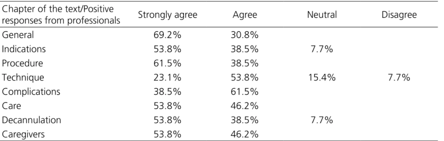
The second questionnaire converted the answers that obtained consensus into text. The text was divided into eight chapters for better analysis. After reading each chapter, the specialist reported his/her impression by ticking an item from the Linkert scale quoted below: 1. strongly disagree; 2. I disagree; 3. neutral; 4. agree; 5. strongly agree. We delivered the second questionnaire to the specialists following the same protocol of the first stage. After analysis of the responses from the second questionnaire, the chapters that obtained the answers agree and strongly agree above 70% consensus integrated the final protocol text. We also prepared a manual for the patient with the data from the protocol.
We calculated descriptive statistics for each question item. We display the answers considered valid in percentage8. The calculation of p-values does not apply to this type of project9. We used the Excel software to record data. We delivered the consensus and the patients' manual to the hospital's clinical director.
RESULTS
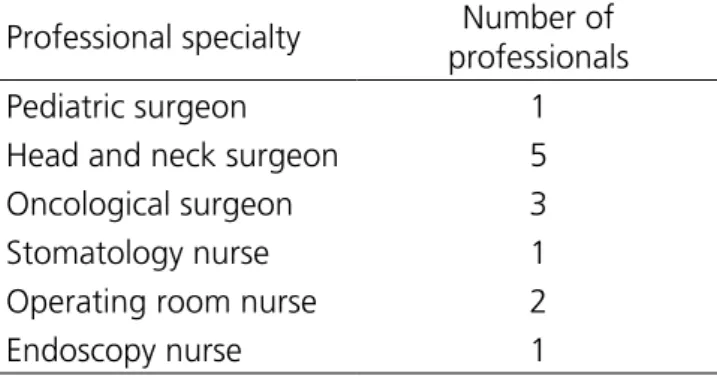
The 20 invited health professionals who accepted to participate in the FCECON study were of several specialties: intensive care physician, oncology emergency physician, physiotherapist, intensive care nurse, cancer surgeon, head and neck surgeon, thoracic surgeon, operating room nurse, stomatotherapist nurse, endoscopy nurse. Thirteen (65%) professionals participated in the two series of questionnaires and we analyzed only their responses in the protocol. Table 1 shows the profile of participants who completed all project steps.
Table 1. Profile of the professionals who fully participated in the questionnaires of the tracheostomy protocol.
Professional specialty Number of
professionals
Pediatric surgeon 1
Head and neck surgeon 5
Oncological surgeon 3
Stomatology nurse 1
Operating room nurse 2
Endoscopy nurse 1
a 65% consensus (Table 2). Twelve comments of subjects that were not in the initial text were added.
53.8%, "strongly agree" (Table 3). When analyzing the six re-submitted questions, only one reached 70% acceptance for consensus.
To integrate the protocol, we selected the texts that presented consensus above 70% by the experts, by means of the sum of the answers "agree" and "strongly agree". During the final writing of the protocol, we chose to describe in the final text the questions that did not obtain consensus among the experts using the data that already are consensus in the literature, since they are of great importance and could not be discarded from the final protocol.
With the data resulting from the consensus, we elaborated a tracheostomy protocol for the hospital staff and a manual for the patient and his/ her caregivers with general guidelines (Table 4).
DISCUSSION
In 2013, Mitchell developed a TCT consensus using the Delphi method, with 110 initial questions. At the end of the second stage, 77 sentences were approved and 36 discarded. For the elaboration of this study's consensus, nine specialists participated, whose specialties were adult and pediatric surgeons, nurses, physiotherapists and emergency physicians10. Another published consensus on TCT brought together professional experts (19 professionals) who carried out a joint review of the literature of 40 articles, and from these data, they elaborated the consensus. These professionals also sought to adapt the literature to their local reality. It was also performed only by doctors and nurses11.
In the present study, there was no consensus among the professionals regarding the ideal time of orotracheal intubation to indicate tracheostomy. Recent studies advocate the early
Table 2. Results of the first stage of the Delphi method. Profile of the questions Number of questions
Questions presented 92
Questions selected by
consensus 53
Table 3. Results of the second stage of the Delphi method. Chapter of the text/Positive
responses from professionals Strongly agree Agree Neutral Disagree
General 69.2% 30.8%
Indications 53.8% 38.5% 7.7%
Procedure 61.5% 38.5%
Technique 23.1% 53.8% 15.4% 7.7%
Complications 38.5% 61.5%
Care 53.8% 46.2%
Decannulation 53.8% 38.5% 7.7%
Caregivers 53.8% 46.2%
Table 4. Summary of the protocol obtained by using the answers from the experts. Main
tracheotomy indications in the FCECON
• Obstruction of high airways due to cancers or swelling caused by radiotherapy. • Laryngo-tracheal stenosis.
• Prophylaxis of airway obstruction in the postoperative period of extensive head and neck surgery.
• Prolonged orotracheal intubation, need of mechanical ventilation. Adults: between seven and 15 days of orotracheal intubation, without possibility of ventilation weaning during this period. Children: one can wait for up to four weeks, scheduling an early procedure if there is no foreseen resolution of the cause.
• Difficult airways.
Procedure • Explain to the patient and family in advance about the procedure, submit the consent form for signature. A copy is in the possession of the family and the other copy of the form must be in the medical record.
• Define the location of the procedure: the tracheotomy is a routine procedure performed in the operating room. In special situations, it can be performed in ICU, or at the bedside in the event of emergencies without transport possibility. Tracheostomy in the operating room without setting the patient on the operating table, doing the procedure on the bed of the patient, is possible in special cases (patients with morbid obesity, in cases in which the mobilization offers risks, such as fractures).
• Team for the procedure: surgeon, auxiliary, scrub nurse, anesthesiologist, nurse. • Tracheostomy under general anesthesia: patient in IOT and children. Tracheostomy under local anesthesia: patient with airway obstruction, without possibility of IOT. • Materials needed for a tracheotomy: equipment for monitoring of vital signs (blood pressure, ECG, SpO2, capnograph), lights, goggles, surgical scrubs, surgical gowns, sterile fields, sterile gloves, antiseptics, Kelly clamp, curved scissors, Farabeuf retractors, electric scalpel, anatomic tweezers, toothed tweezers, scalpel handle n# 3 and 15 blade, 3.0 needle nylon suture, suction catheter, vacuum, tracheostomy cannula: when performing a tracheotomy in adults, evaluate the availability of the chosen cannula (plastic with cuff, plastic cuff-free, metallic). Always provide one cannula of the estimated number for the patient, one bigger and one smaller.
Technique • The preferred position of the patient: supine, in cervical hyperextension, cushion between the shoulder blades and round pad under the head. Always assess if there is any contraindication for this position.
• In case of impossibility of cervical hyperextension, the neck can be set in neutral position. The team must be prepared for a higher technical difficulty in this position. • Antisepsis should be made from the anterior edge of the jaw to the upper third of the thorax. Laterally, until the lateral edges of the sternocleidomastoid muscles.
• The incision of the skin in adults can be transverse or longitudinal. The choice of incision should be the surgeon’s, according to each patient. In children, the preferred incision is the transverse.
• After the retraction of sterno-hyoid and thyroid muscles, one identifies the thyroid isthmus. Cranial retraction of the isthmus is indicated when possible, but isthmotomy may be carried out if necessary.
• Avoid lateral dissection to the trachea (avoid injury of recurrent laryngeal nerve, internal jugular vein, vagus nerve, carotid artery).
• Avoid inferior dissection (avoid injury of brachycephalic artery and innominate vein). • The opening of the trachea should ideally be made between the second and fourth tracheal rings.
• Prudent identification of tracheal rings to avoid injury to cricoid cartilage and to first tracheal ring.
• The surgeon defines the technique used in the tracheotomy according to the needs of the patient: transverse opening between the tracheal rings, resection of the anterior portion of the tracheal ring, resection of the upper and lateral flap, maintaining a fixed lower border (Bjork flap). In children, the transverse opening between the tracheal rings is indicated.
• In cases of difficult tracheostomy, the suggestion of a technical approach is the placement of repair sutures in the trachea, externalizing to the skin, aiming to identify the path in cases of decannulation.
• When in children, the use repair sutures in the trachea is routine, even in tracheostomies without technical difficulties, since the repositioning in accidental decannulation are always more difficult in this age group.
• When positioning the tracheostomy cannula, care must be taken not to injure the posterior wall of the trachea.
• Evaluation of the correct positioning of the cannula after its passage is made by capnography or pulmonary auscultation.
• The fixation of the cannula after placement in the trachea is through ribbons around the neck.
• Initial dressing should be done using gauze around the cannula.
Complications • Materials considered indispensable for the treatment of complications of tracheostomies should be available in places where there are patients with tracheostomy, as well as hospital admission, emergency and ICU sectors: tracheostomy tubes of all sizes (in the adult ward: 5-0, 5-5, 6-0, 6 -5, 7-0, 7-5, 8-0, 8-5; in the pediatric ward: 3-0, 3-5, 4-0, 4-5, 5-0, 5-5), aspiration hose, (in an adult ward: 8, 10, 12, 14; in the pediatric ward: 4, 6, 8), aspirator, small surgery kit, monitoring, Oxygen source, AMBU bag.
• Accidental decannulation: call for help of a professional with experience; if there is no contraindication, place a cushion between the shoulder blades and try to reposition the cannula; if it is not possible to use a cushion, try to reposition in a neutral position; if there are repair sutures, exert a light traction on it and reposition the cannula; in case of repositioning failure, check if there is a professional with experience in the unit and call for help again; monitoring and oxygen supply; make sure all the emergency materials mentioned above are close.
• Post-tracheostomy bleeding: initial evaluation by the currently available physician if the surgeon is not immediately reachable; early evaluation of the surgeon; assess whether bleeding originates around the cannula or in its interior; provide monitoring and oxygenation to the patient; aspirate cannula in case of bleeding originating inside the cannula; evaluate the patient's medical chart if the patient is using anticoagulant drugs.
• Respiratory insufficiency by secretion plug in the cannula: suspect if the patient with tracheostomy has respiratory difficulties; if the patient has a plastic cannula, aspirate it; if there is no improvement or there is resistance in the passage of the tube when trying to aspirate the cannula, nebulize the patient with saline 0.9% and try again the aspiration; if the patient has a metal cannula, remove the inner cannula immediately and clean the cannula; if there is no improvement in the breathing pattern after aspiration and cleaning, it is necessary to change the cannula. If one does not have training in cannula exchange, call for help; provide monitoring and oxygen source immediately, while calling for help. Make available all urgency list materials.
• Pneumothorax and pneumomediastinum after procedure: perform chest X-ray after the procedure, in cases of dyspnea; in children chest radiography is performed routinely in all tracheostomies; if there is pneumothorax, perform closed drainage of the thorax in water seal.
• Tracheo-cutaneous fistula: epithelialization of the path from the orifice of the trachea to the skin, which remains patent after removal of the cannula; clinically diagnosed by the patient's complaint of persistence of airflow and secretion through the tracheostomy orifice after removal of the cannula; evaluation with the surgeon to correct the fistula.
• Tracheoesophageal fistula: one may suspect when food exits through the tracheostomy cannula, or when there are recurrent pneumonias; bronchoscopy and upper endoscopy are indicated when there is a suspected diagnosis.
• Post-tracheostomy dysphagia: if the patient has a cannula with a cuff, assess whether the cuff is not hyperinflated; check for associated laryngo-tracheal aspiration; assessment of speech therapy.
Routine care and management of the patient with tracheostomy
• Care of the metal cannula: remove the inner cannula; cleaning of the inner cannula can be carried out in running water using a brush, provided that the running water is transparent and without residue; neutral soap may be used in this cleaning, and all residue must be removed; after cleaning the cannula, dry it before repositioning; Daily cleaning frequency: Set cleaning frequency according to the patient's expectoration degree. • Tracheostomy humidification: the presence or absence of crusts and the thickness of the secretion during aspiration of the cannula may help to measure the degree of humidification of the airways; according to thickness and amount of secretion, the patient's nebulization frequency must be defined.
• Aspiration of the cannula. Protective equipment for the professional who will perform the procedure: sterile gloves, mask and goggles, lab coat or disposable apron; inform the procedure to the patient; every patient with tracheostomy should have an aspiration mechanism close to their bed (portable or wall vacuum); disposable sterile aspirating catheter, size selected according to the diameter of the cannula; saline solution for cleaning of aspiration catheter; when aspirating, insert the catheter only until the end of the cannula. • Tracheostomy dressing: cleaning with saline solution; Use of gauze on the sides of the cannula continuously; foam and hydrocolloid to be evaluated by the nursing team; suggested minimum frequency for dressing around tracheostomy: once daily; observe daily if there is hyperemia in the skin around the tracheostomy and if there is drainage of secretions, and their appearance.
• Tracheostomy cannula replacement: sterile glove, mask, goggles, lab coat or disposable apron for the professional who will perform the exchange; material for aspiration available; one cannula of the same size and other one size smaller; material for dressing; inform the patient about the procedure; remove dressings and clothing that may block the field of vision; cushion in the shoulder blades if there is no contraindication for cervical hyperextension; use of lidocaine gel in the portion of the cannula to be introduced; fitting with a ribbon on the neck; if there is no success in passing a cannula of the same number, try a second time with a smaller cannula and if successful, forward patient for evaluation of the surgeon; check for correct positioning of the cannula after exchange: patient maintaining normal breathing pattern, air outlet through the inner bore of the cannula; if the second attempt with the smaller cannula is unsuccessful, provide an O2 source for patient monitoring; a second person present should call for help in the event of a failure to exchange; frequency of metal cannula replacement: every 30 days; plastic cannula replacement frequency: within 14 days. • Evaluation of the possibility of phonation of the patient after the tracheostomy: speech-language assessment.
• Decannulation: should be individualized for each patient in FCECON, according to their underlying disease and staging; evaluate if there are schedules of procedures with anesthesia in the next seven to ten days; resolution of the motive that led the patient to tracheostomy; conscious patient; hemodynamic stability; absence of tracheal or glottic stenosis; no signs of laryngotracheal aspiration; all patients should receive speech-language evaluation prior to the decannulation process; In children, bronchoscopy is necessary for decannulation; after decannulation, there is a need for patient follow-up due to the appearance of early and late complications after withdrawal (bleeding, fistulae, stenosis)
procedure, relating increased mortality with the greater difficulty in ventilatory weaning when associated with late tracheostomies (up to 14 days after intubation)12. When performed in up to seven days, the length of ICU stay is reduced13.
Regarding the prevention of accidental decannulation in tracheostomies considered difficult, there was no consensus on how to approach it. Studies have already suggested repairing the trachea using a suture during the first few days until healing of the trajectory14. Concerning the opening in the trachea, there was no uniform conduct among the specialists. Protocols show that all possibilities of opening the trachea have their indications and complications; in fact, there is no consensus14,15. In children, however, only a longitudinal incision of the trachea with the use of suture repair is suggested16. The conduct in cases of accidental decantation did not obtain consensus, although it was re-presented in the second stage. The conduct in the algorithms of tracheostomy emergencies suggests that, in cases of accidental decantation, one should check if the patient is breathing, deflate the cuff, and call for help. One can try to reposition the cannula only once17. It is of extreme importance that this item is reported in a manual of routines, as it is one of the main causes of death due to tracheostomy complications.
Another issue of great importance that did not obtain consensus was the approach in cases of cannula obstruction. The literature directs to initially verify if the positioning of the cannula is correct, to evaluate if the patient is breathing, to stimulate the patient to cough, to aspire to cannula, and to withdraw the inner cannula and to wash it in cases of metal cannula. If none of these maneuvers work, call for help for cannula replacement or orotracheal intubation17.
Concerning the frequency of aspiration of the tracheal cannula, there was also no consensus among the specialists. In the literature, the consensus is that there is no protocol of aspiration periodicity, as it will depend on the amount and fluidity of the secretion eliminated by each patient. Patients with good cough reflex need fewer aspirations during the day. In the series of questionnaires presented to our specialists, we obtained a similar response, of individualizing the aspiration frequency for each patient. The cleaning frequency of the internal cannula follows the same theory of evaluating the patient to define the frequency17.
The FCECON specialists did not define the minimum time required for the first cannula replacement. Protocols already established recommend that the first exchange of the cannula be performed, in cases of need, between 48 and 72 hours at least18,19. The periodic exchanges of metal and plastic cuff cannulae also did not show consensus, and it is recommended that they be changed every 30 days (metallic) and between seven and 14 days (plastic)18,19. These data are fundamental in the infirmary, ICU and outpatient procedures.
Regarding a decannulation protocol, there was also no consensus. When analyzing the comments reported by the specialists, remains the suggestion not to determine a decannulation protocol, but to customize decannulation according to the reason of the tracheostomy and associated comorbidities.
the importance of the orientation of all the staff as for care of the tracheostomy, and not a single professional20,21.
Once the protocol id established in the FCECON, we expect that it will promote a standardized multidisciplinary care adapted to the particularities of the patients. This form of care allows that eventual changes in the professional framework do not modify the treatment model. In addition to personalized care, patients and their caregivers will be advised on care at home. Consulting the specialists in our region who work
at FCECON was the option for this adaptation, since they know the patients' routine. There will be particular cases where it will be necessary for the specialist to customize the conduct.
When reviewing this protocol, annually or every two years, we suggest to try and select a larger number of professionals and especially to call members of other specialties who did not participate in the elaboration of this initial text, so as to elaborate a broader manual as to multidisciplinarity, so important for the patient who is submitted to a tracheostomy.
R E S U M O
Objetivo: criar um manual de rotina multidisciplinar de condutas em traqueostomias para pacientes adultos e pediátricos da Fundação Centro de Controle de Oncologia do Estado do Amazonas. Métodos: o protocolo foi desenvolvido por meio do método Delphi modificado, que consistiu na aplicação de duas séries de questionários a 20 profissionais da unidade. Resultados: treze profissionais concluíram as duas etapas. Na primeira etapa, 53 de 92 questões apresentadas obtiveram consenso (57,6%). Estas sentenças que obtiveram consenso formaram o texto da segunda etapa, que foi dividido em oito capítulos que foram avaliados por meio da marcação de respostas oferecidas em uma escala Linkert. Todos os capítulos apresentados na segunda etapa obtiveram consenso, significando que a soma das respostas concordo e concordo plenamente foram todas acima de 70%. Conclusão: utilizando os dados obtidos no consenso, foi elaborado um protocolo de condutas em traqueostomias e um manual de orientações de cuidados para os pacientes e seus cuidadores.
Descritores: Protocolos Clínicos. Traqueostomia. Oncologia. Hospitais Universitários.
REFERENCES
1. Ricz HMA, Mello Filho FV, Freitas LCC, Mamede RCM. Traqueostomia. Medicina (Ribeirão Preto). 2011;44(1):63-9.
2. Hammarfjord O, Ekanayake K, Norton J, Stassen LF. Limited dissection and early primary closure of the tracheostomy stoma in head and neck oncology operations: a retrospective study of 158 cases. Int J Oral Maxillofac Surg. 2015;44(3):297-300.
3. Coyle MJ, Shrimpton A, Perkins C, Fasanmade A, Godden D. First do no harm: should routine tracheostomy after oral and maxillofacial oncological operations be abandoned? Br J Oral Maxillofac
4. Salgarelli AC, Collini M, Bellini P, Capparè P. Tracheostomy in maxillofacial surgery: a simple and safe technique for residents in training. J Craniofac Surg. 2011;22(1):243-6.
5. Mogedas-Vegara A, Bescós-Atín C, Gutiérrez-Santamaría J, Masià-Gridilla J, Pamias-Romero J, Sáez-Barba M. Manejo de la vía aérea en oncología de cabeza y cuello. Rev Esp Cir Oral Maxilofac. 2014;36(4):164-8.
7. Mitchell R, Parker V, Giles M. An interprofessional team approach to tracheostomy care: a mixed-method investigation into the mechanisms explaining tracheostomy team effectiveness. Int J Nurs Stud. 2013;50(4):536-42.
8. Hasson F, Keeney S, McKenna H. Research guidelines for the Delphi survey technique. J Adv Nurs. 2000;32(4):1008-15.
9. Hsu CC, Sandford B. The Delphi technique: making sense of consensus. Pract Assess Res Eval (Online). 2007;12(10):1-8.
10. Mitchell RB, Hussey HM, Setzen G, Jacobs IN, Nussenbaum B, Dawson C, et al. Clinical consensus statement: tracheostomy care. Otolaryngol Head Neck Surg. 2013;148(1):6-20.
11. Urrestarazu P, Varón J, Rodríguez A, Ton V, Vila F, Cipriani S, et al. Consenso sobre el cuidado del niño con traqueostomía. Arch Argentina Pediatr. 2016;114(1):89-95.
12. Patel SA, Plowman EK, Halum S, Merati AL, Sardesai MG. Late tracheotomy is associated with higher morbidity and mortality in mechanically ventilated patients. Laryngoscope. 2015;125(9):2134-8. 13. Liu CC, Livingstone D, Dixon E, Dort JC. Early
versus late tracheostomy: a systematic review and meta-analysis. Otolaryngol Head Neck Surg. 2015;152(2):219-27.
14. Scurry WC Jr, McGinn JD. Operative tracheotomy. Oper Tech Otolaryngol Head Neck Surg. 2007;18(2):85-9. 15. Durbin CG Jr. Tracheostomy: why, when, and how?
Respir Care. 2010;55(8):1056-68.
16. Fraga JC, Souza JCK, Kruel J. Traqueostomia na criança. J Pediatr (Rio J). 2009;85(2):97-103.
17. Singapore. Ministry of Health. Moh Nursing Clinical Practice Guidelines 2/2010. Nursing Management of Adult Patients with Tracheostomy. Ministry of Health: Singapore, 2010.
18. Dawson D. Essential principles: tracheostomy care in the adult patient. Nurs Crit Care. 2014;19(2):63-72. 19. National Tracheostomy Safety Project Manual
[Internet]. Manchester: National Tracheostomy Safety Project; 2013. Availbale from: www. tracheostomy.org.uk
20. McCormick ME, Ward E, Roberson DW, Shah RK, Stachler RJ, Brenner MJ. Life after tracheostomy: patient and family perspectives on teaching, transitions, and multidisciplinary teams. Otolaryngol Head Neck Surg. 2015;153(6):914-20.
21. Yelverton JC, Nguyen JH, Wan W, Kenerson MC, Schuman TA. Effectiveness of a standardized education process for tracheostomy care. Laryngoscope. 2015;125(2):342-7.
Received in: 01/30/2018
Accepted for publication: 05/10/2018 Conflict of interest: none.
Source of funding: none.
Mailing address: