Age-speci
fic periictal electroclinical features of generalized
tonic
–clonic seizures and potential risk of sudden unexpected
death in epilepsy (SUDEP)
Joel Freitas
a,b, Gurmeen Kaur
a, Guadalupe Baca-Vaca Fernandez
a, Curtis Tatsuoka
c, Farhad Kaffashi
d,
Kenneth A. Loparo
d, Shyam Rao
a, Jakrin Loplumlert
a, Kitti Kaiboriboon
a, Shahram Amina
a,
Ingrid Tuxhorn
e, Samden D. Lhatoo
a,⁎
aEpilepsy Center, Neurological Institute, University Hospitals Case Medical Center, 11100 Euclid Avenue, Cleveland, OH 44106, USA b
Neurology Department, Hospital de Santo António, Centro Hospitalar do Porto, Largo Professor Abel Salazar, 4099-001 Porto, Portugal
c
Neurological Outcomes Center, Neurological Institute, University Hospitals Case Medical Center, 11100 Euclid Avenue, Cleveland, OH 44106, USA
d
Department of Electrical Engineering and Computer Sciences, Case School of Engineering, Case Western Reserve University, 10900 Euclid Avenue, Cleveland, OH 44106, USA
e
Division of Pediatric Epilepsy, Rainbow Babies and Children's Hospital, University Hospitals Case Medical Center, 11100 Euclid Avenue, Cleveland, OH 44106, USA
a b s t r a c t
a r t i c l e i n f o
Article history: Received 22 July 2013 Revised 7 August 2013 Accepted 9 August 2013 Available online 5 September 2013 Keywords:Generalized tonic–clonic seizures Age-specific
SUDEP
Postictal EEG suppression Children
Adults
Generalized tonic–clonic seizure (GTCS) is the commonest seizure type associated with sudden unexpected death in epilepsy (SUDEP). This study examined the semiological and electroencephalographic differences (EEG) in the GTCSs of adults as compared with those of children. The rationale lies on epidemiological observa-tions that have noted a tenfold higher incidence of SUDEP in adults. We analyzed the video-EEG data of 105 GTCS events in 61 consecutive patients (12 children, 23 seizure events and 49 adults, 82 seizure events) recruited from the Epilepsy Monitoring Unit. Semiological, EEG, and 3-channel EKG features were studied. Periictal seizure phase durations were analyzed including tonic, clonic, total seizure, postictal EEG suppression (PGES), and recovery phases. Heart rate variability (HRV) measures including RMSSD (root mean square successive difference of RR intervals), SDNN (standard deviation of NN intervals), and SDSD (standard deviation of differences) were analyzed (including low frequency/high frequency power ratios) during preictal baseline and ictal and postictal phases. Generalized estimating equations (GEEs) were used tofind associations between electroclinical features. Separate subgroup analyses were carried out on adult and pediatric age groups as well as medication groups (no antiepileptic medication cessation versus unchanged or reduced medication) during admission. Major differ-ences were seen in adult and pediatric seizures with total seizure duration, tonic phase, PGES, and recovery phases being significantly shorter in children (p b 0.01). Generalized estimating equation analysis, using tonic phase duration as the dependent variable, found age to correlate significantly (p b 0.001), and this remained significant during subgroup analysis (adults and children) such that each 0.12-second increase in tonic phase duration correlated with a 1-second increase in PGES duration. Postictal EEG suppression durations were on average 28 s shorter in children. With cessation of medication, total seizure duration was significantly increased by a mean value of 8 s in children and 11 s in adults (pb 0.05). Tonic phase duration also significantly increased with medication cessation, and although PGES durations increased, this was not significant. Root mean square successive difference was negatively correlated with PGES duration (longer PGES durations were associated with decreased vagally mediated heart rate variability; pb 0.05) but not with tonic phase duration. This study clearly points out identifiable electroclinical differences between adult and pediatric GTCSs that may be relevant in explaining lower SUDEP risk in children. Thefindings suggest that some prolonged seizure phases and prolonged PGES duration may be electroclinical markers of SUDEP risk and merit further study.
© 2013 Elsevier Inc. All rights reserved.
1. Introduction
The risk of sudden unexpected death in epilepsy (SUDEP) in children is up to tenfold less than that in adults, comparable with general
population rates, varying between 1.1 and 3.4/10,000 patient-years
[1–3]. Pediatric SUDEP may be phenomenologically different from adult SUDEP[1,4]. Generalized tonic–clonic seizure (GTCS) is the sei-zure type most strongly associated with SUDEP [5–9]. Carefully analyzed video-EEG studies have shown that typical GTCSs are rare in children under 3 years of age [10,11]. Postictal EEG suppression (PGES) is an EEG phenomenon linked to the tonic phase of GTCSs[12]
⁎ Corresponding author. Fax: +1 216 844 3160.
E-mail address:[email protected](S.D. Lhatoo). 1525-5050/$– see front matter © 2013 Elsevier Inc. All rights reserved.
http://dx.doi.org/10.1016/j.yebeh.2013.08.010
Contents lists available atScienceDirect
Epilepsy & Behavior
and has been proposed as a risk marker for SUDEP[13], which in the vast majority of cases is an ictal or a postictal phenomenon[9,14]. Other studies have pointed out an association between GTCSs and PGES[15–18]as well as postictal impairment of respiratory function and arousal[15,16,18]. We set out to examine and compare these periictal clinical (semiological) and electroencephalographic differences between adults and children in a population of patients with Treat-ment-refractory GTCSs, a high-risk group for SUDEP.
2. Methodology
We analyzed the video-EEG data of patients with Treatment-refractory epilepsy from the Epilepsy Monitoring Unit at Rainbow Babies and Children's Hospital and University Hospitals Case Medical Center, Cleveland, USA, monitored during a 9-year period up to January 2012 after obtaining IRB approval. We included all patientsN1 month in age who had at least one GTCS event during monitoring. The pediatric group comprised patients≤16 years of age, whereas older patients were considered adults. Data on age, sex, epilepsy onset, seizure fre-quency, type of epilepsy, comorbidities, etiology, learning disabilities, MRIfindings, localization of the putative epileptogenic zone, current and past antiepileptic drugs (AEDs), and AED status during monitoring (unchanged, reduction, and withdrawal). Electroencephalogram results were recorded on Nihon Kohden EEG acquisition software with a 1000-Hz sampling rate using conventional bipolar and common average referenced 10–20 montages.
2.1. Clinical analysis
Generalized tonic–clonic seizures were defined as seizures resulting in tonic and clonic motor phenomena, regardless of sequence, involving all four limbs and with complete loss of consciousness. Seizure type, lateralizing signs, clinical onset, and duration of tonic and clonic phases of each seizure were studied. Where there was more than one tonic or clonic phase, the sum of both phases was used in statistical analysis. Onset of the tonic phase was defined as the point where there was clear bilateral tonicity and included the“vibratory” or “jittery” phase described by Gastaut (8-Hz EMG artifact)[19]. The onset of the clonic phase was defined as the end of the “vibratory period” (where EMG artifact slowed to 4 Hz). Seizure end was defined as cessation of all clinical manifestations and/or EEG paroxysmal activities.
2.2. Electrophysiological analysis
Electroencephalogram recordings of GTCSs were analyzed. Postictal EEG suppression was defined as the immediate postictal (within 30 s), generalized absence of EEG activityN10 μV in amplitude, allowing for muscle, movement, breathing, and electrode artifacts[13,15–18]. We extended EEG analysis to the“recovery phase”, defined as the period beginning from the end of continuous PGES until normal background resumed. Three channel electrocardiographic recordings were consid-ered in automatic R-wave detection and results of detection visually validated. Afterward, heart rate variability (HRV) measures including RMSSD (root mean square successive difference of RR intervals), SDNN (standard deviation of RR intervals), SDSD (standard deviation of differences) [20], and standard Poincare′ parameters [21] were computed (short-term variability SD1, long-term variability SD2, and the short-term to long-term ratio SD1/SD2) and analyzed during the preictal baseline and ictal and postictal (to 5 min) phases using an in-house validated and automated MATLAB™ HRV program.
3. Statistical analysis
All data were analyzed using STATA 10 for Windows. T-test mean values and analogous two-sample t-test were used to report means, which correspond to nonparametric tests but are more robust to
normality violations. Generalized estimating equation (GEE) model analysis using linear regression models was used tofind associations be-tween electroclinical features. Generalized estimating equation models were employed to account for correlation between more than one sei-zure event in a single subject.
4. Results
A total of 105 seizure events fulfilled the study criteria (12 children with 23 seizure events and 49 adults with 82 seizure events). Clinical characteristics of subjects and seizures are presented inTable 1. Mean ages (with standard deviations) were 11.1 ± 3.4 years for the pediatric population and 35.1 ± 12.2 years for the adult population. All study children were≥5 years old at the time of assessment. No gender differ-ences were found.
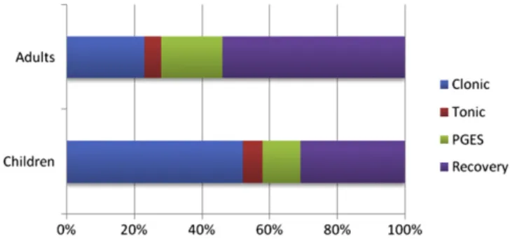
4.1. Periictal seizure phases
Adult and pediatric seizures were different. Postictal EEG suppres-sion was present in 13/23 (57%) GTCS events in 5/12 (42%) pediatric pa-tients, whereas it was present in 77/82 (94%) seizure events in 44/49 (90%) adult patients. Using the independent sample Mann–Whitney U test, total seizure, tonic phase, PGES, and recovery phase durations were all found to be significantly shorter in children (Fig. 1,Table 2). In terms of means, PGES duration was 8 times longer in adults and recovery duration twice as long. A periictal seizure phase versus time plot was constructed to compare groups. In adults, the PGES and re-covery phases contributed to almost three quarters of the periictal period (Fig. 2). Table 1 Patient characteristics. Children (seizures; N = 12) Adults (seizures; N = 49) Mean age with standard deviation at assessment 11.1 ± 3.4 years 35.1 ± 12.2 Etiology Unknown/cryptogenic 6 (50%) 30 (62%) Remote stroke 1 (8.33%) 1 (2%) Meningoencephalitis 1 (8.33%) 2 (4%) Cortical dysplasia 1 (8.33%) 0 Posterior leukomalacia 1 (8.33%) 0 Gliosis caused by previous abscess 1 (8.33%) 3 (6%) Mesial temporal sclerosis 0 7 (14%) Encephalomalacia (unexplained) 0 2 (4%) Post traumatic brain injury 0 1 (2%) Low grade glioma 0 1 (2%)
Cavernoma 0 1 (2%)
Genetic generalized 1 (8.33%) 1 (2%) Ictal localization
Left temporal lobe 1 (8.33%) 16 (32%) Right temporal lobe 1 (8.33%) 4 (8%) Bitemporal lobe 0 11 (23%) Left frontal lobe 1 (8.33%) 6 (13%) Right frontal lobe 0 4 (8%) Left parietal 1 (8.33%) 0 Right occipital 0 2 (4%) Right hemisphere 0 1 (2%) Genetic generalized 6 (50%) 4 (8%) Multifocal 1 (8.33%) 1 (2%) Left insular 1 (8.33%) 0 Number of antiepileptic medications at the time
of seizure [23 seizure events (children) and 82 seizure events (adults)]
One or none 4 (18%) 18 (22%)
Two 12 (52%) 44 (54%)
Three 6 (26%) 17 (21%)
4.2. Primary versus secondary GTCSs
Since 50% of the children had genetic generalized epilepsy, while only 8% of the adults had this diagnosis, we looked at phase durations in the primary GTCSs of genetic generalized epilepsies and the sec-ondary GTCSs of focal epilepsies to clarify whether seizure types were significantly different. We found that (Table 3) total seizure duration, tonic, PGES, and recovery phases were significantly longer in secondary GTCSs as compared with primary GTCSs.
4.3. Tonic phase duration
Using GEE models looking at parametric estimates and tonic phase duration as the dependent variable, we found age to be significantly associated (B = 0.122, 95% CI: 0.075–0.196; p b 0.0001) (Table 4a). In subgroup (adult or child) analysis, this remained significant such that each year, an increase in age increased tonic phase duration by 0.12 s on average. Similarly, PGES duration was significantly increased in direct proportion to tonic phase duration (B = 0.030, 95% CI: 0.002– 0.058; pb 0.05). Each second of PGES correlated with a 0.12-second increase in tonic phase duration. Antiepileptic drug cessation signi fi-cantly prolonged the tonic phase (pb 0.05) by an average of 1.7 s. In subanalysis, this did not hold true in children (p = 0.207).
4.4. PGES duration
With GEE analysis using PGES duration as the dependent variable, children had PGES phases that were on average 28 s shorter. Each year of increase in age at the time of study was associated with a 0.6-second increase in PGES duration. Clonic phase duration was signif-icantly and inversely proportional to PGES duration. Recovery phase duration and decreased HRV were significantly and directly propor-tional to PGES duration (Table 4b). In subgroup analysis (adult or child), only recovery phase duration remained a significant association in children. Conversely, when recovery phase duration (Table 4c) was
taken as the dependent variable, PGES duration increase was the only significant association.
4.5. Medication effects
Medication profiles in terms of AED numbers in both groups were similar (Table 1). With medication as the dependent variable using GEE, no differences in tonic, clonic, PGES, or recovery phases between medication groups were seen. However, in subanalysis, total seizure duration with cessation of medication was significantly increased by a mean value of 8 s in children and 11 s (pb 0.05) in adults. Medication cessation significantly increased PGES duration in adults (p b 0.0001) but not in children (p = 0.385). Similar significances were not seen when clonic phase duration was the dependent variable. The differ-ences in subjects on different classes of AEDs were not analyzed because the numbers were too small for any meaningful analysis.
4.6. Heart rate variability
Generalized estimating equation models looking at RMSSD (to 5 min postictally), SDSD, and SDNN with seizure phase, PGES, and recovery durations did not show any significant results except for RMSSD (at 2 min) which had a negative correlation with PGES duration (longer PGES durations were associated with decreased vagally mediated heart rate variability; pb 0.05) when tonic phase and PGES duration were dependent variables (Table 4).
5. Discussion
Sudden unexpected death in epilepsy is rare in children. One study, assuming a pediatric epilepsy prevalence of 0.59% over a 10-year period, estimated an incidence of 27/138,620 person-years of epilepsy (2 per 10,000 person-years). In comparison with predominantly adult SUDEP estimates of 1 to 2 per 1000 person-years[22,23], this represents a 10-fold lower rate of SUDEP in children[1]. Whether this is due to age-related syndromic, etiologic, electroclinical, or other factors is not clear.
Fig. 1. Comparison of durations in seizure phases, postictal generalized EEG suppression (PGES), and the recovery phase in children and adults. y axis = time in seconds. * signif-icant difference (pb 0.001). PGES = postictal generalized EEG suppression.
Table 2
Comparison of mean durations of seizure phases (in seconds), postictal generalized EEG suppression (PGES), and the recovery phase in children and adults.
Phase (in seconds) Children (N = 23) Adults (N = 82) Significance Total seizure duration 86.2 ± 48.5 110.5 ± 53.6 0.001⁎ Tonic 6.5 ± 2.2 10.5 ± 3.6 0.00⁎ Clonic 53.5 ± 33.6 49.2 ± 26.8 0.464 PGES 11.7 ± 14.4 38.8 ± 24.4 0.00⁎ Recovery 31.5 ± 42.1 113.7 ± 77.3 0.00⁎ PGES = postictal generalized EEG suppression.
⁎ Significant finding at p b 0.05.
Fig. 2. Periictal seizure phases showing differences between children and adults. PGES = postictal generalized EEG suppression.
Table 3
Comparison of mean durations of seizure phases (in seconds), postictal generalized EEG suppression, and recovery phases in the primary GTCSs of genetic generalized epilepsies and secondary GTCSs of focal epilepsies.
Phase (in seconds) Primary GTCSs (N = 19)
Secondary GTCSs (N = 86)
Significance Total seizure duration 76.2 ± 37.94 111.62 ± 54.2 0.008⁎ Tonic 7.42 ± 2.73 10.08 ± 3.78 0.005⁎ Clonic 53.16 ± 37.56 49.48 ± 26.02 0.611 PGES 16.26 ± 21.72 36.51 ± 24.55 0.001⁎ Recovery 19.63 ± 58.01 32.29 ± 43.48 0.028⁎ PGES = postictal generalized EEG suppression.
GTCSs = generalized tonic–clonic seizures. ⁎ Significant finding at p b 0.05.
Generalized tonic–clonic seizures are the commonest seizure type associated with SUDEP. Semiological analyses in pediatric seizures indi-cate differences from adult seizures[10,11,24]that may be relevant in agonal SUDEP phenomenology. One analysis of 109 seizure events in 77 infants did notfind a single typical GTCS[11]. Another study of 296 seizure events in 76 children up to the age of three years similarly reported complete absence of GTCSs[10], suggesting that this is a rare seizure type in children. Few studies have compared electroclinical seizure phases[13], and none have done so comparing adults and pedi-atric GTCSs. In our study, only 23 children inN500 patients monitored over 9 years had true GTCSs, and none were under the age of 5 years. On the other hand, Dravet's syndrome is a pediatric epilepsy syndrome strongly associated with SUDEP. In an analysis of 623 patients with Dravet's syndrome, 59 deaths were examined (a proportional mortality rate ofN10%), of which 53% were sudden death cases[25]. Generalized tonic–clonic seizures are frequently observed in patients with Dravet's syndrome[26,27]in contrast to those without [10,11]and may at least, in part, explain the relatively lower incidence of SUDEP in children without Dravet's syndrome as compared with children and adults with Dravet's syndrome. These observations are of interest because of the consistently strong association between GTCSs and SUDEP in case– control and epidemiological studies in both adults and children[5–9].
In our study, we found several age-related electroclinical differences in GTCSs. Total seizure and tonic phase durations were significantly lon-ger in adults, and AED cessation during monitoring further prolonged these in this higher SUDEP-risk population. Children appear to have a shorter tonic phase that is relatively unaffected by the absence of medica-tion. Why childhood GTCSs are semiologically different from adult GTCSs is unclear. This has been attributed to relative immaturity and lack of organization of developing brains, characterized by variable neuronal excitability, imperfect myelination, and incomplete interhemispheric connections[10,11,28]. Partial seizures in humans are attributed to fore-brain seizure circuitry[29]although some phases of GTCSs may be driven by brainstem seizure circuitry[30,31]. In animals, GTCSs can be induced by electrical stimulation of the brainstem reticular core, despite the removal of the forebrain. Although there is no described mechanism to connect brainstem-driven GTCS phenomena in humans with postictal
autonomic and cardiorespiratory compromise, it is tempting to specu-late that in adults, prolonged tonic phases may conceivably drive pontomedullary autonomic network dysfunction and increase SUDEP risk. The shorter tonic phase in children may reflect immature, poorly established subcortical seizure networks and lesser postictal autonomic dysregulation.
The directly proportional relationship between tonic phase duration (when tonic phase was the dependent variable) and PGES duration seen in our study confirms the findings of one recent report[12]; the chronology of these phenomena seems to suggest that prolonged tonic phases are reflected in greater disturbances of cortical function in our patients, regardless of age. The recovery phase was not similarly affected, suggesting that the tonic phase's main effect is on the early postictal period when the patient is presumed most vulnerable to SUDEP. Prolonged PGES has been shown to indicate increased SUDEP risk in refractory epilepsy in one study[13]where it was significantly longer in the GTCSs of patients with SUDEP. With PGES durations ofN50 s, SUDEP odds were significantly increased, with a quadrupled risk with PGESN80 s. Another study which examined the EEG records of 17 SUDEP cases and matched controls questioned this association although this may be explained by methodological differences[18]. Patients undergoing presurgical evaluations for temporal lobectomy were predominantly those with temporal lobe epilepsy. The matched surviving controls are likely to have become seizure-free with surgery and the risk of SUDEP artificially removed but, in essence, are potential-ly biologicalpotential-ly indistinct from cases. Postictal EEG suppression also ap-pears to inversely correlate with clonic phase duration, regardless of tonic phase and total seizure duration. This effect is difficult to explain unless seizures with long clonic phases result in less obtundation. It may be relevant that in clinical practice, generalized clonic seizures (without a tonic component) sometimes occur without loss of con-sciousness. Overall, PGES was three times longer in adults.
Postictal EEG suppression occurs in between 8% of pediatric pa-tients with seizure[32]to 65% or more of adult patients with GTCSs
[13]and has been reported in several monitored SUDEP/near SUDEP cases[13,33–37]where some authors have used the term“cerebral shut-down”[35]. The increased incidence and duration of PGES in adults are
Table 4
Estimated generalized linear models using generalized estimating equations and different dependent variables.
Total sample (N = 105 seizure events) Estimated B-value estimate Standard error of interval 95% Wald confidence p value a) Tonic phase duration in seconds as the dependent variable
PGES duration 0.030 0.0143 0.002 to 0.058 0.034⁎
Clonic phase duration 0.003 0.024 −0.36 to 0.043 0.864
Total seizure duration 0.006 0.0046 −0.03 to 0.16 0.167 Effect of medication −1.704 0.7578 2.565 to 8.644 0.025⁎
Recovery phase −0.004 0.0081 −0.020 to 0.012 0.625
RMSSD 0.000 0.0001 0.000 to 0.000 0.159
Intercept 5.605 1.5507 2.565 to 8.644 0.000⁎
b) Postictal generalized EEG suppression duration in seconds as the dependent variable
Tonic phase duration 1.477 0.908 −0.301 to 3.26 0.104
Clonic phase duration −0.155 0.0586 −0.270 to −0.041 0.008⁎ Total seizure duration −0.016 0.0243 −0.063 to 0.032 0.516 Effect of medication reduction 11.689 6.281 −0.62 to 2400 0.063
Recovery phase 0.228 0.0300 0.169 to 0.287 0.000⁎
RMSSD 0.002 0.0007 0.000 to 0.003 0.009⁎
Intercept 45.472 12.037 2.1.88 to 69.07 0.000⁎
c) Recovery phase as the dependent variable
Tonic phase duration −3.07 1.631 −3.504 to 2.889 0.851
Clonic phase duration 0.214 0.2523 −.0281 to 0.708 0.397 Total seizure duration −0.123 0.1789 −0.474 to 0.228 0.492 Effect of medication reduction 13.9 13.878 −13.3 to 41.1 0.317
RMSSD 2.581 0.2421 2.107 to 3.056 0.000⁎
PGES 0.003 0.0016 −1.983 to 0.006 0.051
Intercept −53.292 25.824 −103.9 to −2.678 .039⁎
PGES = postictal generalized EEG suppression.
Effect of medication = no or some reduction versus no medication. RMSSD = root mean square successive difference of RR intervals.
noteworthy as they are a higher risk population than children. Several studies highlight the possible significance of PGES. In one study of 48 pa-tients, those with PGES were significantly more likely to be motionless postictally and to have simple resuscitative interventions[15]. These ob-servations are indirectly corroborated in another study that analyzed 21 GTCS events with no periictal interventions and 84 with interventions. Earlier interventions were associated with briefer hypoxia and shorter PGES duration[16]. Another study compared secondary GTCSs with and without PGES and found that oxygen desaturation duration and extent as well as peak end-tidal CO2elevation were more marked in
pa-tients with PGES[17]. Thus, PGES appears to indicate a greater degree of postictal obtundation and vulnerability to respiratory compromise. The high incidence of PGES in our patients possibly reflects a high rate of medication cessation during monitoring. Postictal EEG suppression dura-tion appears to directly correlate with recovery phase duradura-tion suggesting a continuum of recovery processes in the postictal period.
In common with at least one more study[18], we found no correla-tion between HRV measures and electroclinical seizure variables, with one exception. Root mean square successive difference measures at 2 min postictally were negatively correlated to PGES suggesting that PGES may be associated with the reduced vagal tone observed in some patients[38].
The effect of AED cessation during monitoring (usually done to induce seizures as part of presurgical assessment) is interesting as this artificially amplifies the refractoriness of a patient's epilepsy or creates a situation akin to noncompliance, another risk factor for SUDEP[14]. Total seizure durations were significantly increased by a mean value of 8 s and 11 s, respectively, in children and adults, and both PGES and tonic phase durations significantly increased in adults when medi-cation was stopped. This indirectly appears to corroborate literature suggesting greater SUDEP risk in patients with refractory epilepsy and, in particular, those who are noncompliant with medication, where sei-zure frequency and severity can be expected to be worse than on treatment.
Our study has limitations. Patient records were retrospectively ana-lyzed with its attendant biases. A much smaller number of pediatric GTCSs reflect the relative rarity of this seizure type in this age group and limit statistical power. The medication tapering protocols and total duration of hospital stay is, in general, shorter in children. Antiepileptic drugs have different half-lives which may influence the seizure duration, but our AED groups are too small to look for these dif-ferences. We also considered the adult population to beN16 years of age rather than the≥20-year figure used in some SUDEP studies for pragmatic reasons. All 4 patients in the 16- to 20-year bracket were aged 19 years and, in biological terms, were more suited to be analyzed as adults. Additionally, we did not have respiratory measurements to determine the presence and influence of hypoxia, bradypnea, and apnea, phenomena that are potential SUDEP mechanisms and that are known to occur in pediatric seizures[39].
Since there is no forward surveillance of patients, the true incidence of SUDEP in both groups cannot be known, and, hence, there is no gold standard for validation of the observed results. However, our data clearly point out identifiable electroclinical differences between the adult and pediatric population which may at least, in part, explain differences in SUDEP incidence. It also highlights the importance of careful characterization of seizure semiology and EEG, particularly PGES. There is a gathering body of evidence that PGES is an impor-tant postictal phenomenon; its pathophysiology requires further, careful elucidation. Overall, however, prolonged PGES may be best seen as a potential risk“marker” of SUDEP rather than a risk “factor”; the latter implies a causal role which is as yet uncharacterized and unproven.
We confirm that we have read the Journal's position on issues involved in ethical publication and affirm that this report is consistent with those guidelines.
Disclosure
None of the authors has any conflict of interest to declare. Acknowledgments
This study was supported in part by NINDS grant NS076965-01— The Prevention and Risk Identification of SUDEP Mortality (PRISM) Project.
References
[1]Donner EJ, Smith CR, Snead III OC. Sudden unexplained death in children with epilepsy. Neurology 2001;57:430–4.
[2]Milroy CM. Sudden unexpected death in epilepsy in childhood. Forensic Sci Med Pathol 2011;7:336–40.
[3]Nickels KC, Grossardt BR, Wirrell EC. Epilepsy-related mortality is low in children: A 30-year population-based study in Olmsted County, MN. Epilepsia 2012;53:2164–71.
[4]Rodriguez ML, McMillan K, Crandall LA, Minter ME, Grafe MR, Poduri A, et al. Hippo-campal asymmetry and sudden unexpected death in infancy: a case report. Forensic Sci Med Pathol 2012;8:441–6.
[5]Hesdorffer DC, Tomson T, Benn E, Sander JW, Nilsson L, Langan Y, et al. Combined analysis of risk factors for SUDEP. Epilepsia 2011;52:1150–9.
[6]Hesdorffer DC, Tomson T, Benn E, Sander JW, Nilsson L, Langan Y, et al. Do antiepileptic drugs or generalized tonic-clonic seizure frequency increase SUDEP risk? A combined analysis. Epilepsia 2012;53:249–52.
[7]Langan Y, Nashef L, Sander JW. Case–control study of SUDEP. Neurology 2005;64: 1131–3.
[8]Nilsson L, Farahmand BY, Persson PG, Thiblin I, Tomson T. Risk factors for sudden unexpected death in epilepsy: a case-control study. Lancet 1999;353:888–93.
[9]Shorvon S, Tomson T. Sudden unexpected death in epilepsy. Lancet 2011;378:2028–38.
[10]Hamer HM, Wyllie E, Luders HO, Kotagal P, Acharya J. Symptomatology of epileptic seizures in thefirst three years of life. Epilepsia 1999;40:837–44.
[11]Korff C, Nordli Jr DR. Do generalized tonic–clonic seizures in infancy exist? Neurology 2005;65:1750–3.
[12]Tao J, Yung I, Lee A, Rose S, Jacobsen J, Ebersole JS. Tonic phase of a generalized convulsive seizure is an independent predictor of post-ictal generalized EEG suppression. Presented at American Epilepsy Society, San Diego, CA; 2012.
[13]Lhatoo SD, Faulkner HJ, Dembny K, Trippick K, Johnson C, Bird JM. An electroclinical case–control study of sudden unexpected death in epilepsy. Ann Neurol 2010;68(6): 787–96.
[14]Surges R, Sander JW. Sudden unexpected death in epilepsy: mechanisms, preva-lence, and prevention. Curr Opin Neurol 2012;25:201–7.
[15]Semmelroch M, Elwes RD, Lozsadi DA, Nashef L. Retrospective audit of postictal generalized EEG suppression in telemetry. Epilepsia 2012;53:e21–4.
[16]Seyal M, Bateman LM, Li CS. Impact of periictal interventions on respiratory dysfunc-tion, postictal EEG suppression, and postictal immobility. Epilepsia 2012;53(5): 825–31.
[17]Seyal M, Hardin KA, Bateman LM. Postictal generalized EEG suppression is linked to seizure-associated respiratory dysfunction but not postictal apnea. Epilepsia 2012;53:825–31.
[18]Surges R, Strzelczyk A, Scott CA, Walker MC, Sander JW. Postictal generalized elec-troencephalographic suppression is associated with generalized seizures. Epilepsy Behav 2011;21:271–4.
[19]Gastaut H. Semiology and physiopathogenesis of generalized epileptic seizures. Helv Med Acta 1963;30:319–37.
[20]NASPE. Heart rate variability. Standards of measurement, physiological interpretation, and clinical use. Task Force of the European Society of Cardiology and the North American Society of Pacing and Electrophysiology. Eur Heart J 1996;17:354–81.
[21]Fishman M, Jacono FJ, Park S, Jamasebi R, Thungtong A, Loparo KA, et al. A method for analyzing temporal patterns of variability of a time series from Poincare plots. J Appl Physiol 2012;113:297–306.
[22]Derby LE, Tennis P, Jick H. Sudden unexplained death among subjects with refractory epilepsy. Epilepsia 1996;37:931–5.
[23]Tennis P, Cole TB, Annegers JF, Leestma JE, McNutt M, Rajput A. Cohort study of incidence of sudden unexplained death in persons with seizure disorder treated with antiepileptic drugs in Saskatchewan, Canada. Epilepsia 1995;36:29–36.
[24]Loddenkemper T, Wyllie E, Neme S, Kotagal P, Luders HO. Lateralizing signs during seizures in infants. J Neurol 2004;251:1075–9.
[25]Sakauchi M, Oguni H, Kato I, Osawa M, Hirose S, Kaneko S, et al. Mortality in Dravet syn-drome: search for risk factors in Japanese patients. Epilepsia 2011;52(Suppl. 2):50–4.
[26]Bureau M, Dalla Bernardina B. Electroencephalographic characteristics of Dravet syndrome. Epilepsia 2011;52(Suppl. 2):13–23.
[27]Dravet C. The core Dravet syndrome phenotype. Epilepsia 2011;52(Suppl. 2):3–9.
[28]Arzimanoglou A, Guerrini R, Aicardi J. Epilepsy in infants. In: Arzimanoglou A, Guerrini R, Aicardi J, editors. Aicardi's epilepsy in children. Philadelphia: Lippincott and Williams; 2004. p. 210–9.
[29]Jobe PC, Mishra PK, Dailey JW, Ko KH, Reith MA. Genetic predisposition to partial (focal) seizures and to generalized tonic/clonic seizures: Interactions between sei-zure circuitry of the forebrain and brainstem. In: Berkovic SF, Genton P, Hirsch E, Picard F, editors. Genetics of focal epilepsies. Avignon, France: John Libbey & Company Ltd.; 1999. p. 251–60.
[30]Coffey LL, Reith ME, Chen NH, Mishra PK, Jobe PC. Amygdala kindling of forebrain seizures and the occurrence of brainstem seizures in genetically epilepsy-prone rats. Epilepsia 1996;37:188–97.
[31]Jobe PC, Browning RA. Mammalian models of genetic epilepsy characterized by sensory evoked seizures and generalized seizure susceptibility. In: Pitkanen A, Schwartzkroin PA, Moshe S, editors. Models of seizures and epilepsy. Amsterdam: Academic Press; 2006. p. 261–71.
[32]Kim AJ, Kuroda MM, Nordli Jr DR. Abruptly attenuated terminal ictal pattern in pediatrics. J Clin Neurophysiol 2006;23:532–50.
[33]Bateman LM, Spitz M, Seyal M. Ictal hypoventilation contributes to cardiac arrhythmia and SUDEP: report on two deaths in video-EEG-monitored patients. Epilepsia 2010;51:916–20.
[34]Bird JMD, K.A.T., Sandeman D, Butler S. Sudden unexplained death in epilepsy. Epilepsia 1997;38(Suppl. 11):S52–6.
[35]McLean BN, Wimalaratna S. Sudden death in epilepsy recorded in ambulatory EEG. J Neurol Neurosurg Psychiatry 2007;78:1395–7.
[36]So EL, Sam MC, Lagerlund TL. Postictal central apnea as a cause of SUDEP: evidence from near-SUDEP incident. Epilepsia 2000;41:1494–7.
[37]Tao JX, Qian S, Baldwin M, Chen XJ, Rose S, Ebersole SH, et al. SUDEP, suspected po-sitional airway obstruction, and hypoventilation in postictal coma. Epilepsia 2010;51:2344–7.
[38]Poh MZ, Loddenkemper T, Reinsberger C, Swenson NC, Goyal S, et al. Autonomic changes with seizures correlate with postictal EEG suppression. Neurology 2012;78:1868–76.
[39]Singh K, Katz ES, Zarowski M, Loddenkemper T, Llewellyn N, Manganaro S, et al. Car-diopulmonary complications during pediatric seizures: a prelude to understanding SUDEP. Epilepsia 2013;54:1083–91.