2016
UNIVERSIDADE DE LISBOA FACULDADE DE CIÊNCIAS QUÍMICA E BIOQUÍMICA
Dealing with copper toxicity: New insights into copper
detoxification in yeast
Ana Carolina Rodrigues Gaspar Cordeiro
Mestrado em Bioquímica Especialização em Bioquímica
Dissertação orientada por:
Professor Doutor Francisco Rodrigues Pinto Doutora Catarina Ribeiro Pimentel
v
Copper, an essential metal, is a double-edged sword, as its vital nature is counterbalanced by the toxic effect that it can exert on cells when not properly controlled. As such, organisms have evolved defence mechanisms against copper toxicity and, in the yeast Saccharomyces cerevisiae, the transcription factor Ace1 orchestrates several of those mechanisms, by activating copper-detoxification genes.
In Saccharomyces cerevisiae iron uptake is partially dependent on copper, as the membrane multicopper-oxidase Fet3, which is part of the high-affinity iron uptake system, requires copper as a cofactor. Aft1, the low iron-sensing transcription factor in yeast, is known to regulate the expression of FET3 gene. However, we found that a strain lacking FET3 is more sensitive to copper surplus conditions than a strain carrying the AFT1 gene disrupted. This finding suggests that under such conditions another regulator came into play and controls FET3 gene expression.
We next evaluated whether Ace1 could be the alternative regulator of FET3 under copper excess. To test this hypothesis, the expression of FET3 gene in cells lacking ACE1, AFT1 or both was monitored, using yeast-one hybrid and qRT-PCR approaches. A decrease of FET3 transcripts is observed, under copper loading, when ACE1 is deleted, suggesting that Ace1 is involved in FET3 regulation. Remarkably, in the absence of AFT1 we observed a copper-dependent induction of FET3 gene that was, however, completely abrogated when ACE1 was deleted from the aft1 mutant background. Our data clearly indicates that FET3 expression is dependent on Ace1 when copper is overly abundant.
In this study, the link between iron and copper homeostasis was further established. Indeed, we found that ace1 cells display marked upregulation of Aft1’s targets, such as CTH2, and accumulate higher amounts of iron and copper, in a mechanism dependent on Aft1.
Altogether, this work unveiled a novel protection mechanism against copper toxicity mediated by Ace1, which relies in the activations of FET3 and results in the restriction of Aft1 activity as to prevent excessive copper accumulation.
vi
O cobre é um metal essencial para a vasta maioria dos organismos, maioritariamente devido às suas propriedades de oxidação-redução, que o tornam ideal para ser cofactor de inúmeras proteínas. Nos humanos, o cobre é essencial para inúmeras funções celulares, nomeadamente para o metabolismo energético, produção de melanina e catecolaminas, destoxificação de espécies reactivas de oxigénio e homeostase de ferro. No entanto, a sua natureza essencial é contrabalançada pelos efeitos tóxicos causados quando este metal está presente em níveis acima dos considerados fisiológicos. De facto, sabe-se que o excesso de cobre potencia a produção de espécies reactivas de oxigénio (ROS), através da reacção de Fenton, e que este metal pode mesmo danificar os centros metálicos de enzimas e proteínas, levando à perda de função. Assim, os organismos desenvolveram inúmeros mecanismos de defesa contra a toxicidade provocada pelo cobre. Na levedura Saccharomyces cerevisiae, o factor de transcrição Ace1 é o responsável pela regulação destes mecanismos, ao activar genes de destoxificação do cobre, tais como genes codificantes de metalotioneínas CUP1 e CRS5, que actuam como quelantes de iões de cobre, e o gene SOD1, que codifica o superóxido dismutase, enzima que além de conter cobre no seu centro activo, é essencial na resposta ao stress oxidativo causado por este metal.
Em Saccharomyces cerevisiae a entrada de ferro (Fe) na célula é parcialmente dependente do cobre, uma vez que o Fet3, uma ferroxidase de membrana que faz parte do complexo de transporte de alta afinidade de ferro, requer cobre como cofactor. O Aft1, o principal factor de transcrição envolvido na resposta à deficiência de ferro em levedura, é o conhecido regulador da expressão do gene FET3. No entanto, diversos resultados apresentados neste trabalho demonstram claramente que uma estirpe sem o gene FET3 é mais sensível ao excesso de cobre do que uma estirpe mutada no gene AFT1. Esta evidência sugere que, em condições de excesso de cobre, um outro regulador controla a expressão do gene FET3. Deste modo, foi avaliada a possibilidade de o factor de transcrição Ace1 ser o regulador alternativo do gene FET3 em condições de excesso de cobre. De facto, a região promotora do gene FET3 contém uma possível sequência de ligação do Ace1, localizada a -578 bp a montante do codão de iniciação ATG (+1). Através da monitorização dos níveis de expressão do FET3 na estirpe-selvagem e na estirpe mutada no gene ACE1 (mutante ace1), foi possível concluir que o factor de
vii
transcrição Ace1 está envolvido na regulação do gene FET3. Foi ainda observado que mesmo na ausência do regulador Aft1, ocorre uma indução da expressão do FET3, dependente do cobre que, no entanto, é totalmente suprimida quando se elimina o gene ACE1.
Através da monitorização da expressão de um dos genes alvo do factor de transcrição Aft1, CTH2, na estirpe selvagem e na estirpe ace1, em condições de excesso de cobre, através da técnica de qRT-PCR, observou-se que, na ausência do Ace1, há um aumento drástico dos níveis dos transcritos de CTH2. Estes dados sugerem que, na estirpe mutante ace1, a homeostase de ferro encontra-se desregulada, pelo que ocorrerá uma super-activação do factor Aft1. De acordo com esta hipótese, foi observado que a estirpe ace1 contém níveis de ferro e de cobre intracelulares marcadamente elevados quando comparados com os da estirpe-selvagem. Esta acumulação é dependente da actividade do regulador Aft1, uma vez que a sua eliminação (na estirpe mutante aft1ace1) repõe os níveis dos dois metais para valores semelhantes aos da estirpe-selvagem.
O Fet3 é um enzima importante para a resposta celular ao excesso de cobre. Foi descrito que a estirpe mutante fet3 acumula grandes quantidades de cobre. Neste estudo, observou-se que esta acumulação ocorre através de um mecanismo dependente do factor de transcrição Aft1, dado que o duplo mutante fet3aft1 reverte os níveis de cobre intracelulares para níveis semelhantes aos da estirpe selvagem. Através da análise dos genes alvo do Aft1, foram seleccionados possíveis proteínas envolvidas neste mecanismo. A deleção do genes FET4, que codifica um transportador de ferro de baixa afinidade, que também tem a capacidade de transportar cobre, e do ARN4, que codifica um transportador de sideróforos de ferro, cujo substrato é a enterobactina, levou a um ganho de resistência ao cobre quando deletados de uma estirpe fet3. Resultados semelhantes foram registados após a deleção do gene FRE1, que codifica uma metaloreductase de membrana, envolvida no transporte de ferro e cobre. Estes resultados evidenciaram que a acção combinada de várias proteínas envolvidas no transporte de ferro contribuem para a acumulação de cobre observada numa estirpe mutante fet3. Assim a deficiência de ferro observada na estirpe mutante fet3, conduzirá à activação do factor de transcrição Aft1 que induzirá a expressão dos genes dessas proteínas, numa tentativa de colmatar a falta de ferro consequente da perda do sistema de transporte de ferro de alta afinidade.
Neste estudo, foi ainda possível observar que a estirpe mutante aft1, que apresenta um fenótipo de sensibilidade em condições normais crescimento, recupera o seu défice
viii
de crescimento quando cobre é adicionado ao meio de cultura. Contrariamente ao esperado, a atenuação observada não é devida a um aumento do conteúdo intracelular de ferro, quando as células são crescidas na presença de cobre, uma vez que os níveis de ferro da estirpe aft1 são inferiores aos níveis registados quando cultivada em condições normais de crescimento. Com o objectivo de compreender o mecanismo subjacente a este fenómeno, testaram-se outras vias dependentes de cobre poderiam estar afectadas em condições normais de crescimento no mutante aft1, nomeadamente a respiração mitocondrial, (dado que o cobre é um cofactor do citocromo c oxidase), e a resposta ao stress oxidativo (uma vez que o cobre faz parte do Cu,Zn-Superóxido dismutase). Através da análise da sensibilidade do mutante aft1 em meio YPDGE (meio de cultura rico, contendo quase exclusivamente fontes de carbono não fermentáveis), concluiu-se que a adição de cobre melhora a capacidade respiratória da estirpe aft1, dado que foi observado uma significativa recuperação da sensibilidade deste mutante. Além disso, a análise fenotípica do mutante aft1 cultivado em meio contendo paraquat (um composto gerador de anião superóxido), suplementado ou não com cobre, permite concluir que a adição de cobre alivia também a sensibilidade desta estirpe ao stress oxidativo provocado pelo paraquat. Assim, os resultados obtidos sugerem que numa estirpe aft1 a adição de cobre promove o metabolismo respiratório e fortalece a resposta ao stress oxidativo, contribuindo desta forma para o aliviar do défice de crescimento exibido por esta estirpe.
Assim, no seu conjunto, os dados apresentados nesta dissertação permitem propor uma nova via de destoxificação de cobre mediada pelo factor de transcrição Ace1. Em condições de excesso de cobre, as células de levedura activam este regulador, que por sua vez irá induzir a expressão do gene FET3. Sendo uma proteína que contém cobre no seu centro activo, o enzima Fet3 contribui não só para o tamponamento dos iões de cobre livres presentes no citosol, como também para a entrada de ferro na célula. A entrada de ferro irá prevenir a activação excessiva do Aft1, e consequente expressão dos seus genes alvo, limitando assim a entrada de mais cobre através de transportadores de baixa afinidade para ferro, o que eventualmente, levaria à morte celular. Assim, neste estudo foi pela primeira vez demonstrado que além do seu papel essencial na destoxificação do cobre intracelular, o Ace1 possui também um papel fundamental na prevenção da entrada de cobre extracelular.
ix
Acknowledgments ... iv
Abstract ... v
Resumo ... vi
List of Figures ... xi
List of Tables ... xiii
List of Acronyms ... xiv
I. Introduction ... 1
1.1 Copper in biological systems ... 2
1.1.1 Copper and disease ... 2
1.1.2 Copper at the host pathogen interface ... 3
1.2 Yeast as a model organism for metal homeostasis studies ... 5
1.3 Copper homeostasis in the yeast cell ... 6
1.3.1 Copper depletion ... 6
1.3.2 Copper overload ... 9
1.4 Transcriptional Regulation of Copper homeostasis ... 11
1.5 Crosstalk between copper and iron homeostasis ... 12
Objectives ... 13
II. Materials and Methods ... 14
2.1 Yeast strains and Growth conditions ... 15
2.2 Plasmids ... 17
2.3 Yeast competent cells preparation ... 17
2.4 Yeast transformation ... 17
2.5 Western blot assays ... 18
2.6 Measurements of ß-galactosidase activity ... 20
2.7 Measurement of total iron and copper intracellular content ... 21
2.8 qRT-PCR ... 22
III. Results ... 23
3.1 Aft1 is not the major regulator of FET3 gene under copper excess conditions ... 24 3.2 Copper accumulation observed in the fet3 mutant strain is dependent on Aft1 25
x
3.3 Ace1 regulates FET3 expression under high-copper conditions ... 27
3.4 Ace1 restricts Aft1 activation under copper excess conditions ... 29
3.5 Copper rescues the growth defect exhibited by the aft1 strain ... 31
IV. Discussion ... 33
V. Conclusions and Future Perspectives ... 36
VI. Appendix ... 39
xi
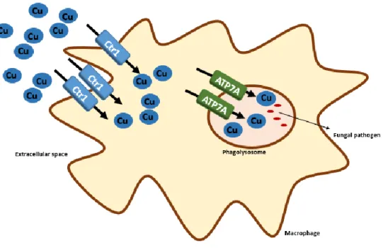
Figure 1.1 – Copper role in pathogen killing by macrophages. Activated macrophages exploit copper toxic properties to promote pathogen killing by concentrating copper (Cu) inside the phagolysosome. Upregulation of copper transporters Ctr1 and ATP7A is observed upon infection... 4
Figure 1.2 - Key player in the yeast response to copper depletion. In low-copper conditions, Mac1, a copper-responsive transcription factor, activates the expression of copper-uptake genes, such CTR1 and CTR3, but also of metalloreductases, as FRE1/2/7. Vacuole copper stores can also be mobilised, in this conditions, through Ctr2.In the cell, through the action of metallochaperones, copper can be incorporated in several enzymes, such as superoxide dismutase (SOD1), Fet3 and cytochrome c oxidase (CcO). ... 7
Figure 1.3 - The yeast response to toxic levels of copper. When cells are exposed to copper surplus Ace1 activates the expression of copper-detoxification genes that allow cells to cope with high copper levels. Metallothioneins, encoded by CUP1 and CRS5 genes, act as intracellular copper chelators and SOD1 neutralises copper-generated ROS. ... 10
Figure 3.1 - Under copper overload, Aft1 is not the major regulator of FET3 gene. A:Growth exhibited by the WT, aft1, fet3 and aft1fet3 strains in SC media was recorded over a period of 24 h. Closed symbols and open symbols represent strains grown in media containing or not 2mM of CuSO4, respectively. Each colour represents one of the used
strains: WT is represented in black, aft1 in blue, fet3 in green and aft1fet3 in orange. . 24
Figure 3.2 - fet3 copper accumulation is dependent on Aft1. A: Copper (Cu) content of WT, aft1, fet3 and aft1fet3 strains were determined by ICP-AES, in SC medium after treatment with 2mM CuSO4 for 15 h. B: Iron (Fe) content of WT, aft1, fet3 and aft1fet3
strains were determined by ICP-AES, in SC medium after treatment with 2mM CuSO4
for 15 h. C: Growth sensitivity of WT, fet3, fe4, arn4, fre1, fet3fet4, fet3arn4 and fet3fre1 strains in SC plates and SC plates containing 3 or 5mM CuSO4 D: Growth sensitivity of
WT and fet3, in SC plates and SC plates containing 2mM of CuSO4 supplemented or not
with 250µM ZnSO4. E: Growth sensitivity of WT and fet3, in SC plates and SC plates
containing 2mM of CuSO4 supplemented or not with 500µM FeSO4. ... 26
Figure 3.3 - Ace1 regulates FET3 expression under copper excess conditions. A: Schematic representation of the FET3 promoter region containing the one putative Ace1-binding site located at -578 upstream of the ATG. B: WT and ace1 mutant strains were transformed with the pFET3-lacZ plasmid. Cells were grown exponentially in SD medium and challenged with 0.5mM CuSO4 for 30 min. β-Galactosidase activity was
xii
challenged with 2mM CuSO4 and harvested at the indicated time points. The expression
of FET3 was monitored by qRT-PCR. D: aft1 and aft1ace1 strains were challenged with 2mM CuSO4 and harvested at the indicated time points. The expression of FET3 was
monitored by qRT-PCR. E: The WT Fet3-TAP and the ace1 Fet3-TAP strains were incubated with 2 mM CuSO4 or the indicated times. Fet3 protein levels were analysed by
Western-blot with an anti-TAP antibody. Fet3 protein levels were normalized to Pgk1. Significance of differences was calculated with the t-test (* p<0.05). ... 28
Figure 3.4 - Ace1 prevents Aft1 hyper-activation under copper excess conditions. A: WT (wild-type) and ace1 strains were challenged with 2mM CuSO4 and harvested at the
indicated time points. The expression of CTH2 was monitored by qRT-PCR. B: Copper content of WT, ace1 and ace1aft1 strains, after treatment with 2mM CuSO4 (+Cu) for 15
was determined by ICP-AES. C: Fe content of WT, ace1 and ace1aft1 strains were determined by ICP-AES, in SC medium or after treatment with 2mM CuSO4 (+Cu) for
15 h. Significance of differences was calculated with the t-test (* p<0.05) ... 30
Figure 3.5 - Copper rescues the growth defect exhibited by the aft1 strain. A: Exponentially growing WT and aft1 were serially diluted and spotted onto SC plates and SC plates supplemented with 2, 3 or 5 mM CuSO4. Growth was recorded at 48hours
incubation at 30ºC. B: Exponentially growing WT and aft1 were serially diluted and spotted onto SC plates and SC plates supplemented with 500µM FeSO4. Growth was
recorded at 48hours incubation at 30ºC. C: Fe content of WT and aft1 strains were determined by ICP-AES, in SC medium or after treatment with 2mM CuSO4 (+Cu) for
15 h. D: Copper content of WT and aft1 strains were determined by ICP-AES, in SC medium. E: Growth sensitivity of WT and aft1 in YPD and YPDGE plates containing 2mM CuSO4 .(+Cu) F: Growth sensitivity of WT and aft1 in YPD containing 500 µM of
Paraquat, supplemented with 2mM CuSO4 (+Cu). Significance of differences was
calculated with the t-test (* p<0.05). ... 32
Figure 5.1 - Model for the role of Ace1 in copper detoxification. Under copper (Cu) overload conditions, Ace1, the transcription factor involved in the detoxification of copper, activates the expression of copper detoxification genes such as SOD1, CUP1 and CRS5. In this study, we have showed that Ace1 regulates FET3, a multicopper oxidase essential for Fe uptake. This regulation serves as to restrict Aft1 hyper-activation, whose target expression, can lead to cell death. The novel pathway for Ace1-mediated copper detoxification proposed in this study is depicted in red. ... 38
xiii
Table 1 - Strains used in this study ... 15 Table 2 - Antibodies used in this study ... 19 Table S3 - List of oligonucleotides (primers) used in this study. Highlighted in bold are the sequences complementary to the histidine marker (HIS) or kanamycin (KAN). ... 40
xiv
APS Ammonium persulphate
ATP Adenosine triphosphate
CuSO4 Copper sulphate
DMSO Dimethyl Sulphoxide
DNA Deoxyribonucleic acid
FeSO4 Iron(II) Sulphate
LiAc Lithium Acetate
M Molar
mM Mili Molar
Na2HPO4 Sodium phosphate dibasic
NaH2PO4 Sodium phosphate monobasic
nm Nanometres
OD600 Optical Density at 600 nanometres
ORF Open Reading Frame
PBS Phosphate Buffer Saline
PCR Polymerase Chain Reaction
PEG Polyethylene glycol
PMSF Phenylmethilsulfonyl fluoride
RNA Ribonucleic acid
ROS Reactive Oxygen Species
rpm Revolutions per minute
RT Reverse Transcriptase
SC Synthetic Complete Medium
SDS sodium dodecyl sulphate
SDS-PAGE SDS- Polyacrylamide gel electrophoresis
SOD Superoxide Dismutase
TE Tris-EDTA
TEMED N,N,N',N-Tetramethylethylenediamine\
TGN Trans-Golgi network
µM Micro molar
V Volt
YPD Yeast Peptone Dextrose growth medium
2
Copper is a transition metal that is present in nature predominantly in two oxidation states, Cu (I) and Cu (II). Given its ability to bind organic ligands and its electron transfer properties, this metal has a high potential to be utilized by organisms, mainly as a cofactor for proteins. In humans, copper is essential for oxidative phosphorylation, melanin and catecholamine production (through tyrosinase and dopamine-β-hydroxylase, respectively), superoxide dismutase activity and iron homeostasis [1].
Wilson and Menkes diseases are rare genetic conditions caused by mutations in Cu-ATP-Binding-Cassette transporters, ATP7B and ATP7A, respectively [2, 3]. Within the cell, both proteins co-localize to the trans-Golgi network (TGN), where they import copper into TGN’s lumen, to be then incorporated in copper-containing proteins. However, both proteins appear to translocate to intracellular vesicles and to the plasma membrane in response to copper overload [4, 5]. These proteins have different localization patterns, with ATP7B being present mainly in the liver, and ATP7A distributed across the whole organism, with the exception of the liver. The phenotype of Wilson disease, due to a deficient biliary excretion of copper that causes an accumulation of copper in tissues, is a failure of liver function and neurological impairment. Menkes disease is characterized by systemic copper deficiency, as copper fails to reach circulation, which leads to the loss of activity of copper-containing proteins, and has as consequence a delay in neuronal development and a reduction of muscle tone [2, 3, 6].
There is now increasing evidence suggesting that neurodegenerative disorders, such as Alzheimer, Parkinson and Creutzfeldt-Jakob disease, are associated with an imbalance of copper homeostasis [7]. Copper is known to promote protein aggregation and oxidative stress, which are determinant factors for disease progression [7, 8]. Although copper levels above the homeostatic threshold have been observed in these and
3
other diseases, such as cancer, the mechanisms underlying this phenomenon are not yet clearly understood [7, 9] .
Copper-chelation therapies are currently used to treat Wilson disease and are considered an attractive strategy to prevent the onset of neurodegenerative diseases [9].
The treatment of Menkes disease relies on copper-supplementation therapy, through copper-histidine complex formulations, that can increase copper in ceruloplasmin and in organs, such as the liver [9–11].
In addition to their major role as protein cofactors, metals, such as Cu, Fe and Zn play active functions in infection biology. The use of copper as an antimicrobial compound dates from the early 19th century, when it begun to be incorporated in the Bourdeaux and Burgundy mixtures, the vineyard fungicides [12, 13]. In fact copper is now thought to have a protective role in infection, as Menkes disease carriers, that have systemic copper deficiency, are more susceptible to infections [10]. Also, mice fed on a low-copper diet have higher mortality rates upon infection with C. albicans [14].
One of the host mechanisms to fight microbial pathogens is the withdrawal of essential metals, such as iron and calcium, from the pathogen’s vicinity, as to limit its growth [15, 16]. In contrast, upon infection, host copper-stores are mobilized, and innate immunity cells accumulate copper ions next to the pathogen in an attempt to increase the efficacy of pathogen killing (Figure 1.1) [17]. Macrophages constitute the first line of defence against microbial pathogens, such as fungi. These innate immunity cells have the ability to phagocyte the pathogen. In the phagolysosome environment, copper toxicity is maximized, as the low pH and the high concentrations of ROS potentiate copper-driven Fenton chemistry [13]. This was further corroborated by the observation that activated macrophages show marked upregulation of the copper-transporter, hCtr1, and of ATP7A at the phagolysosome membrane, which suggest that copper is being compartmentalized into the phagolysosome, after entering the macrophage (Figure 1.1) [18]. Interestingly, the macrophage-activating cytokine interferon-γ (IFN-γ) and lipopolysaccharide (LPS) also mediate the accumulation of copper inside the phagosome [18]. This regulated mechanism of copper compartmentalization ensures copper-damage only to the pathogen. Although accumulation of copper inside activated macrophages occur, the source of this
4
metal is unknown. Copper is thought to be mobilised from circulating ceruloplasmin, which contains over 90% of Cu in the plasma and, indeed, ceruloplasmin is increased during the acute phase of infection. However there are no direct evidences that ceruloplasmin is the copper-donor to activated macrophages [13, 19].
Figure 1.1 – Copper role in pathogen killing by macrophages. Activated macrophages exploit copper toxic properties to promote pathogen killing by concentrating copper (Cu) inside the phagolysosome. Upregulation of copper transporters Ctr1 and ATP7A is observed upon infection
5
In 1996, the first complete sequence of a eukaryote genome, the Saccharomyces cerevisiae genome, was published [20, 21]. The yeast’s 12.8 Mb genome, composed of 16 chromosomes, was revealed to contain more than 5.800 protein-coding genes, 85% of which are presently assigned to a biological function. Although twenty years have passed since the release of the genome, the baker’s yeast is still one of the most widely used eukaryotic model organism. This is due to the fact that it is genetically amenable, allowing genes to be deleted, altered or inserted in the genome, it has a short generation time and, importantly, it has many genes which are conserved among species, including plants, fungal pathogens and mammals [22]. A few years after the sequencing of S. cerevisiae genome, a library with almost each open-reading frame (ORF) mutated was generated [23]. The study of the effect of each deletion has allowed to uncover the biological function of many genes and to shed light on the molecular mechanisms of several human diseases [24–27].
Toxic metals (Cd, As, Hg, etc.) are major environmental and human health-threatening hazards. On the other hand, essential metals (Zn, Fe and Cu), present in virtually all biological systems, are a double-edge sword, as its essential nature is counterbalanced by the toxic effect that it can exert on both eukaryotic and prokaryotic cells when not properly controlled (i.e., when scarce or in excess). To maintain adequate cellular metal levels and avoid metal-loading, cells have developed sophisticated systems to assure the balanced cellular metal homeostasis. The single celled yeast S. cerevisiae has proven to be an invaluable model organism to study metal homeostasis in higher eukaryotes, as a consequence of conservations across species of several of the key players involved in those processes [26].
6
In the budding yeast Saccharomyces cerevisiae copper is involved in three major biological processes: mitochondrial oxidative phosphorylation, superoxide anion detoxification and iron homeostasis [28, 29]. However, the same redox properties that make it ideal for electron transfer reactions also render it susceptible to generate reactive oxygen species (ROS), causing oxidative damage to cells [30].
To counteract sudden fluctuations in copper bioavailability, Saccharomyces cerevisiae cells have evolved mechanisms to maintain a tight control of copper homeostasis. These mechanisms are involved on the one hand in the upregulation of copper-uptake and mobilization systems, which enable copper to be utilized in crucial cellular processes, and on the other hand in the activation of copper detoxification systems, in order to prevent toxicity and repair Cu-induced damages [31–33].
When the intracellular copper content is low, yeast cells import copper mainly via the high affinity copper transport system, composed of two functionally redundant proteins: Ctr1 and Ctr3 [34] (Figure 1.2). Yeast cells deleted for CTR1 gene (ctr1 mutant strains) grow poorly in copper-depleted medium, have low Cu,Zn-SOD1 activity (and consequently higher oxidative stress), cannot grow in non-fermentable carbon sources and exhibit impaired high-affinity iron uptake [35]. CTR3 was identified during a screen for mutations allowing growth in low copper conditions, in the absence of CTR1 [34]. In most yeast laboratory strains, however, CTR3 gene is disrupted by a Ty2 transposable element and affinity copper transport is only conveyed by Ctr1 [36, 37]. The high-affinity copper uptake system transports copper in the reduced, cuprous, state. As such, copper uptake is dependent on the activity of membrane metalloreductases, such as Fre1 and Fre2, which reduce copper that, in the extracellular environment, is mainly in the oxidised, cupric, state (Figure 1.2) [38]. Fre1/2 are also involved in the reduction of extracellular iron prior to its uptake [39].
7
Figure 1.2 - Key player in the yeast response to copper depletion. In low-copper conditions, Mac1, a copper-responsive transcription factor, activates the expression of copper-uptake genes, such CTR1 and CTR3, but also of metalloreductases, as FRE1/2/7. Vacuole copper stores can also be mobilised, in this conditions, through Ctr2.In the cell, through the action of metallochaperones, copper can be incorporated in several enzymes, such as superoxide dismutase (SOD1), Fet3 and cytochrome c oxidase (CcO).
8
Besides the high-affinity transporters, copper can enter cells through low-affinity metal transporters, such as Fet4, Smf1 and Pho84 [40]. Fet4 is a cell surface low affinity iron transporter also involved in the uptake of other metals such as cobalt, cadmium, and copper [41]. Other transporters such as Smf1 and Pho84 are also thought to be able to transport copper to cells, however their significance is low, since Smf1, a manganese transporter, and Pho84, a phosphate transporter, are only active in manganese- and phosphate-starvation conditions, respectively [42, 43]. Furthermore, it was demonstrated that copper transported by Smf1 and Pho84 is not bioavailable, since ctr1ctr3fet4 mutants are defective in acquiring copper for SOD1 [44].
In the cytosol, free copper has to be minimized to avoid inadequate interactions that could lead to generation of reactive oxygen species (ROS). For this reason, under normal growth conditions, the yeast cell is thought to have less than one free copper atom, suggesting the existence of mechanisms that effectively bind and distribute intercellular copper [45]. This observation led to the discovery of a class of proteins, named metallochaperones, that bind and deliver copper to organelles and proteins [46]. In yeast several classes of metallochaperones have been identified. CCS (Cu Chaperone for SOD1) delivers copper to the Cu,Zn-SOD1 [47]. Cox17 binds and delivers copper to Sco1 and Cox11, that promote the insertion of copper into the active sites of CcO, CuA and CuB, respectively [48]. Atx1 delivers copper to Ccc2 a Cu-P-type-ATPase, orthologue of the human proteins ATP7A and ATP7B [26, 49, 50], that pumps copper into the TGN’s lumen, where apoFet3 is loaded with copper [51, 52]
In conditions of copper deficiency, yeast cells can also mobilize intracellular copper stores. Copper is predominantly stored in the vacuole through a mechanism yet to be investigated, as no vacuole copper importer has been identified so far. Copper mobilization from the vacuole is known to be mediated by Ctr2 [31].
9
Copper accumulation beyond homeostatic levels is highly toxic. Copper cycling between reduced and oxidised state allow it to undergo Fenton reactions, generating hydroxyl radical, an extremely labile molecule that reacts with biomolecules [30]. Additionally, metals, when in excess, can bind metal-sites in metalloproteins causing mismetallation, and consequently protein loss of function.
In 2009, another possible mechanism of copper-mediated cellular toxicity was suggested by Imlay and Macomber [53]. Studies conducted in E.coli unravelled that copper is able to interfere with the [4Fe-4S] iron sulphur clusters present in dehydratases. Later, in 2014 Tan et al. [54] expanded this concept by showing that copper can also interact with [4Fe-4S]-assembly machinery, in particular with the bacterial proteins IscA and SufA. Isa1 and Isa2 are the yeast orthologues to the aforementioned bacterial proteins [55] and the interference of copper with the iron-sulphur assembly machinery was also observed in Saccharomyces cerevisiae [56].
The response to copper-overload is mediated by Cu-chelator proteins, named metallothioneins, a class of small cysteine-rich proteins that have the ability to bind metals, thereby diminishing the toxic intracellular pool of free Cu(I) ions [57]. In addition, superoxide dismutase (SOD1) plays a dual role in detoxifying copper: on the one hand it helps the cell dealing with copper-generated oxidative stress, and one the other hand, being a copper containing protein, it contributes to the decrease of cellular free copper [31, 58] (Figure 1.3).
Excess of copper also triggers the post-translational regulation of the metal high-affinity uptake system, by a mechanism involving Cu-binding to the C-terminal of Ctr1 [59]. This interaction triggers degradation of Ctr1 at the plasma membrane [60]. Indeed a C-terminus truncated version of Ctr1 is functional in transporting copper but displays increased sensitivity to growth in Cu-enriched medium [59]
To fight the toxic effects of copper accumulation, fungal pathogens, such as Candida albicans, besides the mechanisms above mentioned also use Cu-export pumps (Crp1, in C. albicans) to transport copper out of the cell [61, 62].
10
Figure 1.3 - The yeast response to toxic levels of copper. When cells are exposed to copper surplus Ace1 activates the expression of copper-detoxification genes that allow cells to cope with high copper levels. Metallothioneins, encoded by CUP1 and CRS5 genes, act as intracellular copper chelators and SOD1 neutralises copper-generated ROS.
11
The transcriptional regulation of copper homeostasis is orchestrated by two copper-responsive transcription factors: Ace1 and Mac1 [39, 63].
When cells are subjected to copper-depletion, Mac1 activates the expression of genes involved in copper uptake, such as the high-affinity copper transporters, CTR1/CTR3 and the metalloreductases, encoded by FRE1, FRE2 and FRE7 genes [64] [39, 65]. Mac1 binds to two tandem TTTGCTCA sequences present in the promoter region of these genes [64]. When copper levels are adequate, Mac1 transcriptional activity is inhibited, by the binding of this metal to the copper regulatory domain of Mac1, causing its release from the DNA with consequent inhibition of gene expression [66].
Yeast cells exposed to high concentrations of copper (above 1 µM) activate Ace1, a transcription factor involved in the regulation of copper detoxification genes [32, 58]. When copper binds to Ace1, the protein undergoes a conformational change that allows it to bind the cis-element TTNNGCTG. Four Cu(I) ions bind to the cysteine-rich regulatory domain of this transcription factor [67]. Ace1 activates the expression of metallothioneins encoded by the CUP1 and CRS5 genes [58, 68]. The yeast genome contains 15 tandem copies of CUP1 [69] and Jensen et al. have demonstrated that Cup1 plays the major role in copper detoxification, as cells harbouring a single copy of this gene are more effective in detoxifying copper than cells with one copy of CRS5 [33]. Interestingly, the tandem amplification of the CUP1 gene is related with an evolutionary gain of tolerance to copper [70]. In addition to copper-chelators, Ace1 also plays a role in activating the expression of the cytosolic Cu, Zn-SOD1, encoded by SOD1 [58].
12
Several interconnections between iron and copper metabolism are known. In humans, copper is present in multicopper oxidases with ferroxidase activity, ceruloplasmin and hephaestin, two proteins essential for systemic iron homeostasis. Ceruloplasmin mobilises and oxidises iron prior to its association with transferrin, and hephaestin, mainly produced in enterocytes, transfers iron across the basolateral membrane into the circulation [71, 72].
Also in the yeast Saccharomyces cerevisiae copper is necessary for the oxidase activity of Fet3, a membrane ferroxidase and the yeast ortholog of ceruloplasmin, that together with Ftr1 mediates the high-affinity uptake of Fe(III) [73]. The assembly of the Fet3-Ftr1 complex occurs in the TGN. Ccc2 a membrane P-type ATPase is responsible for the transport of copper into TGN’s lumen [26] Yeast mutant strains lacking Ccc2 have no functional Fet3, as without copper the apoFet3-Ftr1 maturation is impaired and the complex does not reach the plasma membrane [74]
Interestingly, FET3 gene expression is induced by copper [63, 75]. Indeed, Fet3 appears to have a protective role in copper-detoxification, since deletion of FET3 renders cells highly sensitive to copper excess [75, 76]. Li and Kaplan proposed that yeast cells lacking Fet3 try to re-establish iron homeostasis by upregulating low-affinity iron transporters, which transport iron but also copper, thereby leading to toxicity [77]. Later, Shi et al, demonstrated that Fet3 can use copper as a substrate, and proposed that the oxidation of Cu (I) to the less toxic species Cu(II), by Fet3 may constitute another line of protection mediated by this enzyme against copper toxicity [76].
In yeast, Aft1 is the major regulator of the transcriptional response to iron deficiency. Aft1 is constitutively expressed and in low-Fe conditions accumulates in the nucleus where it binds the DNA consensus sequence PyPyCACCC of its target genes [78]. When intracellular iron levels rise, Aft1 is inactivated by an ISC-intermediate, in a mechanism not yet fully understood, causing the nuclear export of this transcription factor [79]. In conditions of iron deficiency, Aft1 is the main regulator of FET3 gene expression [78] and Gross et al. suggested that under copper surplus conditions Aft1 also upregulates FET3 [63]. Moreover, the role Aft1 as a regulator of genes involved in copper homeostasis extends beyond FET3 regulation, as it also controls the expression of CCC2, CTR2 and FRE1 genes [80, 78].
13
In the yeast Saccharomyces cerevisiae, the multicopper oxidase Fet3 appears to have a pivotal role in the crosstalk between copper and iron homeostasis, as it is required to overcome both iron deficiency and copper overload. Accordingly, FET3 expression is induced in those conditions in a way dependent on Aft1 [63, 78]. At an initial stage of this work, however, while screening for metal-sensitive mutant strains, we found that the mutant fet3 is more sensitive to copper than the mutant aft1. This data indicates that a yet unidentified transcription factor is regulating FET3 expression under copper surplus conditions. In this context, the main aims of this study were to:
1. Identify the transcription factor that regulates FET3 expression under copper excess growth conditions.
15
Saccharomyces cerevisiae strains used in this study are listed on table 1. The deletion mutants constructed in this study were generated by the micro-homology PCR
Strain Genotype Source
By4742 MATα his3Δ1 leu2Δ0 lys2Δ0 uraΔ0 Euroscarf
By4742 aft1 MATα his3Δ1 leu2Δ0 lys2Δ0 uraΔ0; YGL071W:: HIS Unpublished
data
By4742 fet3 MATα his3Δ1 leu2Δ0 lys2Δ0 uraΔ0; YMR058W::kanMX4 Euroscarf
By4742 ace1 MATα his3Δ1 leu2Δ0 lys2Δ0 uraΔ0; YGL166W::kanMX4 This study
By4742 arn4 MATα his3Δ1 leu2Δ0 lys2Δ0 uraΔ0; YOL158C::HIS This study
BY4742 fet4 MATα his3Δ1 leu2Δ0 lys2Δ0 uraΔ0; YMR319C::HIS Unpublished
data
By4742 fre1 MATα his3Δ1 leu2Δ0 lys2Δ0 uraΔ0; YLR214W::HIS This study
By4742 aft1fet3 MATα his3Δ1 leu2Δ0 lys2Δ0 uraΔ0; YGL071W:: HIS; YMR058W::kanMX4
This study
By4742 aft1ace1
MATα his3Δ1 leu2Δ0 lys2Δ0 uraΔ0; YGL071W::HIS YGL166W::kanMX4;
This study
By4742 fet3fet4 MATα his3Δ1 leu2Δ0 lys2Δ0 uraΔ0; YMR058W::kanMX4; YMR319C::HIS
This study
By4742 fet3arn4
MATα his3Δ1 leu2Δ0 lys2Δ0 uraΔ0; YMR058W::kanMX4; YOL158C::HIS
This study
By4742 fet3fre1 MATα his3Δ1 leu2Δ0 lys2Δ0 uraΔ0; YMR058W::kanMX4; YLR214W::HIS
This study
Fet3-TAPTag BY4741 MATa his3Δ1 leu2Δ0 met15Δ0 ura3Δ0;YMR058W-TAP::HIS Yeast Collection (Thermo
Scientific-Open Biosystems)
Fet3-TAPTag BY4741 MATa his3Δ1 leu2Δ0 met15Δ0 ura3Δ0; YMR058W-TAP::HIS YGL166W::kanMX4
This study Table 1 - Strains used in this study
16
method [81], using primers listed in supplementary table S1. All the deletion mutants were confirmed by PCR analysis of genomic DNA using upstream and downstream specific primers, generally A1 and A4, which are listed in supplementary table S1. Strains were grown at 30°C in synthetic complete media (SC) (SC: 0.67% ammonium sulphate-yeast nitrogen base without amino acids (Difco), 0.60% Bacto™ Casamino Acids (DIFCO) 2% D-glucose, supplemented with the appropriate amino acids, according to the strains auxotrophic markers) or SC lacking specific components (SD), to ensure plasmid retention or after transformation to select positive mutants.
For long term storage, yeast strains were maintained at -80°C in 43.5% glycerol stock. For continued use strains were plated in YPD (0.3% (w/v) yeast extract, 1% peptone, 1% glucose/dextrose, 2% agar, dissolved in bi-distillated sterile water), grown for 2 days at 30ºC and stored at 4ºC.
Phenotypic growth assays were carried out by growing cells overnight and inoculating them at 0.1 OD600/mL. Cells were grown until they reached early exponential
phase (OD600 = 0.4-0.6). Five 10-fold serial dilutions were performed to obtain samples
ranging from, approximately, 5x10³ to 10 cells. 5 µL of each dilution were spotted in SC or YPD medium containing the concentrations of the mentioned drugs. Growth was recorded after up to 3 days at 30ºC. YPDGE was used to assay mitochondrial function, this medium contains almost exclusively non-fermentable carbon sources [82].
To perform the growth curves, 4 biological replicates of each strain were pre-inoculated overnight in 3mL of SC media at 30ºC. Cells were pre-inoculated at OD600=0.1
with or without 2 mM of CuSO4. Optical densities at 600nm (OD600) were recorded for
24hours.
Escherichia coli strain XL1-Blue recA1 edA1 gyrA96 thi-1 hsdR17 supE44 relA1 lac [F9proAB lacIqZDM15 Tn10 (Tetr)] (Stratagene) was used as the host for routine cloning purposes. Standard methods were used for genetic analysis, cloning and transformation.
17
To construct the pFET3-LacZ plasmid, FET3 promoter sequence was amplified by PCR with specific primers Fw_pFET3 and Rv_pFET3, which contain XbaI and KpnI restriction sites, respectively. The PCR product was digested with XbaI and KpnI. The resulting fragment was inserted into XbaI and KpnI digested yEP356R vector, which contains the lacZ reporter gene, using T4 DNA ligase (New England BioLabs). For successful ligations 15ng of vector were used and ideally 1µg of insert fragment. The ligation product was used to transform E. coli competent cells and transformants were selected by plating cells in in LB agar plates supplemented with Ampicillin 100µg/mL
The procedure used for the competent yeast cells preparation was the one described by Gietz et al. [83], with few modifications.
Cells were grown overnight in YPD, to reach stationary phase, and then inoculated at 0.2 OD600/mL, in 25mL YPD media. Cells were then grown 5 hours at 30ºC until
OD600=0.7-0.8. The cell culture was centrifuged at 3600 rpm for 2 minutes, supernatant
was discarded and the pellet was washed with bi-distillated sterile water and a similar centrifugation was performed. Cells were resuspended in 1 mL LiAc/TE solution (100 mM Lithium acetate, TE 1X) and transferred to a 2 mL eppendorf. After centrifugation, cells were resuspended in 400 µL of LiAc/TE solution and stored at 4ºC for up to 5 days.
Approximately, 1 µg of the disruption cassette DNA or 300 ng of plasmid DNA were mixed with 5 µg of carrier DNA (salmon sperm DNA [SIGMA]) and 300 µL of PEG3350 solution (40% PEG3350; 1X TE; 100 mM lithium acetate), and added to 50 µL of competent cells. The mixture was incubated for 30 minutes at 30ºC, 40µL of DMSO was added, followed by heat shock at 42ºC for 15 minutes. 800 µL of bi-distillated sterile water was added followed by centrifugation at 5000 rpm for 1 minute. Cells were resuspended in 1 mL of YPD, and incubated for 3 hours at 30ºC. Cells were then collected by centrifugation using the same conditions as above stated, resuspended in 200 µL of bi-distillated sterile water and plated in appropriate selective media.
18
Western blot analyses were performed using early exponential phase cells (Fet3-TAPTag and ace1 Fet3-(Fet3-TAPTag, challenged with the indicated concentrations of CuSO4
and harvested at the indicated time points. Total protein extracts were obtained from 30 mL of cell culture using Cell Lysis Buffer (50 mM HEPES , pH7.5; 100 mM KCl; 1mM EDTA;: 0.1% NP-40; 1 % glycerol) supplemented with 1mM PMSF and 1X protease inhibitors (ROCHE) and glass beads, as follows:
After harvesting, cells were washed with 800 µL of cold bi-distillated sterile water and centrifuged at 14000 rpm for 1 minute. The supernatant was discarded and cells were re-washed with 800 µL Cell Lysis Buffer and centrifuged in the same conditions. Glass beads and 200 µL Cell Lysis Buffer were added to the remaining pellet and the mixture was vortexed for 15 minutes a 4ºC. After centrifugation at 14000 rpm for 30 minutes at 4ºC, the supernatant (the protein extract) was collected to a new tube. The protein extracts were quantified using the Bradford method (BRADFORD BIORAD), and the absorbance was read at 595nm, in a SmartSpec™ 3000 spectrophotometer (BIORAD).
Approximately, 100 µg of each protein sample was resolved by SDS-PAGE. The resolving 12% poly-acrylamide gel was prepared with 1.2mL acrylamide/Bis-Acrylamide (37.5:1), 1mL Tris-HCl 1.5 M pH8.8, 1.75 mL H2O, 20 µL SDS 20%, 40 µL APS 10% and 4µL TEMED. After polymerization of the resolving gel, the stacking 5% poly-acrylamide gel was added (0.625 mL Acrylamide/Bis-poly-acrylamide mix 40% (39:1); 0.63 mL Tris-HCl 1M pH608, H2O 3.645 mL, 25 µL SDS 20%, 50 µL PSA 10% and 5 µL TEMED). Each sample was loaded into a lane along with a molecular-weight marker (Precision Plus Protein™ Standards: Dual Colour [BIORAD]). The electrophoresis was performed at 100 V in a BIORAD Mini-Protean® II with running buffer 1 X (STOCK 10X: Tris 250 mM, glycine 2M and 1% (w/v) SDS), for 2hours or until the samples reached the bottom of the gel.
After the run, the gel and the nitrocellulose membrane were readily placed in Transfer Buffer 1X for 10 minutes (20 mL Transfer Buffer Stock: 20mL methanol; 185µL SDS 20%; make up with bi-distillated water to a total of 100mL). The transfer system was assembled in the following way: 6 sheets of Hybond™ blotting paper (Amersham™) paper wet in buffer, nitrocellulose membrane (Amersham), SDS-page gel and 6 sheets of
19
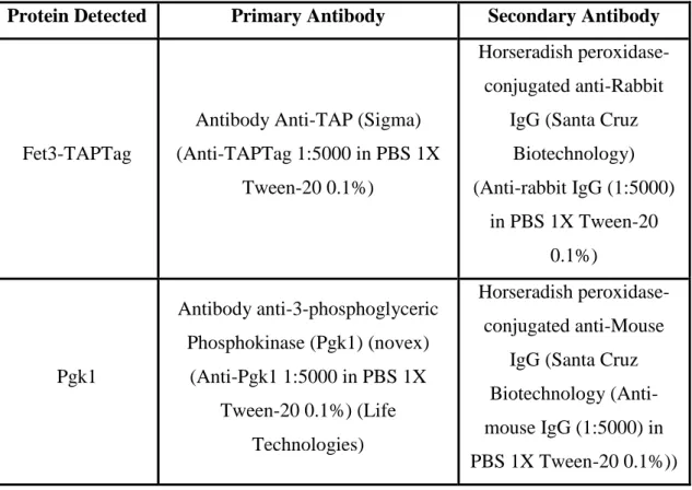
Hybond™ blotting paper. The transfer to the membrane was performed during 50 minutes at 15 V in a in a semi-dry system (ATTA Trans-Blot SD Transfer Cell [BIORAD]). The success of the protein transfer to the nitrocellulose membrane was monitored by membrane incubation with Ponceau S solution (30% (w/v) Ponceau S and 30% (w/v) sulphosalicylic acid) diluted 1:10 in bi-distillated water. The membrane was washed and then incubated with 30mL of a solution of PBS 1X, Tween-20 0.1% with 5% low-fat milk, for 45 minutes at room temperature, to block unspecific interactions with the antibody. Protein levels were detected by incubation with the primary antibody for 1hour at room temperature and with the Horseradish peroxidase-conjugated secondary antibody in the same conditions. The antibodies used in this study are listed in Table 2. Between antibodies’ incubation and after the incubation with the secondary antibody, the membrane was washed 3 times during 10 minutes with PBS 1X Tween-20 0.1% solution. The detection system used was the chemo-luminescent SuperSignal® West Fento Maximum Sensitivity Substrate (Thermo). The membrane was then exposed to an autoradiograph film (Hyperfilm MP, Amersham). 3-PhosphoGlycerate Kinase (Pgk1) was used as loading control. Western-blots were repeated at least twice with different protein extracts.
Table 2 - Antibodies used in this study
Protein Detected Primary Antibody Secondary Antibody
Fet3-TAPTag
Antibody Anti-TAP (Sigma) (Anti-TAPTag 1:5000 in PBS 1X
Tween-20 0.1%)
Horseradish peroxidase-conjugated anti-Rabbit
IgG (Santa Cruz Biotechnology) (Anti-rabbit IgG (1:5000) in PBS 1X Tween-20 0.1%) Pgk1 Antibody anti-3-phosphoglyceric Phosphokinase (Pgk1) (novex) (Anti-Pgk1 1:5000 in PBS 1X Tween-20 0.1%) (Life Technologies) Horseradish peroxidase-conjugated anti-Mouse
IgG (Santa Cruz Biotechnology (Anti-mouse IgG (1:5000) in PBS 1X Tween-20 0.1%))
20
The procedure used for the measurement of β-galactosidase activity was the one described by Möckli et al.[84], with few modifications.
WT(BY4742) and ace1 strains were transformed with pFET3-LacZ plasmid. The transformed cells were grown in SD medium (without uracil) to early exponential phase (OD600 =0.5), and incubated with 0.5 mM of CuSO4 for 30 minutes. Cells were harvested
by centrifugation resuspended in LacZ Buffer (10.6 mg/mL Na2HPO4.2H2O; 5.5 mg/mL
NaH2PO4.H2O; 0.75mg/mL KCl and 0.25 mg/mL MgSO4.7H2O; pH 7, dissolved in H2O)
supplemented with 270 µL β-mercaptoethanol per 100mL LacZ Buffer. Cells were permeabilized by adding 60µL of chloroform and vortexing for 45 seconds. 100 µl of ortho-Nitrophenyl-β-galactoside (ONPG) solution (4mg/mL dissolved in KPO4 Buffer
0.1M (0.1M: 61mL K2HPO4 0.2M; 39 mL KH2PO4 0.2M) sterile water was added to cells
and enzymatic activity was assayed by following the degradation of the colorimetric substrate ONPG at A420m, using the following equation:
𝛽 − 𝐺𝑎𝑙𝑎𝑐𝑡𝑜𝑠𝑖𝑑𝑎𝑠𝑒 𝑎𝑐𝑡𝑖𝑣𝑖𝑡𝑦 = (𝐴𝑏𝑠420−(1.75×𝐴𝑏𝑠550))×1000 𝑡×𝑉×𝑂𝐷600
Where t stands for time in minutes and V for the total reaction volume. Enzyme activity was normalized against optical density (OD600). Absorbance measurements were
21
Triplicates of the indicated strains were grown in 3 mL of SC medium and cultured until reach stationary phase. Cells were then diluted to a final OD600= 0.1 in 40
mL of SC and left untreated or treated with 2mM of CuSO4 for 15 hours. Cells were then
collected by centrifugation at 4000 rpm for 5 minutes. The pellet was washed with 5 mL EDTA 10mM pH8, and twice with miliQ water (Q-POD® MiliQwater – Milipore). The pellet was resuspended with 5mL miliQ water and OD600 was measured. 4mL of the cell
resuspensions were transferred to HS23204N tubes (Heathrow Scientific® LLC) and centrifuged at 4000g for 5 minutes. Supernatant was discarded and 1mL of Nitric Acid 10% (v/v) was added to the cells. The samples were digested for 18h at 80ºC. Samples were centrifuged at 1200rpm for 5 minutes and 2mL of the supernatant were sent to analysis. Samples were analysed by inductively coupled plasma atomic emission spectroscopy (ICP-AES) at the service of Mass Spectrometry of REQUIMTE. Data obtained were normalized against OD600.
22
Biological triplicates of the indicated strains were grown until early exponential phase (OD600 = 0.4 -0.5), induced with 2mM of CuSO4 and harvested (30mL) at the
indicated time points, by centrifugation at 3000 rpm for 2 minutes. Total cellular RNA content was extracted using the phenol-chloroform method. Cell were collected by centrifugation and the pellet was resuspended in 1 mL of cold sterile bi-distillated water, transferred to a microcentrifuge tube and again centrifuged in the same conditions, to remove the supernatant. 350 µL of Acidic phenol: chloroform (1:1) and 350 µL of LETS Buffer (LETS Buffer: 0.1 M LiCl; 0.01M Na2EDTA; 0.01M Tris-HCl pH 7.4; 0.2% SDS,
dissolved in bi-distillated sterile water) was added to the samples. Samples were then vortexed 5 minutes at 4ºC, and centrifugation at 14000rpm to separate the organic from the aqueous fraction. The aqueous fraction was transferred to a microcentrifuge tube, 1 mL of ethanol 100% was added and RNA was precipitated overnight at -20ºC. RNA was collected by centrifugation at 14000 rpm for 15 minutes and the pellet was resuspended in 200µL of ethanol 70% and centrifuged at 14000 rpm for 30 minutes. The pellet was air-dried and then resuspended in 100µL of RNAse free water and incubated 15 minutes at 65ºC to help the solubilisation of the pellet. RNA samples were treated with DNAse (TURBO DNAse [Ambion®]) according to manufacturer’s instructions, at 37ºC during 40minutes. RNA samples were then purified using RNeasy® Mini Kit (QIAGEN). cDNA synthesis was performed using “Transcriptor Reverse Transcriptase” (ROCHE). qRT-PCR reactions were prepared using the oligonucleotides described in table S1. Detection of the qRT-PCR products was done using the DNA intercalating compound SYBR Green (LightCycler LC-Faststart DNa MASTER SYBR Green I, (ROCHE)) and the qRT-PCR reaction was performed in a LightCycler® 480 equipment (ROCHE). Data was analysed with the software LightCycler® Software 4.1. Gene expression was normalized relatively to ACTIN mRNA levels. Two technical replicates were performed for each sample.
24
Gross et al. while identifying the copper regulon in Saccharomyces cerevisiae by DNA microarrays noticed that elevated copper levels also induced the expression of the high affinity iron uptake system composed of Fet3 and Ftr1 [63]. The authors further suggested that copper-induced expression of FET3 and FTR1 arose from an indirect copper effect on cellular iron pools and was mediated by Aft1 [63].
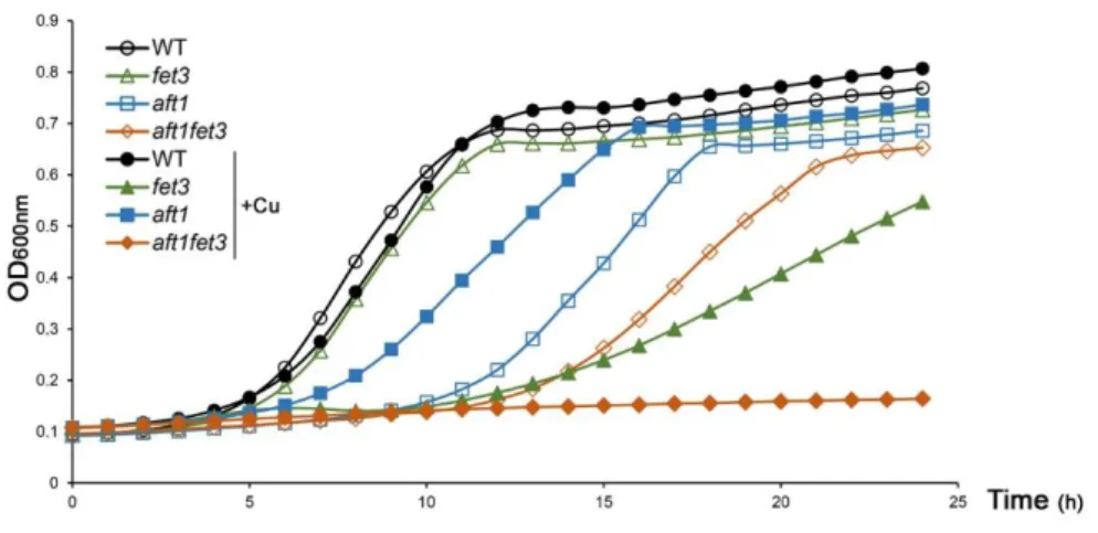
In this work we have examined the growth phenotype of wild-type, (WT) aft1 and fet3 strains in the presence of copper excess (Figure 3.1). Unexpectedly, we found that a strain lacking Fet3 is more sensitive to copper surplus conditions than a strain carrying the AFT1 gene disrupted (Figure 3.1). If Aft1 is the main regulator of FET3 expression under such conditions, one would expect that the aft1 mutant to be more sensitive than, or as sensitive as, the fet3 mutant. This finding suggests that FET3 has another yet unidentified regulator under copper loading conditions. Moreover, an epistatic analysis revealed that Fet3 cannot be downstream of Aft1 as the double mutant aft1fet3 has a more pronounced growth defect in copper surplus compared to fet3 and aft1 single mutants (Figure 3.1).
Figure 3.1 - Under copper overload, Aft1 is not the major regulator of FET3 gene. A:Growth exhibited by the WT, aft1, fet3 and aft1fet3 strains in SC media was recorded over a period of 24 h. Closed symbols and open symbols represent strains grown in media containing or not 2mM of CuSO4,
respectively. Each colour represents one of the used strains: WT is represented in black, aft1 in blue,
25
Aiming at understanding why the fet3 mutant is more sensitive to copper excess than the aft1 strain, we measured, by inductively coupled plasma atomic emission spectroscopy (ICP-AES), the intracellular copper and iron concentrations of both strains in copper overload conditions (Figure 3.2 A and B, respectively). We observed that cells lacking FET3 are iron depleted (Figure 3.2 B) and accumulate over 80% more copper than aft1 or wild-type cells (Figure 3.2 A), which is consistent with fet3 sensitivity to high copper. This finding has been reported by Li et al. [77], but here we show that fet3 excessive accumulation of copper is mediated by Aft1. Indeed, deletion of this transcription factor from the fet3 background, avoids copper accumulation (Figure 3.2A). The activation of Aft1 in the fet3 mutant is probably due to the iron depletion observed in this strain, after copper treatment (Figure 3.2B).
In an attempt to identify the players involved in the above mechanism, we search in the literature for Aft1 targets known to encode membrane proteins and previously associated with copper transport. We found FET4, FRE1 and ARN4 as promising candidates. Fet4 is a cell surface low affinity iron transporter able to transport other divalent metals, including copper [40, 85]. Fre1 is a membrane metalloreductases involved in the uptake of iron and copper [64, 78]. Arn4 is a siderophore-iron transporter, whose substrate is enterobactin [86]. ARN4 expression increases in high copper and its deletion renders cells resistant to copper [87]. To assess the involvement of these proteins in copper accumulation of fet3 mutant, FET4, FRE1 and ARN4 genes were deleted from the fet3 background, and phenotypic growth assays were performed under copper loading conditions (Figure 3.2C). Deletion of each of the gene increase the resistance to copper compared with fet3, with the effect of FRE1 being the most pronounced (Figure 3.2C). None of the deletions, however, was able to fully restore cells growth in high copper. Interestingly we observed that supplementation of the growth medium with zinc or iron attenuates fet3 sensitivity to copper (Figure 3.2 D and E, respectively), which is consistent with the involvement of low-affinity metal transporters in this phenomenon.
26
Figure 3.2 - fet3 copper accumulation is dependent on Aft1. A: Copper (Cu) content of WT, aft1, fet3 and
aft1fet3 strains were determined by ICP-AES, in SC medium after treatment with 2mM CuSO4 for 15 h.
B: Iron (Fe) content of WT, aft1, fet3 and aft1fet3 strains were determined by ICP-AES, in SC medium after treatment with 2mM CuSO4 for 15 h. C: Growth sensitivity of WT, fet3, fe4, arn4, fre1, fet3fet4,
fet3arn4 and fet3fre1 strains in SC plates and SC plates containing 3 or 5mM CuSO4 D: Growth sensitivity
of WT and fet3, in SC plates and SC plates containing 2mM of CuSO4 supplemented or not with 250µM
ZnSO4. E: Growth sensitivity of WT and fet3, in SC plates and SC plates containing 2mM of CuSO4
supplemented or not with 500µM FeSO4.
27
Because the transcriptional response to copper overload is dependent on Ace1 [32, 58], we put forward the hypothesis that Ace1 could play as well a role in regulating the expression of FET3. To test this, we first analysed FET3 promoter sequence using the online tool YEASTRACT [88] and found a putative Ace1 binding-site located at -578 bp from the ATG (+1) (Figure 3.3A). Next, a plasmid including the promoter region of FET3 fused to the lacZ reporter gene (pFET3-lacZ) was generated and used to transform the WT and ace1 mutant strains. We observed that β-Galactosidase activity was higher in the WT compared to the mutant strain (Figure 3.3B). The dependence of FET3 on Ace1 was confirmed by the analysis of the levels of FET3 transcripts in both strains, either in the absence or presence of copper, by qRT-PCR (Figure 3.3C). Under non-stressed conditions (time point 0) both strains exhibit similar levels of FET3 mRNA, which is in agreement with the hypothesis that Ace1 only regulates FET3 under copper overload. To further demonstrate an Aft1-idependent role of Ace1 in FET3 regulation, AFT1 was deleted from the ace1 background and FET3 mRNA levels were followed by qRT-PCR. As depicted in Figure 3.3D, in the absence of Aft1, it was possible to observe a marked induction of FET3 expression that was, however, completely abrogated upon deletion of ACE1.
Finally, we have also assessed the protein expression of a TAP-tagged version of Fet3 driven by its native promoter. To this end, the ACE1 gene was deleted from a strain with a TAP-tagged genomic version of FET3 (WT) and Fet3 expression was monitored by Western blot using an anti-TAPtag antibody. In agreement with the gene expression data, we observed that protein levels were consistently higher in the WT compared to the ace1 mutant strain (Figure 3.3 E).
28
Figure 3.3 - Ace1 regulates FET3 expressionunder copper excess conditions. A: Schematic representation of the FET3 promoter region containing the one putative Ace1-binding site located at -578 upstream of the ATG. B: WT and ace1 mutant strains were transformed with the pFET3-lacZ plasmid. Cells were grown exponentially in SD medium and challenged with 0.5mM CuSO4 for 30 min. β-Galactosidase activity was
assayed as detailed in Materials and Methods. C: WT (wild-type) and ace1 strains were challenged with 2mM CuSO4 and harvested at the indicated time points. The expression of FET3 was monitored by
qRT-PCR. D: aft1 and aft1ace1 strains were challenged with 2mM CuSO4 and harvested at the indicated time
points. The expression of FET3 was monitored by qRT-PCR. E: The WT TAP and the ace1 Fet3-TAP strains were incubated with 2 mM CuSO4 or the indicated times. Fet3 protein levels were analysed by
Western-blot with an anti-TAP antibody. Fet3 protein levels were normalized to Pgk1. Significance of differences was calculated with the t-test (* p<0.05).
29
In this work we show that in the absence of FET3 cells accumulate high amounts of copper in an Aft1 dependent manner (Figure 3.2A), which is deleterious to the cell. We suggest that iron depletion caused by deletion of FET3 leads to the activation of Aft1 that in turn activates genes involved in low-affinity metal uptake. In this context it is reasonable to hypothesize that Ace1 activation of FET3 under copper loading serves to prevent cells from sensing iron depletion, which would activate Aft1 and, consequently, low-affinity iron transporter that would transport copper to the cell.
As a first approach to test this hypothesis, Aft1 activity in WT and ace1 strains was monitored by evaluating the expression of CTH2, a target gene of Aft1 [89], by qRT-PCR (Figure 3.4 A). After copper treatment, the expression of this gene is upregulated in both strains, but a highly pronounced increased in CTH2 mRNA levels (about 9-fold) was observed in ace1 cells 60 minutes after copper exposure, suggesting that Aft1 is more active in this strain (Figure 3.4A). In this sense, the unexpected increase of FET3 mRNA levels in ace1 cells observed after 60 minutes of induction with copper (Figure 3.2 C) may also be mediated by Aft1. Corroborating our hypothesis we found that ace1 cells exposed to a prolonged copper treatment (15 hours) exhibit higher intracellular copper and iron levels compared to WT, as measured by ICP-AES. (Figure 3.4 B and C). As expected, copper and iron contents in the ace1 strain were dependent on Aft1, suggesting a role of Ace1 in preventing Aft1 activation.
Overall these results suggest that Ace1 regulates FET3 gene to prevent Aft1 hyper-activation.
30
Figure 3.4 - Ace1 prevents Aft1 hyper-activation under copper excess conditions. A: WT (wild-type) and ace1 strains were challenged with 2mM CuSO4 and harvested at the indicated time points. The expression
of CTH2 was monitored by qRT-PCR. B: Copper content of WT, aft1, ace1 and ace1aft1 strains, after treatment with 2mM CuSO4 (+Cu) for 15 was determined by ICP-AES. C: Fe content of WT, aft1, ace1
and ace1aft1 strains were determined by ICP-AES, in SC medium or after treatment with 2mM CuSO4
31
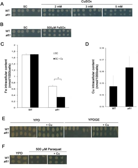
Remarkably, we found that copper alleviates the growth deficiency exhibited by the aft1 mutant under normal growth conditions (Figure 3.1A), being the effect more evident the higher was the concentration of copper tested (Figure 3.5A). As Ace1 regulates FET3 in copper surplus (figure 3.3) and as iron supplementation also attenuates aft1 growth defect (Figure 3.5B), we first reasoned that copper could relieve the iron depletion sensed by the aft1 strain. To test this hypothesis, we measured by ICP-AES the intracellular levels of iron in WT and aft1 strains, after copper treatment. Contrary to our expectation, we observed that the iron content of the aft1 mutant was decreased by approximately 50% in the presence of copper (Figure 3.5C). As Aft1 regulates several genes of copper metabolism we also tested, whether aft1 cells could be copper deficient, before copper treatment. ICP-AES analyses of copper contents led us to rule out this possibility, as intracellular copper levels are similar in both strains (Figure 3.5D).
Because differences in iron and copper levels between aft1 and WT strains could not explain the growth fitness of Aft1 after of copper supplementation, we decided to explore whether cellular processes dependent on copper were affected in the mutant aft1. Copper is an essential player in respiratory metabolism and oxidative stress regulation, as it is a cofactor for cytochrome C oxidase (CcO) and Cu,Zn-Superoxide dismutase (SOD1), respectively [28, 90]. Iron-depleted cells and aft1 mutants are hypersensitive to oxidative stress and defective in respiratory growth [91]. Accordingly, SOD1 is induced in high copper conditions by Ace1 [58] and copper supplementation increases growth fitness when cells are cultured in medium containing a non-fermentable carbon source [36, 92]. As such, we next tested if copper was rescuing aft1 growth defect by promoting the respiratory metabolism and/or strengthening the oxidative stress response.
To examine the respiratory metabolism, the growth of WT and aft1 strains was inspected in medium containing almost exclusively non-fermentable carbon sources (YPDGE), which is commonly used to assess mitochondrial function [82]. We observed that aft1 is sensitive to growth in YPDGE, a finding which is consistent with the knowledge that aft1 strains are defective in mitochondrial function [91], and we found that copper supplementation to the medium rescues growth (Figure 3.5E).
32
Finally, as a first approach to understand whether the oxidative stress response could be enhanced in the aft1 strain upon copper addition, we monitored the growth of WT and aft1 strains in the presence of paraquat and copper, (Figure 3.5F). Paraquat is a redox-cycling drug that produces superoxide anion [93]. We noticed that aft1 is sensitive to the drug, but the growth deficiency is significantly attenuated by copper (Figure 3.5F).
Figure 3.5 - Copper rescues the growth defect exhibited by the aft1 strain. A: Exponentially growing WT and aft1 were serially diluted and spotted onto SC plates and SC plates supplemented with 2, 3 or 5 mM CuSO4. Growth was recorded at 48hours incubation at 30ºC. B: Exponentially growing WT and aft1 were
serially diluted and spotted onto SC plates and SC plates supplemented with 500µM FeSO4. Growth was
recorded at 48hours incubation at 30ºC. C: Fe content of WT and aft1 strains were determined by ICP-AES, in SC medium or after treatment with 2mM CuSO4 (+Cu) for 15 h. D: Copper content of WT and
aft1 strains were determined by ICP-AES, in SC medium. E: Growth sensitivity of WT and aft1 in YPD
and YPDGE plates containing 2mM CuSO4 .(+Cu) F: Growth sensitivity of WT and aft1 in YPD containing
500 µM of Paraquat, supplemented with 2mM CuSO4 (+Cu). Significance of differences was calculated