published on-line: 17/11/2011 www.scionresearch.com/nzjfs
Quercus suber – Phytophthora cinnamomi interaction: a hypothetical
molecular mechanism model
†Ana Cristina Coelho
1,2,*,
Marília Horta
1,3Ghazal Ebadzad
1and Alfredo Cravador
1,3 1 University of Algarve, Penha e Gambelas Campi, 8005-139 Faro, Portugal2 CIQA - Center for Research in Chemistry of Algarve, University of Algarve, Campus de Gambelas, 8005-139 Faro, Portugal 3 IBB/CGB University of Algarve, Portugal
(Received for publication 30 September 2010; accepted in revised form 13 October 2011)
*corresponding author: [email protected]
Abstract
Phytophthora cinnamomi Rands is involved in the decline and mortality of Quercus suber L. and Quercus ilex L. in Southern
Europe, in particular in Portugal and Spain. The presence and spread of P. cinnamomi in these regions is a severe threat to these oak ecosystems leading to expectable severe consequences for the production of cork and acorns in the near future. Molecular mechanisms underlying oomycete-host interactions are poorly understood. As a first step to identify transcripts involved in the Quercus suber – Phytophthora cinnamomi interaction, we applied complementary deoxyribonucleic acid-amplified fragment length polymorphism (cDNA-AFLP) methodology to cork oak seedlings infected with zoospores or mycelium of P. cinnamomi.
Forty-four Quercus suber genes that were differentially expressed when exposed to Phytophthora cinnamomi were selected and sequenced. Several of these genes were fully sequenced and the deduced aminoacid sequences showed consistent homology with proteins involved in the defence mechanism of other plant species. These findings led to the design of a simplified hypothetical model that illustrates the initial events of the interaction between Q. suber and P. cinnamomi.
Keywords: cDNA-AFLP; defense-response pathways; oak/oomycete interaction; pathogenesis related proteins;
Phytophthora cinnamomi; Quercus suber.
† Based on a paper presented at the fifth meeting of the IUFRO working party S07-02-09, Phytophthora Diseases in
Forests and Natural Ecosystems, 7 – 12 March 2010, Auckland and Rotorua, New Zealand.
Introduction
It is broadly documented that the genus Phytophthora accounts for several highly aggressive plant pathogens which have been and continue to be the cause of serious devastation of agricultural crops and native forests worldwide (Erwin & Ribeiro, 1996; Jung, et al., 2000). Phytophthora cinnamomi Rands, specifically,
has a wide host range and causes root rot disease in thousands of plant species (Hardham, 2005). Due to its wide temperature tolerance for growth and survival, it is specifically well adapted to the southern Iberian Peninsula; here it has been consistently associated with the severe decline of oaks that is threatening typical agroforestry ecosystems with an overstorey of cork oak (Quercus suber L.) and holm oak (Quercus ilex
L. subsp. rotundifolia) resulting in significant economic losses (Brasier, et al., 1993; Moreira & Martins, 2005; Sánchez, et al., 2002). In infested sites, trees can suffer in two different ways: a slow deterioration of the crown with a gradual dieback of leaf bearing branches (slow decline) and a sudden mortality. Serra do Caldeirão in the south of Portugal, is one of the regions most affected by the disease where trees showing these contrasting behaviours can be frequently found. The film entitled “The cork oak, Quercus suber L.” http:// w3.ualg.pt/~acoelho/Thecorkoakquercussuber.wmv clearly illustrates the impact of the decline in the Algarve region.
It has been predicted that Phytophthora diseases of trees in Europe could become more severe and might expand to the north and east within the next hundred years (Bergot, et al., 2004; Brasier, 1996; Jung, 2009). Therefore, strategies to control oak decline are urgently needed, and foremost, basic knowledge on the defence mechanisms against infection is required. Oomycetes form a phylogenetically distinct group of eukaryotic microorganisms which includes several of the most devastating pathogens of plants. During infection, plant pathogens secrete numerous peptides and proteins referred to as “effectors” (Schornack et al., 2009). Effectors function at the forefront of the invasion by establishing adhesion, penetration of host tissue, and degradation of cell walls. Moreover, effectors interact with plant cell specialised receptors at the cell wall, plasma membrane or in the cytoplasm thereby reprogramming the host cell to accommodate the needs of the pathogen. The pathogen has to be able to suppress basic defence responses (PAMP triggered immunity) triggered by effectors known as PAMPs (pathogen associated molecular patterns) (Jones & Dangl, 2006). Once these barriers are overcome the pathogen may face another barrier, i.e. the effector triggered immunity (ETI). However, ETI is only triggered when the plant carries R genes (plant resistance genes) that monitor specific effectors (Eitas & Dangl, 2010; Govers & Gijzen, 2006; Tyler, et al., 2006). Molecular mechanisms underlying oomycete-host interactions are poorly understood and knowledge on the Quercus defence mechanisms against biotic stress is scarce. The pathogenic process comprises a sequence of molecular signalling and interaction events. Therefore, our understanding of the Quercus
suber – Phytophthora cinnamomi interaction will
benefit greatly from quantitative measurements, such as gene expression during the infection process. Basic knowledge about the key processes of the defence system provided by this research may contribute to studies aimed at the selection of individuals tolerant to P. cinnamomi.
The present study aimed to identify and characterise
Quercus suber genes implicated in the defence
response against Phytophthora cinnamomi.
Materials and Methods
Methodological approaches
Summary
• In order to reveal the molecular response of
Quercus suber to infection by Phytophthora cinnamomi, the following process was undertaken:
• Complementary deoxyribonucleic acid amplified fragment length polymorphism (cDNA-AFLP) analysis (Money et al., 1996) was performed with messenger ribonucleic acid (mRNA) isolated from
Q. suber roots. The changes in mRNA transcripts
on root cells before and after inoculation with
P. cinnamomi were compared and cDNA
fragments derived from genes showing enhanced expression after fungal infection were isolated and sequenced. The cDNA-AFLP methodology was described in detail by Coelho et al. (2006). • Partial nucleotide sequences of genes coding
for enzymes known to participate in the phenylpropanoid metabolism were established from fragments produced by a polymerase chain reaction (PCR) process using primers of heterologous composition, designed in the most conserved regions of known genes of other species (Coelho, 2004).
• The selected cDNA-AFLP and PCR fragments were used as probes to screen a genomic library of Q. suber prepared in Lambda FIX II vector (Stratagene) and fully characterise these genes. Simultaneously, the full length sequences of the genes were established from the corresponding cDNAs produced by RACE-PCR 3’/5’ (rapid amplification of cDNA ends by PCR) (GeneRacer Kit, Invirogen). • Based on the sequence information, the genes were classified into families as described for other species. Deoxyribonucleic acid and protein similarity (basic local alignment search tool [BLAST]) searches were performed against nucleotide and aminoacid sequence databases at the National Center for Biotechnology Information (NCBI: http://www.ncbi.nlm.nih.gov).
• The genes were classified as genes coding to a cinnamyl alcohol dehydrogenase 1 (QsCAD1); a cinnamyl alcohol dehydrogenase 2 (QsCAD2); a resistance protein against P. cinnamomi (QsRPc); a protein disulphide isomerase (QsPDI); a RelA/ SpoT-like protein (QsRSH); a phenylalanine ammonia-lyase (QsPAL) and peroxidases (QsPOX), and
• •
• Finally the expression of the mRNA of these genes in roots of Q. suber seedlings infected with P. cinnamomi was assessed by Reverse transcription (RT) PCR and hybridisation with specific probes. The experimental procedures are described for one of the studied genes in Coelho et al. (2006).
Biological samples
In our biological system, approximately twenty roots of Quercus suber, with 8 – 10 cm, germinated from seeds, were put in direct contact with Phytophthora
cinnamomi mycelium mats enriched in sporangia
and zoospores. Roots in contact with mycelium mats were placed at 25 oC, in dark, for 24 h. The infected roots were then cut and stored at -80 oC. The infection process started when the motile zoospores released from the mycelium encyst in the root surface, forming walled cysts that germinate and penetrate the plant.
Phytophthora cinnamomi mycelium was routinely
maintained in the dark at 24 oC on semi-solid complete 10% V8 agar. To obtain zoospores, five P. cinnamomi agar plugs were tacked from the edge of the actively growing colony and placed onto a Miracloth disc (Calbiochem), on a fresh 10% V8 Agar plate. The following procedures performed to produce mycelium mats with sporangia and zoospores were described by Robinson and Cahill (2003).
cDNA-AFLP analysis
Total RNA was extracted from 50 mg of Quercus
suber roots infected with Phytophthora cinnamomi
and from non-infected roots with the RNeasy Kit from Qiagen, according to the instructions supplied by the manufacturer. Total RNA was then treated with DNase I (1 U/µL; Gibco), in the presence of 2 µL of RNaseout (40 U/µL; Gibco) (Sambrook & Russell, 2001). The reaction occurred in a final volume of 100 µL, for 30 min at 37 oC. After DNA digestion, the total RNA was purified with the RNeasy Kit. The quality and the quantity of total RNA present in the samples were evaluated by ultra-violet (UV) spectrophotometry and by electrophoresis in denaturing agarose gel. To perform cDNA-AFLP analysis, the synthesis of cDNA was accomplished with the cDNA synthesis system (Roche), with modifications to the original protocol. Synthesis of the first cDNA strand was initiated with the degenerate primer [5’AGTGAATTCT12V3’ (V = A; C; G)], comprised of an equimolar mixture of the three oligonucleotides (Money et al., 1996). Synthesis of the second cDNA strand and digestion of residual RNA were performed according to the kit protocol (Gubler, 1988; Gubler & Hoffmann, 1983). Samples of cDNA (20 µL) were then subjected to the standard AFLP template production (Vos et al., 1995). The cDNA-AFLP procedure was described in detail by Coelho et al. (2006).
Labelled selective amplification products were separated on standard 6% polyacrylamide sequencing gels. After electrophoresis, the gel was dried on filter paper (3MM paper; Whatman) and exposed to X-ray film for 30 h. The cDNA fragments were visualised by autoradiography, after positional marking the gel and the film.
The cDNA-AFLP profiles of Quercus suber roots infected with Phytophthora cinnamomi were compared with the profiles of the non-infected roots. The gene fragments to be excised from the gel were selected based on the increasing of the intensity of the band, present simultaneously in the infected root and in the non-infected root and on the gene fragments only present in the infected roots. A piece of the dried gel, containing the band of interest, was cut out and soaked in 40 µL of H2O for 10 min on ice. The sample was then heated for 15 min at 95 oC and cooled again on ice. After a brief centrifugation (30 s; 13 000 rpm), 5 µL of the supernatant were transferred to another tube. Re-amplification of the recovered fragment was performed under the same conditions used for cDNA-AFLP (Coelho et al., 2006). The re-amplified PCR product was run on a 2% agarose gel, cut out and purified with the Qiaquick PCR Purification Kit (Qiagen) and finally cloned into the pCRII Topo vector, with the TA Cloning Kit (Invitrogen). Manufacturer’s instructions for these kits were followed throughout. The fragments were sequenced with an Applied Biosystems PRISM Ready Reaction DyeDeoxy Terminator cycle sequencing kit and an automated sequencer.
PCR using primers of heterologous composition The nucleotide sequences (ORF-open reading frames) of genes codifying to phenylalanine ammonia-lyases, to cinnamyl alcohol dehydrogenases and peroxidases, from six different plant species, belonging to Arabidopsis, Populus, Eucalyptus,
Pinus, Solanum and Lycopersicum genera, published
in GenBank (NCBI: http://www.ncbi.nlm.nih.gov), were submitted to multiple sequence analysis with CLUSTAL W 1.8 software to find the most conserved regions of these genes. Several pairs of primers of heterologous composition were chosen and tested in PCR reactions for the ability of amplifying fragments of the aforementioned genes in Quercus suber. Details of the composition of the primers and genes used as reference are shown in Table 1. Polymerase chain reactions were performed in a total volume of 25 µL, 120 ng of genomic DNA, 2 µL of primers (10 μM) and 12.5 µL of PCR Master Mix from Promega. Polymerase chain reaction conditions were as follows: 1 min at 94 oC; (30 s at 94 oC; 30 s at 46 oC; 1 min at 72 oC) x 30; 5 min at 72 oC. Genomic DNA was extracted from healthy roots of Q. suber germinated seedlings with DNeazy Kit from Qiagen. The amplified fragments were then cloned into the pCRII Topo vector, with the TA Cloning Kit (Invitrogen) and sequenced.
The sequences of the cDNA-AFLP fragments and the sequences of the PCR fragments amplified with primers of heterologous composition were analysed against NCBI database to search for homology with sequences of genes from other plant species involved in defence systems. These blast searches were performed with Vector NTI 6 bioinformatic software. Construction and screening of genomic DNA library of Quercus suber
Then the cDNA-AFLP and PCR fragments from
Quercus suber showing homology with genes involved
in defence systems in other plant species were selected and used as probes to screen a genomic library of
Q. suber prepared in Lambda FIX II vector (Stratagene)
and fully characterise these genes.
The construction and screening of the genomic library of Quercus suber included several sequential steps like: extraction of genomic DNA (DNeasy Plant Maxi Kit; Qiagen); partial digestion of eukaryotic DNA with Sau3AI restriction enzyme (Biolabs); removal of small fragments of nucleic acid from preparation of genomic DNA by centrifugation through sucrose density gradient; ligation of bacteriophage λ (λ-FIX II; Stratagene) arms to fragments of Q. suber genomic DNA; in vitro packaging reactions; estimation of the total number of recombinant plaques generated and calculation of the depth to which the library covers the size of the target genome; amplification of the genomic library; transfer of bacteriophage DNA from plaques to filters followed by screening to identify the positive clones by DIG nonradioactive hybridisation.
The aforementioned probes used to screen the genomic library were the products of amplification reactions with primers chosen in the cDNA-AFLP and PCR with heterologous primers sequenced fragments labelled with digoxigenin. Hybridisation of digoxigenin-labelled probes, with the target DNA, was achieved at 42 oC in the presence of DIG Easy Hybridisation buffer (Roche). Digoxigenin-labelled nucleic acid hybrids were detected with a Dig Luminescent Detection Kit (Roche).
The experimental procedures for the construction and screening of the genomic library of Quercus suber are bibliographically supported by: Stratagene manual for construction of genomic libraries in Lambda FIX II vector; DIG Application Manual for filter hybridisation from Roche Molecular Biochemicals; and Sambrook and Russell (2001).
RT-PCR and hybridisation
Total RNA (2 µg) was extracted from noninfected roots of cork oak seedlings, and from infected roots
with Phytophthora cinnamomi. Complementary
deoxyribonucleic acid was synthesised with Superscript II RNase H-reverse transcriptase, in a total volume of 40 µL. Two microliters of the cDNA synthesis solution were diluted 50, 100 and 200 times, and 10 µL of these dilutions were used as cDNA template for amplification reactions. Fragments from QsCAD1; QsRPc; QsPDI;
QsPOX; QsCAD2; QsPAL and QsRSH transcripts
were amplified with specific primers. PCR products were separated on 1% agarose gels and blotted to membrane (Hybond-N+) on a Trans-Blot SD semi-dry electrophoretic transfer cell. The specificity of the Quercus suber
Target Gene Nucleotide Sequence Expected Size of the Amplified
Fragment (bp)
NCBI Identification Number of Genes from other Plant Species used as References QsPAL Forward: 5’CAACACYCTSCTYCAAGG3’Reverse:5’CCATCYAAAATRTGYTCCAT3’
Y(T7C); S(G/C); R(G/A) 488
M83314-Lycopersicon D43802-Populus L11747-Populus AF167487-Eucalyptus
QsCAD2 Forward: 5’AYTAYCCWATGGTYCCTGG3’Reverse:5’GGRATTYTCACCACAAAC3’
Y(T7C); R(G/A); W(A/T) 699
Z31715-Arabidopsis AF060491-Pinus AJ295837-Populus AF217957-Populus AF294793-Eucalyptus
QsPOX Forward: 5’GTGCRCACACRKTWGG3’Reverse:5’TYYCCCATYTTKATCAT3’
Y(T7C); R(G/A); W(A/T); K(G/T) 334
X99952-Arabidopsis D11102-Populus X15853-Lycopersicon X15854-Lycopersicon TABLE 1: Characteristics of the primers with heterologous composition.
amplified products was confirmed by hybridisation. The DNA fragments obtained for each one of the referenced genes with number of base pairs ranging from 588 to 1837 bp were labelled with Digoxigenin and used as probes in the hybridisation reaction. Hybridisation of digoxigenin-labelled probes, with the target DNA, was achieved at 42 oC in the presence of DIG Easy Hybridisation buffer (Roche). Digoxigenin-labelled nucleic acid hybrids were detected with a Dig Luminescent Detection Kit (Roche).
Bioinformatics and gene characterisation
The characterisation of the genes was performed using the following bioinformatic software:
• Vector NTI 6, InforMax, Inc;
• GCG (Genetics Computer Group, University of Wisconsin, Madison, 1981);
• EMBL-EBI (European Bioinformatics Institute, www.ebi.ac.uk);
• Coilscan (GCG);
• PSORT (Klein et al., 1985);
• RADAR (http://www.ebi.ac.uk/radar); • PLACE (Higo et al., 1999);
• GeneMark, version 2.2a (Borodovsky & McIninch, 1993; http://dixie.biology.gatech. edu); and
• PROF (http://cubic.bioc.columbia.edu/ predictprotein).
Results and Discussion
Identification and characterisation of Quercus
suber defence genes
Seven Quercus suber genes coding for:
1. a cinnamyl alcohol dehydrogenase 1 (QsCAD1); 2. a protein disulphide isomerase (QsPDI);
3. a RelA/SpoT protein (QsRSH); 4. a NB-LRR resistance protein (QsRPc); 5. a cationic peroxidase (QsPOX1);
6. a cinnamyl alcohol dehydrogenase 2 (QsCAD2); and
7. a phenylalanine ammonia-lyase (QsPAL);
were cloned and characterised. At least four of these genes are potentially involved in the response to infection by Phytophthora cinnamomi, as it was shown that the expression of QsCAD1, QsRPc, QsPDI and
QsPOX1 increased in the first 24 hours of infection.
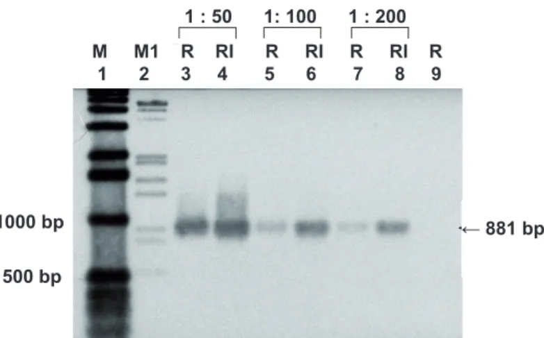
The results were not conclusive for the other genes. Figure 1 illustrates the increased expression of QsPDI gene in roots of Q. suber infected with P. cinnamomi analysed by RT-PCR and hybridisation, as example of the expression pattern observed to the mentioned genes.
The characterisation of the seven genes was performed using bioinformatic software; the results of the theoretical analysis are summarised in Table 2.
FIGURE 1: Expression of QsPDI gene in non-infected (R) roots of Quercus suber and in roots exposed to Phytophthora cinnamomi (RI) after 24 h of interaction, analysed by RT-PCR and hybridisation.
Lanes 1 and 2: Molecular markers;
Lanes 3 – 8: cDNA synthesis solutions were diluted 50, 100 and 200 times as indicated and 10 uL of these dilutions were used as cDNA template for amplification reactions; Lane 9: actin cDNA, used as negative control to evaluate the efficiency of PCR and absence of contamination. ← 881 bp 1000 bp 500 bp 1 : 50 1: 100 1 : 200 M M1 R RI R RI R RI R 1 2 3 4 5 6 7 8 9
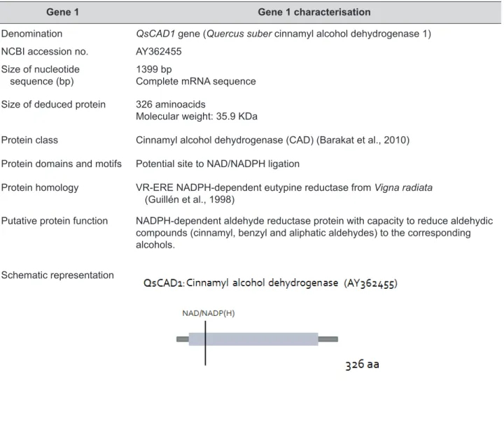
TABLE 2: Identification and characterisation of Quercus suber defence genes. Empirical data characterising the genes and deduced
proteins include: the type and size of nucleotide sequence (mRNA or DNA or both); size and molecular weight of the proteins deduced from the nucleotide sequences; the protein class and the motifs or domains justifying their classification; proteins from other plant species with which these proteins share the highest degree of homology, potential cellular function and the protein schematic representation. This is also shown in Table 2.
Quercus suber– Phytophthora cinnamomi
interaction: hypothetical molecular mechanism model
The disclosure of Quercus suber genes involved in defence response to infection by Phytophthora
cinnamomi contributes to a better understanding of
the molecular mechanisms underlying the interaction. Based on the various functions assigned to proteins deduced from these genes (Table 2), it is possible to develop a range of theoretical scenarios representing
the initial events of the interaction between Q. suber and P. cinnamomi and illustrating the molecular pathways during the defence response to infection. The particular theoretical model presented in Figure 2 was designed to allow a simple condensed view of the action of proteins in response to cellular stimuli or effector molecules secreted by the oomycete. It also illustrates how these proteins could interact with each other and how to trigger the cellular defence pathways with activation of genes related to pathogenesis. This model involves all seven genes although the expression of only four of them has been proven to increased in the first 24 hours after infection. In this model, it is postulated that QsCAD1 reduces aldehydic effector compounds to the corresponding alcohols, while QsRPc will recognise and interact with effector molecules secreted by Phytophthora
cinnamomi and activate downstream signalling
responses.
Gene 1 Gene 1 characterisation
Denomination QsCAD1 gene (Quercus suber cinnamyl alcohol dehydrogenase 1)
NCBI accession no. AY362455
Size of nucleotide
sequence (bp) 1399 bp Complete mRNA sequence
Size of deduced protein 326 aminoacids
Molecular weight: 35.9 KDa
Protein class Cinnamyl alcohol dehydrogenase (CAD) (Barakat et al., 2010)
Protein domains and motifs Potential site to NAD/NADPH ligation
Protein homology VR-ERE NADPH-dependent eutypine reductase from Vigna radiata
(Guillén et al., 1998)
Putative protein function NADPH-dependent aldehyde reductase protein with capacity to reduce aldehydic
compounds (cinnamyl, benzyl and aliphatic aldehydes) to the corresponding alcohols.
TABLE 2 cont.: Identification and characterisation of Quercus suber defence genes.
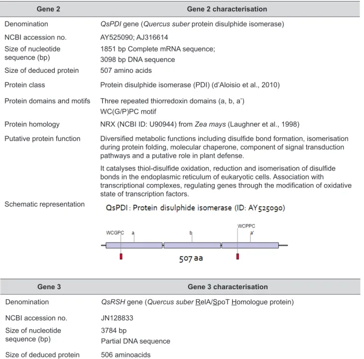
Gene 2 Gene 2 characterisation
Denomination QsPDI gene (Quercus suber protein disulphide isomerase)
NCBI accession no. AY525090; AJ316614
Size of nucleotide
sequence (bp) 1851 bp Complete mRNA sequence; 3098 bp DNA sequence
Size of deduced protein 507 amino acids
Protein class Protein disulphide isomerase (PDI) (d’Aloisio et al., 2010)
Protein domains and motifs Three repeated thiorredoxin domains (a, b, a’) WC(G/P)PC motif
Protein homology NRX (NCBI ID: U90944) from Zea mays (Laughner et al., 1998)
Putative protein function Diversified metabolic functions including disulfide bond formation, isomerisation during protein folding, molecular chaperone, component of signal transduction pathways and a putative role in plant defense.
It catalyses thiol-disulfide oxidation, reduction and isomerisation of disulfide bonds in the endoplasmic reticulum of eukaryotic cells. Association with
transcriptional complexes, regulating genes through the modification of oxidative state of transcription factors.
Schematic representation
Gene 3 Gene 3 characterisation
Denomination QsRSH gene (Quercus suber RelA/SpoT Homologue protein)
NCBI accession no. JN128833
Size of nucleotide
sequence (bp) 3784 bpPartial DNA sequence
Size of deduced protein 506 aminoacids
Protein class RSH: RelA/SpoT-like proteins (Kim et al., 2009)
Protein domains and motifs Synthethase domain of (p)ppGpp (guanosine tetra and pentaphosphate)
Protein homology AtRSH2 and AtRSH3 from Arabidopsis thaliana (Van der Biezen et al., 2000)
Putative protein function Synthesis and degradation of guanosine tetra and pentaphosphate
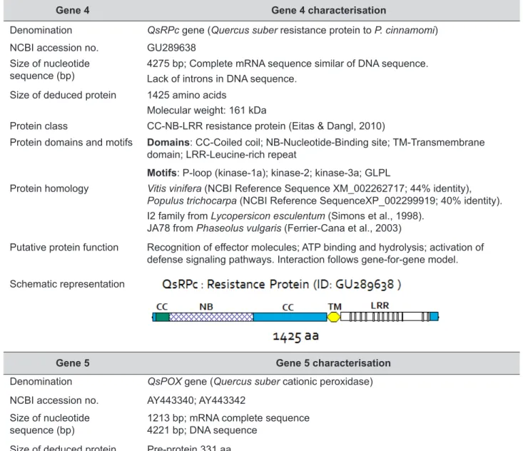
Gene 4 Gene 4 characterisation
Denomination QsRPc gene (Quercus suber resistance protein to P. cinnamomi)
NCBI accession no. GU289638
Size of nucleotide
sequence (bp) 4275 bp; Complete mRNA sequence similar of DNA sequence.Lack of introns in DNA sequence.
Size of deduced protein 1425 amino acids
Molecular weight: 161 kDa
Protein class CC-NB-LRR resistance protein (Eitas & Dangl, 2010)
Protein domains and motifs Domains: CC-Coiled coil; NB-Nucleotide-Binding site; TM-Transmembrane
domain; LRR-Leucine-rich repeat
Motifs: P-loop (kinase-1a); kinase-2; kinase-3a; GLPL
Protein homology Vitis vinifera (NCBI Reference Sequence XM_002262717; 44% identity),
Populus trichocarpa (NCBI Reference SequenceXP_002299919; 40% identity).
I2 family from Lycopersicon esculentum (Simons et al., 1998). JA78 from Phaseolus vulgaris (Ferrier-Cana et al., 2003)
Putative protein function Recognition of effector molecules; ATP binding and hydrolysis; activation of defense signaling pathways. Interaction follows gene-for-gene model. Schematic representation
TABLE 2 cont.: Identification and characterisation of Quercus suber defence genes.
Gene 5 Gene 5 characterisation
Denomination QsPOX gene (Quercus suber cationic peroxidase)
NCBI accession no. AY443340; AY443342
Size of nucleotide
sequence (bp) 1213 bp; mRNA complete sequence 4221 bp; DNA sequence
Size of deduced protein Pre-protein 331 aa
Protein 300 aa; MW: 32.8 KDa; Isoelectric point: 9.55
Protein class Class III Cationic peroxidase (Elfstrand et al., 2002; Faurie et al., 2009)
Protein domains and motifs Motifs: GASLLRLHFHDC and DMVALSGAHTV
Proximal and distal histidines
Protein homology Plant class III proteins (POX, EC 1.11.1.7)
1SCH A from Arachis hypogeal (Buffard et al., 1990)
Putative protein function Catalysis of the oxidation of a diverse group of organic compounds using hydrogen peroxide as the ultimate electron acceptor. Involved in various metabolic steps such as the oxidation of cinnamyl alcohols prior to their polymerisation during lignin and suberin formation.
Gene 6 Gene 6 characterisation
Denomination QsCAD2 gene (Quercus suber cinnamyl alcohol dehydrogenase 2)
NCBI accession no. JN132134; JN132135
Size of nucleotide sequence (bp) 760 bp; mRNA partial sequence
Size of deduced protein 289 aa
Protein class Cinnamyl alcohol dehydrogenase (CAD) (Saballos et al., 2009)
Protein domains and motifs Catalytic domain for binding of zinc ion (GHEVVGEVVEVGSDV)
Binding domain for NADP cofactor (GLGGVG)
Protein homology EgCAD2 from Eucalyptus gunnii (Grima-Pettenati et al., 1993)
Putative protein function Implicated in the synthesis of lignin and/or suberin.
Responsible for the NADPH-dependent reduction of cinnamyl aldehydes (p-coumaryl, coniferyl and sinapyl) to the corresponding alcohols. Schematic representation
TABLE 2 cont.: Identification and characterisation of Quercus suber defence genes.
Gene 7 Gene 7 characterisation
Denomination QsPAL gene (Quercus suber phenylalanine ammonia-lyase)
NCBI accession no. AY443341
Size of nucleotide sequence (bp) 2499 bp; Complete mRNA sequence
Size of deduced protein 710 aa
Protein class Phenylalanine ammonia-lyase (PAL) (Milcevicova et al., 2010;
Chandra et al., 2007)
Protein domains and motifs
-Protein homology PAL from Populus balsamifera subsp. Trichocarpa x Populus deltoids
(ID: AAQ74878)
Putative protein function Catalysis of the first step in the metabolism of phenylpropanoids, the
deamination of l-phenylalanine to transform into trans-cinnamic acid.
The interaction of the effector molecules with QsRPc protein induces the activation of their ATP binding site (NB) and enables the interaction with proteins implicated in the regulation of transcription factors associated with the coiled coil domain (CC).
It is assumed that QsRSH is one of the proteins associated with QsRPc that is activated after recognition of the effector molecules by the resistance protein. In response to biotic stress the level of the signalling molecule (p)ppGpp in plant is regulated by the protein QsRSH. As in prokaryotes, it is expected that (p)ppGpp is going to mediate cellular processes like protein synthesis, regulation of defence genes and transcriptional reprograming. Furthermore, the protein disulphide isomerase (QsPDI) activates by oxidation or reduction and/or isomerisation of cysteine residues, transcription factors and other proteins involved in the cellular response to the oxidative burst that occurs after pathogen invasion. The proteins QsCAD2, QsPAL and QsPOX have their functions associated to lignin and suberin biosynthesis, two poly (phenolic) heteropolymers who play a central role in plant structure and defense against pathogens. Lignin and suberin act as a physical barrier blocking the entry of the pathogen.
We are not aware of any theoretical model depicting the molecular events associated with the interaction between Quercus suber and Phytophthora cinnamomi. However, the model is based on the concepts depicted for other types of interactions between plants and parasites described by several authors (Birch et al., 2009; Dodds et al., 2009; Ellis et al., 2009; Torto-Alalibo et al., 2009). Among these concepts, it emphasises the concept of effector molecule and the concept of host plant with or without cognate R protein and also, the concept of translocation of effector proteins into host cells (Hogenhout et al., 2009). The hypothetical model created for the events observed by Lebrun-Garcia et al. (1999) and Blein (2002) on tobacco cells challenged with the effector molecule cryptogein was also used as reference.
Research in this field is hampered by the lack of information on genome sequences of the involved host and pathogen species and by the difficulty to genetically transform them by conventional methods reported for other plant species and oomycetes. The hypotheses presented in this model related
to the Quercus suber – Phytophthora cinnamomi
pathosystem offers new opportunities to research in order of understanding how this oomycete is able to manipulate host cells and to establish a parasitic relationship with the host. The model anticipates, in a small-scale, the knowledge that will result from sequencing and annotation of the genomes of Q. suber and P. cinnamomi which will create the opportunity to look for typical motifs present in oomycete effectors
and to find the corresponding host proteins targeted by the effectors.
In fact, the infection of Quercus suber by Phytophthora
cinnamomi has a mixed profile of compatible and
incompatible interaction. The pathogen induces in the host the expression of genes involved in both the deactivation of virulent effector molecules (toxins) and the defence response after recognition of effector factors.
The expression of genes involved in compatible and incompatible interactions is coincident with the contrasting phenotypic behavior exhibit by cork oak trees in areas affected by Phytophthora cinnamomi. Some cork oak trees exhibit a range of symptoms and a variable rate of disease expression demonstrative of the profile of an incompatible interaction and of the activation of the defense system. Other cork oak trees die suddenly being the process characterised by a quick drying of the leaves and death of the tree, within a few months, demonstrative of the profile of a compatible interaction (Coelho et al., 2006).
Definition of compatible and incompatible interaction profile follows the traditional models for the interaction between effectors and effector targets in susceptible plants or in resistant ones (Hogenhout et al., 2009; Torto-Alalibo et al., 2009).
Therefore, we propose that during infection
Phytophthora cinnamomi produces effector molecules,
which can be either proteins or smaller molecules. For instance, as shown in Figure 2, aldehyde aromatic compounds, similar to the eutypine toxin, cross the plasmatic membrane and are reduced to alcohols by the QsCAD1 protein (Quercus suber cinnamyl alcohol dehydrogenase 1; Coelho et al., 2006). Simultaneously, the domain LRR of the resistance protein (QsRPc), located in the apoplasm, recognises other effector molecules (proteins) secreted by the oomycete. This interaction is transmitted to the intracellular NB domain where ATP binding and/or hydrolysis occur and activates self proteins that interact with this domain and CC domain (Figure 2). Recognition of the effector results in an influx of calcium into the cell promoting the activation of both pathogenesis related proteins and proteins associated with the plasma membrane (chloride ion channel; NADPH oxidase). It can also contribute to the activation of potassium channel and calcium dependent kinases in cascade (Figure 2). Faurie et al. (2009) showed in grapevine cell cultures that calcium, phosphorylation and active oxygen species are implicated in methyl jasmonate-induced defence responses.
Proteins belonging to the RSH protein family, represented in the model by QsRSH protein, can interact with the NB domain of the resistance protein as it was observed with RPP5 in Arabidopsis (Van
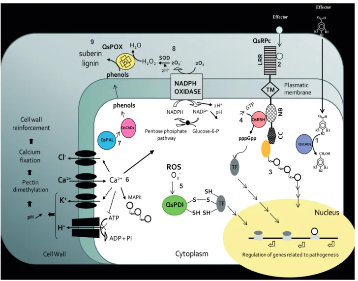
FIGURE 2: Hypothetical molecular mechanism model. Cellular events:
(1) Deactivation of virulence factors secreted by the pathogen performed by the QsCAD1 (Quercus suber cinnamyl alcohol dehydrogenase 1) protein. QsCAD1 reduces aldehydic effector compounds to the corresponding alcohols.
(2)Interaction between effectors molecules and the domain LRR of the QsRPc protein (Q. suber resistance protein to Phytophthora cinnamomi) and signal transduction to the intracellular domains NB and CC.
(3) Activation of proteins and TF (represented by white circles) that interact with the CC and NB domains of QsRPc protein, triggered by conformational changes resulting from the hydrolysis of ATP into ADP or GTP.
(4) Synthesis of (p)ppGpp catalysed by the QsRSH (Q. suber RelA/SpoT homologue) protein and activation of TF involved in the regulation of genes in the nucleus related to pathogenesis.
(5) Activation of transcription factors and other proteins, by oxidation or reduction and/or isomerization of cysteine residues, catalysed by QsPDI protein (Q. suber disulfide isomerase), in response to the production of ROS in the cell.
(6) Cascade of events which include protein phosphorylation, calcium influx (Ca2+ →), potassium and chloride effluxes (K+ and Cl- ←),
inhibition of the plasma membrane H+ -ATPase (ATP→X ADP+PI), activation of a NADPH oxidase responsible for ROS production
and cytosol acidification (pH↓), activation of the pentose phosphate pathway with consumption of Glucose-6-P and activation of mitogen-activated protein kinase (MAPK, represented by white circles) homologues. These events must occur after the recognition of effector molecules by the protein QsRPc as was observed in tobacco plants challenged with cryptogein (Lebrun-Gracia et al., 1999). (7) Activation of protein QsCAD2 (Q. suber cinnamyl alcohol dehydrogenase2) and QsPAL (Q. suber phenylalanine ammonia lyase) involved in the synthesis of phenolic compounds, precursors of lignin and suberin, needed to strengthen the cell walls. (8) Transformation of O2- produced by NADPH oxidase into H
2O2 . The reaction is catalysed by the extracellular SOD.
(9) Polymerisation of phenols in the matrix of lignin and suberin catalysed by the peroxidase QsPOX in a process dependent on H2O2.
Abbreviations: ADP (Adenosine diphosphate); ATP (Adenosine triphosphate); CC (coiled coil domain); Glucose-6-P (Glucose-6-phosphate); GDP (Guanosine diphosphate); H2O2 (hydrogen peroxide); LRR (Leucine-rich repeat); MAPK (mitogen-activated protein kinases); NADPH (nicotinamide adenine dinucleotide phosphate); NB (Nucleotide binding site); PI (inorganic phosphate); (p)ppGpp (guanosine tetra and pentaphosphate); ROS (Reactive oxygen species); SOD (superoxide dismutase); TF (transcription factor); and TM (transmembrane domain). 1 3 8 2 9 5 7 6 4
der Biezen et al., 2000). The result of this interaction is the synthesis of guanosine pentaphosphate [(p) ppGpp] that functions as a cofactor for the activation of the transcription factors (TF, Figure 2) involved in the regulation of genes related to pathogenesis.
During the interaction the production of reactive oxygen species alters the redox state of the cell which is recognised by the protein QsPDI that activates the transcription factors (TF, Figure 2) by isomerisation of cysteine residues.
It is known that the defence system in plants has multiple forms; one of these forms is the activation, during infection, of the transcription of genes involved in the synthesis of phenolic compounds as precursors for suberin and lignin production (Chandra et al., 2007; Hammond-Kosack & Jones 1997; Milcevicova et al., 2010). Enzymes like peroxidases, cinnamyl alcohol dehydrogenases and phenylalanine ammonia-lyases participate in the phenylpropanoid metabolism. The metabolic pathways for the production of salicylic acid and phenolic compounds are dependent on the enzymes QsPAL and QsCAD2 through the phenylpropanoids metabolism (Figure 2). The oxidative polymerisation of phenols in the plasma membrane is catalysed by peroxidases (QsPOX) with the production of lignin and suberin to reinforce the cells wall and prevent colonisation by the oomycete (Figure 2).
Conclusions
The outcome of this study was the identification of seven genes that encode: a cinnamyl alcohol dehydrogenase 1 (QsCAD1); a protein disulphide isomerase (QsPDI); a RelA/SpoT protein (QsRSH); a NB-LRR resistance protein (QsRPc); two cationic peroxidases (QsPOX1 and QsPOX2); a cinnamyl alcohol dehydrogenase 2 (QsCAD2) and a phenylalanine ammonia-lyase (QsPAL).
The consistent homology found between the aminoacid sequences deduced from DNA or cDNA of all the genes identified in Quercus suber, and those from proteins involved in the defence response in other plants allowed the conception of a hypothetical model that illustrates the initial events of the interaction between Q. suber and Phytophthora cinnamomi and the molecular pathways that may be followed during the defence response to infection. The products of some of these genes will probably interact with effector molecules produced by P. cinnamomi during the first 24 hours of infection.
Acknowledgements
G. Ebadzad would like to acknowledge to the Programme Erasmus Mundus EM8 (Iran, Iraq, Yemen) for her PhD fellowship.
M. Horta thanks “Fundação para a Ciência e Tecnologia” for her grant SFRH/BPD/63213/2009. This research was in part financed by Portuguese Ministério da Ciência e do Ensino Superior (MCES) “Fundação para a Ciência e Tecnologia (FCT), Project PTDC/AGR-AAM/67628/2006”.
References
Brasier, C. M., (1996). Phytophthora cinnamomi and oak decline in southern Europe. Environmental constraints including climate change. Annales
des Sciences Forestières, 53, 347-358.
Barakat, A., Bagniewska-Zadworna, A., Frost, C. J., & Carlson, J. E. (2010). Phylogeny and expression profiling of CAD and CAD-like genes in hybrid Populus (P. deltoides × P.
nigra): evidence from herbivore damage
for subfunctionalization and functional divergence. BMC Plant Biology, 10, 100. Bergot, M., Cloppet, E., Pérarnaud, V., Déqué, M.,
Marçais, B., & Desprez-Loustau, M.-L. (2004). Simulation of potential range expansion of oak disease caused by Phytophthora cinnamomi under climate change. Global Change
Biology, 10, 1539-1552.
Birch, P. R. J., Armstrong, M., Bos, J., Boevink, P., Gilroy, E.M., Taylor, R.M., Wawra, S., Pritchard, L., Conti, L., Ewan, R., Whisson, S.C., van West, P., Sadanandom, A., & Kamoun, S. (2009). Towards understanding the virulence functions of RXLR effectors of the oomycete plant pathogen Phytophthora
infestans. Journal of Experimental Botany, 60(4), 1133-1140.
Blein, J. P. (2002). Involvement of lipid-protein complexes in plant-microorganism inter-actions. OCL-Oliagineux Corps Gras Lipids,
9(1), 31-36.
Borodovsky, M., & McIninch, J. (1993). GeneMark: parallel gene recognition for both DNA strands. Computers & Chemistry, 17(19), 123-133.
Brasier, C. M., Robredo, F., & Ferraz, J. F. P.(1993). Evidence for Phytophthora cinnamomi involvement in Iberian oak decline. Plant
Buffard, D., Breda, C., van Huystee, R. B., Asemota, O., Pierre, M., Dang Ha, D. B., & Esnault, R. (1990). Molecular cloning of complementary DNAs encoding two cationic peroxidases from cultivated peanut cells. Proceedings of
the National Academy of Sciences USA, 87,
8874-8878.
Chandra, A., Saxena, R., Dubey, A., & Saxena, P. (2007). Change in phenylalanine ammonia lyase activity and isozyme patterns of polyphenol oxidase and peroxidase by salicylic acid leading to enhance resistance in cowpea against Rhizoctonia solani. Acta
Physiologiae Plantarum, 29, 361-367.
Coelho, A. C., Horta, M., Neves, D., & Cravador, A. (2006). Involvement of a cinnamyl alcohol dehydrogenase of Quercus suber in the defence response to infection by Phytophthora
cinnamomi. Physiological and Molecular Plant Pathology, 69(1-3), 62-72.
Coelho, A. C. (2004). Estudo da biodiversidade
molecular de Quercus suber e caracterização de genes envolvidos na resposta de defesa à infecção por Phytophthora cinnamomi (Study
of the molecular biodiversity of Quercus
suber and characterisation of genes involved
in the defense response to the infection by Phytophthora cinnamomi). PhD Thesis. University of Algarve. Published in 2009 by Editorial Novembro, Colecção Nexus, Portugal. ISBN 978-989-8136-23-7.
d’Aloisio, E., Paolacci, A. R., Dhanapal, A. P., Tanzarella, O. A., Porceddu, E., & Ciaffi, M. (2010). The protein Disulfide Isomerase gene family in bread wheat (T. aestivum L.). BMC
Plant Biology, 10, 101.
Dodds, P. N., Rafiqi, M., Gan, P. H. P., Hardham, A. R., Jones, D. A., & Ellis, J. G. (2009). Effectors of biotrophic fungi and oomycetes:pathogenicity factors and triggers of host resistance. New
Phytologist, 183, 993-1000.
Eitas, T. K., & Dangl, J. L. (2010). NB-LRR proteins: pairs, pieces, perception, partners, and pathways. Current Opinion in Plant Biology,
13, 1-6.
Elfstrand, M., Sitbon, F., Lapierre, C., Bottin, A., & von Arnold, S. (2002). Altered lignin structure and resistance to pathogens in spi 2-expressing tobacco plants. Planta, 214, 708-716.
Ellis, J. G., Rafiqi, M., Gan, P., Chakrabarti, A., & Dodds, P. N. (2009). Recent progress in discovery and functional analysis of effector proteins of fungal and oomycete plant pathogens.
Current Opinion in Plant Biology, 12, 399-405.
Erwin, D. C., & Ribeiro O. K. (1996). Phytophthora
diseases worldwide. St Paul, MI, USA:
American Phytopathological Society Press. Faurie, B., Cluzet, S., & Mérillon, J.-M. (2009).
Implication of signaling pathways involving calcium, phosphorylation and active oxygen species in methyl jasmonate-induced defense responses in grapevine cell cultures. Journal
of Plant Physiology, 166, 1863-1877.
Ferrier-Cana, E., Geffroy, V., Macadré, C., Creusot, F., Imbert-Bolloré, P., Sévignac, M., & Langin, T. (2003). Characterisation of expressed NBS-LRRresistance gene candidates from common bean. Theoretical and Applied
Genetics, 106, 251-261.
Gubler, U. (1988). A one tube reaction for the synthesis of blunt-ended double stranded cDNA. Nucleic
Acids Research, 16, 2726.
Gubler, U., & Hoffmann, B. J. (1983). A simple and very efficient method for generating cDNA libraries. Gene, 25, 263-269.
Govers, F., & Gijzen, M. (2006). Phytophthora genomics: the plant destroyer’s genome
decoded. Molecular Plant-Microbe
Interactions, 19(12), 1295-1301.
Grima-Pettenati, J., Feuillet, C., Goffner, D., Borderies, G., & Boudet, A. M. (1993). Molecular cloning and expression of a Eucalyptus gunnii cDNA clone encoding cinnamyl alcohol dehydrogenase. Plant Molecular Biology, 21, 1085-1095.
Guillén, P., Guis, M., Martínez-Reina, G., Colrat, S., Dalmayrac, S., Deswarte, C., Bouzayen, M., Roustan, J.-P., Fallot, J., Pech, J.-C., & Latché, A. (1998). A novel NADPH-dependent aldehyde reductase gene from Vigna radiate confers resistance to the grapevine fungal toxin eutypine. The Plant Journal, 16(3), 335-343.
Hammond-Kosack, K. E., & Jones, J. D. G. (1997). Plant disease resistance genes. Annual
Review of Plant Physiology and Plant Molecular Biology, 48, 575-607.
Hardham, A. R. (2005). Pathogen profile: Phytophthora
cinnamomi. Molecular Plant Pathology, 6,
589-604.
Higo, K., Ugawa, Y., Iwamoto, M., & Korenaga, T. (1999). Plant cis-acting regulatory DNA elements (PLACE) database: 1999. Nucleic
Acids Research, 27(1), 297-300.
Hogenhout, S. A., Van der Hoorn, R. A. L., Terauchi, R., & Kamoun, S. (2009). Emerging concepts in
effector biology of plant-associated organisms.
Molecular Plant-Microbe Interactions, 22(2),
115-122.
Jones, J. D. G., & Dangl, J. L. (2006). The Plant Immune System. Nature, 444, 323-329. Jung, T. (2009). Beech decline in Central Europe driven
by the interaction between Phytophthora infections and climatic extremes. Forest
Pathology, 39, 73-94.
Jung, T., Blaschke, H., & Oßwald, W. (2000). Involvement of Phytophthora species in Central European oak decline and the effect of site factors on the disease. Plant Pathology,
49, 706-718.
Kim, T.-H., Ok, S. H., Kim, D., Suh, S.-C., Byun, M. O., & Shin, J. S. (2009). Molecular characterization of a biotic and abiotic stress resistance-related gene RelA/SpoT homologue (PepRSH) from pepper. Plant Science, 176, 635-642.
Klein, P., Kanehisa, M., & DeLisi, C. (1985). The detection and classification of membrane-spaning proteins. Biochimica and Biophysica
Acta, 815(3), 468-476.
Laughner, B. J., Sehnke, P. C., & Ferl, R. J. (1998). A novel nuclear member of the thioredoxin superfamily. Plant Physiology, 118, 987-996. Lebrun-Garcia, A., Bourque, S., Binet, M.-N., Ouaked,
F., Wendehenne, D., Chiltz, A., Schaffner, A., & Pugin, A. (1999). Involvement of plasma membrane proteins in plant defense responses. Analysis of the cryptogein signal transduction in tobacco. Biochimie, 81, 663-668.
Milcevicovaa, R., Goschb C., Halbwirthb, H., Stichb, K., Hankec, M-V., Peil, A., Flachowskyc, H., Rozhond, W., Jonake, C., Oufirf, M., Hausmanf, J. F., Matusikovaa, I., Flucha, S., & Wilhelma, E. (2010). Erwinia amylovora-induced defense mechanisms of two apple species that differ in susceptibility to fire blight. Plant Science, 179, 60-67.
Money, T., Reader, S., Qu, L. J., Dunford, R. P., & Moore, G. (1996). AFLP-based Mrna fingerprinting.
Nucleic Acids Research, 24(13), 2616-2617.
Moreira, A., & Martins, J. (2005). Influence of site factors on the impact of Phytophthora
cinnamomi in cork oak stands in Portugal. Forest Pathology, 35, 145-162.
Robinson, L. H., & Cahill, D.,M. (2003). Ecotypic variation in the response of Arabidopsis
thaliana to Phytophthora cinnamomi. Australasian Plant Pathology, 32, 53-64.
Saballos, A., Ejeta, G., Sanchez, E., Kang, C., & Vermerris, W. (2009). A Genomewide Analysis of the Cinnamyl Alcohol Dehydrogenase Family in Sorghum [Sorghum bicolor (L.) Moench] Identifies SbCAD2 as the Brown
midrib6 Gene. Genetics, 181, 783-795.
Sambrook, J., & Russell, D. (2001). Molecular cloning:
a laboratory manual. 3rd ed. New York, USA:
Cold Spring Harbor Laboratory Press.
Sánchez, M. E., Caetano, P., Ferraz, J., & Trapero, A. (2002). Phytophthora disease of Quercus ilex in south-western Spain. Forest Pathology, 32, 5-18.
Schornack, S., Huitema, E., Cano, L. M., Bozkurt, T. O., Oliva, R., Damme, M. V., Schwizer, S., Raffaele, S., Chaparro-Garcia, A., Farrer, R., Secretin, M. E., Bos, J., Haas, B. J., Zody, M. C., Nusbaum, C., Win, J., Thines, M., & Kamoun, S. (2009). Ten things to know about oomycete effectors. Molecular Plant
Pathology, 10(6), 795-803.
Simons, G., Groenebdijk, J., Wijbrandi, J., Reijans, M., Groenen, J., Diergaarde, P., van der Lee, T., Bleeker, M., Onstenk, J., de Both, M., Haring, M., Mes, J., Cornelissen, B., Zabeau, M., & Vos, P. (1998). Dissecation of the Fusarium I2 gene cluster in tomato reveals six homologs and one active gene copy. The Plant Cell, 10, 1055-1068.
Torto-Alalibo, T., Collmer, C., Lindeberg, M., Bird, D., Collmer, A., & Tyler, B. M. (2009). Common and contrasting themes in host cell-targeted effectors from bacterial, fungal, oomycete and nematode plant symbionts described using the gene ontology. BMC Microbiology,
9 (suppl 1), S3.
Tyler, B. M., Tripathy, S., Zhang, X., Dehal, P., Jiang, R. H. Y., Aerts, A., Arredondo, F. D., Baxter, L., Bensasson, D., Beynon, J. L., Chapman, J., Damasceno, C. M. B., Dorrance, A. E., Dou, D., Dickerman, A. W., Dubchak, I. L., Garbelotto, M., Gijzen, M., Gordon, S. G., Govers, F., Grunwald, N. J., Huang, W., Ivors, K. L., Jones, R. W., Kamoun, S., Krampis, K., Lamour, K. H., Lee, M.-K., McDonald, W. H., Medina, M., Meijer, H. J. G., Nordberg, E. K., Maclean, D. J., Ospina-Giraldo, M. D., Morris, P. F., Phuntumart, V., Putnam, H. N., Rash, R., Rose, J. K. C., Sakihama, Y., Salamov, A. A., Savidor, A., Scheuring, C. F., Smith, B. M., Sobral, B. W. S., Terry, A., Torto-Alalibo, T. A., Win, J., Xu, Z., Zhang, H., Grigoriev, I. V., Rokhsar, D. S., & Boore, J. L. (2006) Phytophthora genome sequences uncover evolutionary origins and mechanisms of
pathogenesis. Science, 313(5791), 1261-1266.
Van der Biezen, E. A., Sun, J., Coleman, M. J., Bibb, M. J., & Jones, J. D. G. (2000). Arabidopsis RelA/SpoT homologs implicate (p)ppGpp in plant signaling. Proceedings of the National
Academy of Sciences USA, 97, 3747-3752.
Vos, P., Hogers, R., Bleeker, M., Reijans, M., van de Lee, T., Hornes, M., Frijters, A., Pot, J., Peleman, J., Kuiper, M., & Zabeau, M. (1995). AFLP: a new technique for DNA fingerprinting.