2018
UNIVERSIDADE DE LISBOA
FACULDADE DE CIÊNCIAS
Characterization of mRNA Dysfunctional Mechanisms Associated with the
Genetic Disease Cystic Fibrosis
Doutoramento em Bioquímica
Especialidade de Genética Molecular
Verónica Manuela Rôxo Felício
Tese orientada por:
Professora Doutora Margarida Amaral
2018
UNIVERSIDADE DE LISBOA
FACULDADE DE CIÊNCIAS
Characterization of mRNA Dysfunctional Mechanisms Associated with the Genetic
Disease Cystic Fibrosis
Doutoramento em Bioquímica
Especialidade de Genética Molecular
Verónica Manuela Rôxo Felício
Tese orientada por:
Professora Doutora Margarida Amaral
Júri: Presidente:
● Doutora Amélia Pilar Grases dos Santos Silva Rauter, Professora Catedrática e Presidente do Departamento de Química e Bioquímica da Faculdade de Ciências da Universidade de Lisboa (FCUL)
Vogais:
● Doutora Luísa Maria Ferreira Romão Loison, Investigadora Principal do Instituto Nacional de Saúde Doutor Ricardo Jorge;
● Doutora Luísa Maria Quental Mota Vieira, Investigadora Principal da Unidade de Genética e Patologia Molecular do Hospital do Divino Espírito Santo de Ponta Delgada
● Doutra Margarida Sofia Pereira Duarte Amaral, Professora Catedrática da Faculdade de Ciências da Universidade de Lisboa;
● Doutora Rita Maria Pulido Garcia Zilhão Aranha Moreira, Professora Auxiliar da Faculdade de Ciências da Universidade de Lisboa;
● Doutora Margarida Henriques da Gama Carvalho, Professora Auxiliar da Faculdade de Ciências da Universidade de Lisboa;
Documento especialmente elaborado para a obtenção do grau de doutor
Fundação para a Ciência e Tecnologia do Ministério da Ciência, Tecnologia e Ensino
Superior, SFRH/BD/87478/2012
I
Aos meus filhos
Francisco e Xavier
E ao meu marido
Gonçalo
OS três pilares da minha vida
III
Acknowledgments/Agradecimentos
Agradeço a todos os que contribuíram, de forma direta ou indireta, para a realização deste trabalho. Salientando o meu agradecimento:Em especial à minha orientadora Professora Doutora Margarida Amaral, por todo o apoio e suporte, pela transmissão de conhecimentos, por me ter proporcionado a excelente oportunidade de trabalhar no seu laboratório e por toda a confiança em mim depositada ao longo destes anos. Agradeço toda a paciência, motivação, desafios propostos e a oportunidade de trabalhar com alguém com tanta dedicação, entusiasmo, determinação e rigor.
À Doutora Anabela Ramalho, uma grande impulsionadora do meu doutoramento e que me deu todas as bases para o mesmo. Os seus ensinamentos de trabalho de bancada foram uma mais valia na realização do trabalho laboratorial, tudo o que aprendi sobre genotipagem, PCR, mini-genes, mutações é a si que devo. Obrigada pela sua disponibilidade para as minhas dúvidas e pela sua ajuda na resolução de problemas.
Ao ministério da Ciência e Ensino Superior e à Fundação para a Ciência e a Tecnologia, por terem possibilitado a realização deste trabalho, através da concessão da bolsa de doutoramento de que fui recipiente. Ao Departamento de Química e Bioquímica da Faculdade de Ciências da Universidade de Lisboa e ao BioISI - Biosystems & Integrative Sciences Institute por me acolherem durante o meu trabalho de doutoramento.
Quero agradecer à Dra. Celeste Barreto, à Dra. Luísa Pereira, ao Dr. Carlos Lopes, e ao Dr. José Cavaco pela colaboração preciosa com pacientes de FQ.
Comecei este doutoramento rodeada de pessoas maravilhosas que foram seguindo as suas vidas ao longo do tempo, mas a quem devo muito. Marta Palma, Marisa Sousa, Inna Uliyakina e Simão Luz não estiveram presentes fisicamente até ao fim, mas mesmo longe ajudaram. Muito obrigada pelo constante apoio, amizade e disponibilidade para ajudar em qualquer questão. Os vossos conselhos e sugestões foram absolutamente fulcrais para a minha evolução enquanto pessoa e cientista.
Quero agradecer todos os meus colegas de laboratório: À Susana Igreja pela ajuda e apoio nas experiências, projetos, relatórios e na minha tese de doutoramento; ao Hugo Botelho muito obrigada por todos os ensinamentos de microscopia, análise de resultados, pelas folhas de excel super cromas para os ensaios de efluxo e pela maravilhosa capacidade que tens para tornares tudo mais fácil de perceber. Mas principalmente, muito obrigada pelas pausas,
IV pelos momentos de desanuviar a cabeça e organizar ideias; ao Luka Clarke pela ajuda, partilha de conhecimentos e companhia no trabalho com pacientes. ao Professor Carlos Farinha pelos ensinamentos; ao Luís Marques pela paciência no microscópio e pelas pausas para café; à Sofia Correia, à Madalena, à Margarida, à Sofia Ramalho, à Iris Silva, à Ines Pankonien, ao Luís Sousa obrigada pelas conversas de laboratório que ajudaram na resolução de alguns problemas e também proporcionaram um ambiente divertido.
Em último, mas muito importante, pessoas que considero mais que colegas de laboratório, pessoas que considero amigas. Sara Canato, obrigada por tudo, obrigada pelo apoio, pela ajuda, pela cumplicidade, foste mais que uma colega de trabalho. Nikhil Awatade, thank you for the teachings, for the opportunity to work closer to the patients, for the conversations, for the long hours of work always with good disposition. Remain the memories of days and nights making lungs, that October will never be forgotten. João Santos, Joana Lérias, Arsénia Massinga, Iris Silva e José Múrias vocês foram suportes importantes no laboratório, proporcionaram momentos maravilhosos no trabalho, mas principalmente estiveram presentes na minha vida. Todos vocês deixaram marca em mim como cientista pelas conversas e sugestões, mas marcaram-me muito mais como pessoa, obrigada por isso.
Não posso deixar de agradecer às três da vida airada, Ana Luísa e Sara Fernandes, pelas conversas, pelas fugidas ao café, pelo apoio, carinho e amizade que sempre transmitiram. Pelas loucuras e pelos momentos sérios, um muito obrigado, foram um grande pilar nesta fase da minha vida, em muitas alturas pensei que não aguentava, mas vocês mostraram que tudo se faz e que em grupo é muito mais fácil.
Á minha família devo muito mais que um simples obrigado. Mãe e pai vocês sempre foram a minha inspiração, a minha força ajudaram-me a remar em direção aos meus objetivos. A vocês devo ter chegado ao fim desta etapa. Ao meu irmão agradeço as vezes que me deu na cabeça e me deu força para chegar ao fim. Às minhas avós, aos meus tios, aos meus padrinhos, aos meus priminhos e afilhada, à minha cunhada e sobrinha agradeço os momentos de distração e alegrias que me ajudaram a superar momentos mais difíceis deste doutoramento. Aos meus sogros agradeço a motivação e a insistência e ajuda para terminar esta fase da minha vida. Foram uma grande ajuda neste processo final, sem vocês não teria conseguido.
Aos meus filhos, Francisco e Xavier e ao meu marido Gonçalo, os pilares da minha vida, que me fazem levantar todos os dias com um sorriso e me mostram que eu sou capaz, aos meus 3 homens a quem eu devo tudo…OBRIGADA por compreenderem as minhas ausências, a minha falta de tempo e por vezes de paciência. Gonçalo sem o teu encorajamento e compreensão, teria sido impossível para mim terminar o meu doutoramento.
V
Verónica Manuela Rôxo Felício foi bolseira de doutoramento da Fundação para a
Ciência e Tecnologia do Ministério da Ciência, Tecnologia e Ensino Superior.
VII
SUMMARY
Cystic Fibrosis (CF) is the most common autosomal disease in Caucasians, with an estimated incidence of 1:6000 births in Portugal. The most relevant clinical aspect of its classic manifestation is chronic lung disease, which is the main cause of morbidity and mortality. Other symptoms include, pancreatic dysfunction, male infertility and high concentrations of chloride (Cl-) in sweat. However, even though the classic form of the disease is well defined, its pathophysiology is not completely understood with this pleiotropic disease having highly variable manifestations of clinical phenotypes. Novel therapies aim to correct the basic defect, specifically focusing on the rescue of Cystic fibrosis transmembrane conductance regulator (CFTR) function in CF airways. Most of these CFTR modulator strategies target the F508del, the most common mutation.Nevertheless, widespread evidence has demonstrated that a significant number of CF-causing mutations affect splicing efficiency and the stability of mRNA molecules. Here, we propose to elucidate the regulatory mechanisms underlying these CF-associated mutations. To this end, our aims are: 1) to identify CFTR gene mutations in individuals with non-CF chronic lung diseases, namely chronic obstructive pulmonary disease (COPD), asthma and disseminated bronchiectasis (DB); 2) to identify CFTR gene mutations in individuals with a suspicion of CF, followed by the analysis of CFTR expression in their native tissues to characterize the impact of CFTR splicing or premature termination codon (PTC) mutations in the structure and levels of mRNA; 3) to identify key factors in the nonsense/mediated decay (NMD) pathway by automated microscopy screens using a cell model expressing a novel CFTR NMD-PTC/read-through mini-gene reporter; and 4) to screen for novel compounds suppressing PTC mutations by automated microscopy screens using the previous cell-based model, as potential corrective therapies for CF. The expected results will provide knowledge on RNA-processing dysfunction and on the efficacy of novel RNA modulator compounds towards a "personalised medicine" approach.
Regarding the first objective, our data show that 7 (out of 136) patients with non-CF respiratory diseases presented CFTR mutations in one allele, in contrast with the control group, in which no mutations were detected. We analysed the association of CFTR gene mutations with each of the three respiratory diseases considered. For asthma our data did not show an increase in mutation frequency when compared to the control group. For DB, we found an increase in the frequency of CFTR gene mutations, albeit with no statistical significance, which is in agreement with previous reports. For COPD however, we found a statistically significant increase in CFTR gene mutation incidence, relative to the control group.
VIII Our data reinforce the importance of characterizing CFTR gene mutations on non-CF respiratory diseases in Portuguese patients, to gain a better understanding of the epidemiology and etiology of these diseases. The results also lead to the identification of groups of patients who may benefit from the new therapeutic compounds currently under development to correct the basic CFTR protein defect in CF.
Concerning the second objective, we have developed a novel RNA-based approach to detect unknown CFTR mutations [Felício et al (2017) Clin Genet 91: 476-481]. We are currently using this protocol for patients with a suspicion of CF and none or just one CFTR mutation identified. With this method we identified one mutation (711+3A>T) which had been previously reported but had not been characterized. We can conclude that this is a rapid, robust and inexpensive method to detect rare mutations, and therefore a method that can be easily used after a first screen.
Regarding the third objective, we used this CFTR-NMD reporter to identify novel NMD factors by screening a previously validated shRNA library – a subset of The RNAi Consortium (TRC) – which is enriched in shRNAs targeting genes with a known or predicted involvement in transcript processing (425 genes), using HT microscopy. We selected the 24 top hits for the confirmation: 11 genes with NMD score ≥ 2 and more than 2 shRNAs with the same phenotype; 2 genes from the screen that showed read-through activity; and 11 other genes resulting from a high-throughput screen (HTS) aimed at the identification of CFTR splicing regulators (unpublished data). We chose these genes related to splicing because this is a process known to be required for NMD to occur and thus the knock-down of such genes can lead to NMD inhibition. The confirmation screen was performed using a library of siRNAs targeting the previously selected genes, however the results were inconclusive due to the low NMD score obtained. We have identified 4 genes with higher values but NMD score ≤ 1, three (eIF4A3, SREK1 and RPS19) are related to splicing, elF4A3 is directly and RPS19 is indirectly related to NMD, SREK1 is related only to splicing, with the fourth gene, ADIRF, having unknown functional properties. Some of the hits identified within this screen may be potential drug targets by their effects in inhibiting NMD, when knocked-down. With the results obtained in the confirmation screen we decided to follow the 24 genes identified in the primary screen for validation studies using two different techniques: WB and qRT-PCR (study in progress).
Lastly, the fourth objective was to restore functional protein production to PTC mutations using read-through compounds, with the ultimate aim of CF patient treatment. The CFTR-NMD construct used has the G542X nonsense mutation, mCherry at the N terminus and eGFP fused at the CFTR C-terminus. Through the screen of the library, we identified new
small-IX molecule compounds that induced PTC read-through. To confirm the read-through efficiency future experiments are needed using additional techniques, such as, WB and transcript analysis by semi-quantitative PCR and qRT-PCR. Finally, to validate the top hit compounds, it is necessary to test them in patient’s materials, including nasal primary cells for functional activity and intestinal organoids to determine a dose response and to test these compounds in combination with potentiators and correctors.
The studies presented in this dissertation had the overall aims of advancing the current knowledge on RNA-processing dysfunction and of identifying novel RNA modulator compounds towards a "personalised medicine" approach. The results obtained have indeed provided new insights into: 1) the relationship of other respiratory diseases with the presence of CFTR mutations; 2) new approaches to detect CFTR mutations in DNA and RNA; and 3) our understanding of key factors in NMD and read-through activity in relation to CFTR nonsense mutations.
Keywords: CF diagnosis; genotyping; intronic mutations; rare mutations; splicing; nonsense mutations; premature termination codons (PTC); Nonsense mediated decay (NMD): Read-through activity
XI
RESUMO
A Fibrose Quística (FQ) é a doença autossómica recessiva letal mais comum na população Caucasiana, afetando cerca de 1 em 2500-6000 nados vivos, dependendo da região geográfica do globo, e com uma frequência de portadores de 1 em cada 25-40 indivíduos. Esta doença é causada por mutações no gene CFTR (do inglês Cystic Fibrosis Transmembrane Conductance Regulator) localizado no cromossoma 7. A proteína CFTR é expressa na membrana apical das células epiteliais onde funciona como um canal de cloreto (Cl-), regulando o transporte iónico e de água. Clinicamente, a FQ é caracterizada por múltiplas manifestações em diferentes órgãos, sendo, contudo, a doença a nível pulmonar a principal causa de morbilidade e mortalidade. Outros sintomas clássicos da FQ incluem insuficiência pancreática exócrina, obstrução intestinal, infertilidade masculina e concentração elevada de eletrólitos no suor (característica utilizada no principal teste de diagnóstico).Foram já descritas mais de 2000 mutações no gene CFTR, supondo-se ser grande parte delas causadoras de FQ. Assume-se que cerca de 13% das variantes não sejam patogénicas, mas para a grande maioria (~85%) ainda não estão definidas as respetivas consequências funcionais e clínicas. Existem estudos que sugerem estarem as mutações no gene CFTR também associadas a outras doenças respiratórias, como Bronquiectasias difusas (BD), Doença Pulmonar Obstrutiva Crónica (DPOC) ou Asma Brônquica. A elucidação dos efeitos moleculares e funcionais das mutações é uma importante fonte de informação para prognóstico clínico, mas também sobre a estrutura e função da proteína CFTR, nomeadamente para desenvolver compostos terapêuticos para correção do defeito específico de cada mutação. Assim, a classificação de mutações CFTR de acordo com seu defeito funcional permite conceber uma estratégia terapêutica comum para mutações na mesma classe, tendo sido propostas sete classes de mutações, que mais recentemente evoluíram para "teratipos".
Porém, muitos desafios permanecem na conceção de terapias para pacientes com FQ que apresentam mutações raras e que não podem ser tratadas com as terapêuticas aprovadas. Uma fração significativa (~ 24%) gera um codão de terminação prematura ou PTC (do inglês, "Premature Termination Codon"), incluindo: 8,4% nonsense e 15,7% mutações frameshift. As mutações PTC levam em geral a uma redução significativa, ou ausência total, de expressão de mRNA e proteína normais, e estão normalmente associadas a um fenótipo clínico grave. A redução nos níveis de transcritos ocorre através do mecanismo de degradação do mRNA denominado NMD (do inglês "Nonsense-Mediated Decay") que degrada seletivamente os transcritos portadores de PTCs. Até ao momento, não há terapias para tratar especificamente
XII pacientes portadores de mutações PTC. O NMD é um mecanismo essencial de regulação genética conservado em eucariotas, desempenhando um papel importante na modulação dos fenótipos clínicos, bem como na resposta a terapias. Espera-se que os pacientes que expressam níveis mais altos de mRNA contendo PTC devido a NMD menos eficiente, respondam de forma mais robusta às terapias de supressão / read-through.
Assim, o NMD representa assim um potencial alvo terapêutico para doenças causadas por PTCs, sendo de grande relevância identificar drogas alvo que inibam/ reduzam o NMD, levando ao amento dos níveis de transcritos contendo PTC. Esta redução do NMD acoplada à administração de agentes de read-through podem levar à produção de proteína CFTR completa ("full-length") funcional.
O objetivo geral deste trabalho centrou-se na compreensão do efeito das mutações CFTR a nível fenotípico e posteriormente na caracterização do efeito das mutações que afetam os transcritos quer ao nível splicing quer da estabilidade (NMD). Assim, os objetivos específicos do presente trabalho eram: 1) identificar mutações no gene CFTR em indivíduos com doenças respiratórias não-FQ; 2) identificar mutações CFTR em indivíduos com suspeita de FQ (e apenas com uma ou nenhuma mutação identificada) e analisar tecidos nativos de alguns desses pacientes para caracterizar o impacto de mutações que afetam a estrutura/níveis de mRNA; 3) identificar novos fatores de NMD usando um inovador modelo celular expressando um triplo repórter de CFTR com uma mutação PTC baseado na fluorescência e caracterizar os respetivos mecanismos de NMD; e por fim, 4) testar novos compostos quanto à sua atividade de read-through (supressão de mutações de PTC) naquele modelo celular.
O primeiro objetivo do presente trabalho pretendia determinar se a presença de mutações CFTR está associada a um risco elevado de asma, bronquiectasias disseminadas (BD) ou doença pulmonar obstrutiva crónica (DPOC). Foram analisados, para as mutações mais comuns em Portugal, 187 indivíduos portugueses, divididos em dois grupos distintos: o primeiro incluiu indivíduos com doenças respiratórias não-FQ (n=136): asma (n = 54), DPOC (n = 51) ou BD (n = 31); e o segundo incluiu indivíduos controlo saudáveis (n = 51). Os nossos dados mostraram que 7/136 pacientes com doenças respiratórias não-FQ apresentaram mutações CFTR em um alelo vs nenhum no grupo controlo, sendo que não identificámos diferenças significativas na frequência de mutações vs o grupo controlo para a asma, nem para DB (embora aqui tenha sido encontrada uma tendência para um aumento na frequência de mutações). Para DPOC, no entanto, encontramos um aumento estatisticamente significativo na frequência de mutações do gene CFTR vs o grupo controlo. Confirmámos assim a importância das mutações CFTR em DPOC (e uma tendência em BD) em pacientes portugueses, levando não só a uma melhor perceção da epidemiologia e etiologia destas
XIII doenças, mas também à identificação de grupos de pacientes que podem vir a beneficiar das novas terapias moduladoras da proteína CFTR para a FQ.
No estudo acima referido, identificámos numa paciente com DPOC (fumadora) com 67 anos uma mutação FQ grave (G542X) e mais duas variantes no outro alelo (G576A-R668C), uma delas associada a fenótipo FQ variável por splicing alternativo (por skipping do exão 13). Assim, analisamos esta paciente para averiguar os níveis de CFTR funcional em biópsia retal por camara de Ussing e confirmar/excluir um possível diagnóstico de FQ. Estes testes demonstraram níveis de função normais, revelando assim que o alelo complexo de prognóstico desconhecido, não era neste caso causador de FQ, em concordância com o fenótipo clínico, levando à exclusão do diagnóstico de FQ. Mostrou-se assim que em casos selecionados, o estudo funcional da proteína CFTR pode ser uma ajuda preciosa para confirmar / excluir um diagnóstico de FQ.
Quanto ao segundo objetivo, desenvolvemos uma nova abordagem baseada em RNA para deteção de mutações desconhecidas no gene CFTR [Felício et al (2017) Clin Genet 91: 476-481]. Atualmente este protocolo é usado para pacientes com suspeita de FQ e nenhuma ou apenas uma mutação CFTR identificada. Com este método, identificámos uma mutação (711 + 3A> T) já relatada, mas não caracterizada anteriormente. Podemos concluir que este é um método rápido, robusto e barato para detetar mutações raras que podem ser facilmente utilizadas após uma primeira deteção dum painel de mutações mais comuns.
O terceiro objetivo do presente trabalho, consistiu na identificação de novos fatores que modulam o NMD de transcritos CFTR com mutações PTC. Para tal, construímos um novo modelo celular expressando um mini-gene CFTR (isto é, o cDNA CFTR completo contendo ainda os intrões IVS14, 15 e 16) e com a mutação G542X (exão 12), que promove NMD. Dado conter mCherry em N-terminal, permite detetar uma redução no NMD bem como atividade de read-through através do eGFP em C-terminal. Este modelo foi então usado para rastrear uma biblioteca de 425 genes, envolvidos no processamento de transcritos. Foram selecionados 13 top hits para confirmação: 11 genes com de NMD scrore ≥ 2 e mais de 2 shRNA com o mesmo fenótipo. Para além destes 11 genes foram identificados 2 genes que mostraram atividade read-through. Aos 13 genes identificados no screen de NMD foram adicionados 11 genes que demonstram ter impacto no processo de splicing (S Igreja, MD Amaral, resultados não publicados) pelo que considerámos serem assim relevantes para o estudo de fatores que inibem NMD. Em resumo, o estudo de confirmação utilizando uma biblioteca de siRNA foi inconclusivo, visto que os NMD scores foram muito baixos, mas podemos mesmo assim identificar 4 genes com maior relevância nos resultados. Assim, tendo em conta os resultados do screen de confirmação, seguimos para um screen de validação
XIV com os mesmos 24 genes selecionados, utilizando duas técnicas distintas: análise dos transcritos por qRT-PCR e da proteína resultante por Western blotting.
O quarto e último objetivo do presente trabalho, focou-se na identificação de compostos com atividade de read-through para o tratamento final de pacientes com FQ. Para este fim utilizámos o modelo em cima descrito, utilizando a fluorescência de eGFP em C-terminal para quantificar a proteína completa (full-length) produzida. Assim, por HTS analisámos 24 compostos análogos ou derivados do Ataluren/ PTC124, bem como outros não análogos, que foram sintetizados quimicamente por um grupo com quem colaboramos (Prof. Cristina Moiteiro, FCUL, Lisboa). Identificámos nesta biblioteca alguns compostos que levaram ao aparecimento de fluorescência verde, no entanto para serem considerados verdadeiros compostos de read-through, mais estudos têm de ser realizados para confirmar o efeito dos mesmos ao nível do RNA e da proteína.
Os resultados obtidos nesta dissertação contribuíram para uma maior compreensão da relação entre doenças respiratórias não-FQ e a presença de mutações CFTR, bem como para o desenvolvimento duma nova metodologia para detetar mutações a nível do mRNA. Contribuíram ainda para o avanço do conhecimento dos mecanismos de processamento de mRNA com mutações PTC, ao identificar novos fatores moduladores de NMD e compostos potencialmente moduladores deste processo para abordagens terapêuticas inovadoras de "medicina personalizada".
Palavras-chave: Diagnóstico FQ; genotipagem; mutações intrónicas; mutações raras; mutações nonsense; codão stop prematuro (PTC); Nonsense mediated decay (NMD); atividade "read-through".
XV De acordo com o disposto no artigo 31º do Regulamento de Estudos Pós-Graduados da Universidade de Lisboa, Despacho nº 2950/2025, publicado no Diário da República - 2ª Série - nº 57 – 23 de Março de 2015, foram utilizados nesta dissertação resultados incluídos nos seguintes artigos:
1. Felício V, Ramalho AS, Clarke LA, Awatade NT, Lopes C, Barreto C, Amaral MD. Incidence of mutations in the CFTR gene in Portuguese individuals with respiratory diseases. [Manuscript in preparation].
2. Gonska T, Avolio J, Ip W, Griffin K, Dupuis A, Felício V, Souza CG, Dalge S, Ribeiro MA, Ribeiro AF, Amaral MD, Ribeiro JD, Tullis E, Sousa M, Servidoni M. Comparing β–adrenergic sweat secretion to rectal biopsy and nasal potential difference measurements as adjunctive diagnostic test for Cystic fibrosis. Manuscript submitted.
3. Clarke LA, Awatade NT, Felício V, Calucho M, Pereira L, Azevedo P, Cavaco J, Barreto C, Bertuzzo C, Gartner S, Beekman J, Amaral MD. The effect of PTC mutations on CFTR mRNA abundance: A basis for read-through therapies in Cystic Fibrosis. Manuscript submitted. 4. Felício V, Ramalho AS, Igreja S, Amaral MD (2017) mRNA-based Detection of Rare CFTR Mutations Improves Genetic Diagnosis of Cystic Fibrosis in Populations with High Genetic Heterogeneity. Clin Genet 91: 476-481. [PMID: 27174726]
No cumprimento do disposto da referida deliberação, a autora esclarece serem da sua responsabilidade, exceto quando referido em contrário, a execução das experiências que permitiram a elaboração dos resultados apresentados, assim como a interpretação e discussão dos mesmos. Os resultados obtidos por outros autores foram incluídos com autorização dos mesmos para facilitar a compreensão dos trabalhos e estão assinalados nas respetivas figuras e metodologias. Durante o presente doutoramento, foram ainda produzidos resultados incluídos em outros artigos publicados/ submetidos em revistas internacionais, nomeadamente:
1. Ramalho AS, Clarke LA, Sousa M, Felício V, Barreto C, Lopes C, Amaral MD (2016) Comparative ex vivo, in vitro and in silico analyses of a CFTR splicing mutation: Importance of functional studies to establish disease liability of mutations J Cyst Fibros 15: 21-33.
2. Awatade NT, Ramalho S, Silva IAL, Felício V, Botelho HM, De Poel E, Vonk A, Beekman JM, Farinha CM, Amaral MD (2018) R560S: a class II CFTR mutation that is not rescued by current modulators. J Cyst Fibros. Epub 18 July. https://doi.org/10.1016/j.jcf.2018.07.001
XVI 3. Marcão A, Barreto C, Pereira L, Vaz L, Cavaco J, Casimiro A, Felix M, Silva T, Barbosa T, Freitas AC, Nunes S, Felício V, Lopes L, Amaral M, Vilarinho L (2018) Cystic Fibrosis newborn screening: PAP value in populations with stringent rules for genetic studies. Intl J Neonatal Screen. Issue No: Vol. 4, No. 3 (2018) DOI: 10.3390/ijns4030022
4. Uliyakina I, Da Paula AC, Afonso S, Lobo MJ, Felício V, Botelho HM, Farinha CM, Amaral MD (2018) Full Rescue of F508del-CFTR Processing and Function by CFTR Modulators can be Achieved by Removal of Two Unique Regulatory Regions. BioRxiv. Preprint 10.1101/320630 [www.biorxiv.org/content/early/2018/05/11/320630]
XVII
Contents
Acknowledgments/Agradecimentos ... III SUMMARY ... VII RESUMO ... XI ABBREVIATIONS ... XIX I GENERAL INTRODUCTION ... 11 Cystic Fibrosis- Clinical Phenotype, Diagnosis & Prognosis ... 3
1.1 Clinical Phenotype ... 3
1.2 Diagnosis ... 5
2. CFTR: from Gene to Protein and Function: CFTR Mutations & Functional Classes ... 11
2.1 – CFTR: From Gene to Protein and Function ... 11
2.2 – CFTR mutations & Functional Classification ... 13
3 Nonsense-mediated mRNA decay and CFTR nonsense mutations ... 16
3.1 – PTC-definition and read-through compounds ... 21
4 CFTR Modulator Treatments ... 22
5 Objectives of the present work ... 27
II MATERIALS & METHODS ... 29
1 Characterization of Subjects in Study ... 31
1.1 Ethical ... 31
1.2 Subjects ... 31
1.3 Study Design ... 31
2 Analysis of Patients’ Materials ... 32
2.1 Rectal biopsies and Nasal epithelial cells ... 32
3 CFTR NMD-PTC mini-gene model ... 41
3.1 Production of the CFTR G542X mutation by site-directed mutagenesis ... 42
3.2 Production of stable cell lines and cell culture maintenance ... 43
4 HTS – NMD and PTC read-through screens ... 44
4.1 Screen Assay for NMD and PTC read-through ... 44
4.2 Staining ... 45
4.3 Image Acquisition ... 46
4.4 Image Analysis ... 46
III RESULTS & DISCUSSION ... 49
Chapter 1 - Incidence of mutations in the CFTR gene in Portuguese individuals with respiratory diseases. ... 51
XVIII Chapter 2 - mRNA-based Detection of Rare CFTR Mutations Improves Genetic Diagnosis of Cystic
Fibrosis in Populations with High Genetic Heterogeneity ... 71
Chapter 3 - A High-Throughput shRNA Screen to Identify Genes Rescuing CFTR Mutations Bearing Premature Termination Codons (PTCs). ... 83
Chapter 4 - A Small-Scale Screen to Identify Compounds that Correct CFTR Mutations Bearing PTCs. ... 113
IV General Discussion and Perspectives ... 119
Concluding remarks and perspectives for future work. ... 125
References ... 127
XIX
ABBREVIATIONS
aa Amino acid Ab AntibodyABC ATP-binding cassette ASL Airway surface liquid ATP Adenosine 5’-triphosphate
Band B core-glycosylated or immature form of CFTR Band C fully-glycosylated or mature form of CFTR CaCC Calcium-activated chloride channel cAMP cyclic adenosine 5’-monophosphate
CBAVD Congenital bilateral absence of vas deferens CBC CAP-binding complex
CBP CAP-binding protein CCH Carbachol
cDNA complementary DNA CF Cystic fibrosis
CFTR Cystic fibrosis transmembrane conductance regulator CFTR Gene encoding CFTR protein
CFTR-RD CFTR-Related disorders
CFSPID CF screen positive with an inconclusive diagnosis C-terminus Carboxy terminus
CRMS CFTR-related metabolic syndrome DEAD Asp-Glu-Ala-Asp motif
DECID decay-inducing complex DHX34 DEAH box polypeptide 34 DNA Deoxyribonucleic Acid DMSO Dimethyl sulfoxide
EDTA Ethylenediamine tetra acetic acid EJC exon junction complex
eIF eukaryotic initiation factor
elF4A3 eukaryotic translation initiation factor 4A3 EMEM Eagle’s minimum essential medium ENaC Epithelium sodium channel
ER Endoplasmic reticulum eRF eukaryotic release factors ERQC ER quality control
F508del Deletion of phenylalanine residue at position 508 FBS Foetal bovine serum
FDA Food and Drug Administration FEE Fecal Elastase E1
FEV1 Forced expiratory volume in first second Flag tag sequence motif DYKDDDDK
FLP flippase
FRT flippase recombination target Fsk Forskolin
GFP green fluorescent protein
XX HTS High throughput screen
ICM intestinal current measurement IRT Immunoreactive trypsinogen Isc Short-circuit current
IVS intervening sequence, intron MAGOH mago-nashi homolog
MCC mucociliary clearance mRNA messenger ribonucleic acid
mRNP messenger ribonucleoprotein particle MSD Membrane-spanning domain
MVCC mutation of varying clinical consequence N-terminal amino-terminus
NBD Nucleotide binding domain NBS newborn screening
NGS Next-Generation Sequencing NMD Nonsense mediated decay nt nucleotides
NPD Nasal Potential Difference ORF open reading frame PABP poly(A)-binding protein
PABPC1 poly-A binding protein complex 1 PAP Pancreatitis Associate Protein PBS Phosphate buffered saline PCR polymerase chain reaction PD potential difference
PFA Paraformaldehyde PI pancreatic insufficiency
PKA cAMP-dependent protein kinase PKC Protein kinase C
PM Plasma membrane
PTC Premature termination codon
PTC124 (3-[5-(2-fluorophenyl)-[1,2,4]oxadiazol-3-yl]-benzoic acid QC Quality control
RD Regulatory domain
RFLP Restriction Fragment Length Polymorphisms rpm rotations per minute
RT Room temperature
RT-PCR Reverse transcriptase polymerase chain reaction SD standard deviation
SEM Standard error of the mean siRNA small interfering RNA
SMG suppressor of morphological defects on genitalia SMG-1 Serine/threonine-protein kinase 1
sq-RT-PCR semi quantitative reverse transcriptase polymerase chain reaction SURF SMG1-UPF1-eRFs complex
XXI uORF upstream open reading frame
UPF up-frameshift (proteins)
VX-770 N-(2,4-Di-tert-butyl-5-hydroxyphenyl)-4-oxo-1,4-dihydroquinoline-3-carboxamide VX-809 3-[6-[[1-(2,2-difluoro-1,3-benzodioxol-5-yl) cyclopropanecarbonyl]amino]-3-
methylpyridin-2-yl] benzoic acid
% v/v Percentage expressed in volume per volume WB Western blot
wt-CFTR Wild-type CFTR
1
I GENERAL INTRODUCTION
3
1 Cystic Fibrosis- Clinical Phenotype, Diagnosis & Prognosis
1.1 Clinical Phenotype
Cystic Fibrosis (CF, MIM#2109700) is the most common lethal autosomal recessive disorder in the Caucasian population, affecting 1 in 2,500-6,000 newborns in Europe with currently ~34,000 patients listed in the European Registry. The disease frequency is variable among ethnic groups, being highest in North-eastern European populations and quite rare among oriental populations. In Portugal it is estimated that 1 in 6000 newborns are affected (De Boeck and Amaral, 2016; Santos et al., 2017).
The disease was first described in 1938, based on autopsy studies of malnourished infants, who showed a recessive autosomal pattern of inheritance (Andersen, 1938). Although the biochemical basis of CF was not identified for 50 years, the disease was known to be associated with abnormalities of chloride (Cl-) and sodium (Na+) transport in several epithelia (Quinton, 1983). In 1989, using a positional cloning strategy, it was determined that a single gene was responsible for the disease onset (Kerem et al., 1989; Riordan et al., 1989; Rommens et al., 1989). The discovery of the CF gene led to the demonstration that the impaired chloride transport is due to failure of a cAMP regulated Cl- channel. This gene was shown to be expressed at the apical cell membrane in a large number of epithelial tissues, and was cautiously named Cystic Fibrosis Transmembrane conductance Regulator (CFTR; MIM*602421) (Kerem et al., 1989; Riordan et al., 1989; Rommens et al., 1989).
The pathophysiology of CF can be described by the so-called "CF pathogenesis cascade" which starts with the presence of two defective CFTR genes associated with the CF phenotype (Figure 1.1). At the cellular level, this is characterised by the lack of, or reduced, cAMP-mediated Cl-/HCO3- secretion. In other words, the cells from CF patients do not have the ability to transport cAMP-mediated Cl-/HCO3- to the same extent as normal cells (Amaral and Kunzelmann, 2007; Amaral, 2014).
By acting as a negative regulator of the epithelial Na+ channel (ENaC), CFTR is also a general regulator of ion and water transport in epithelia (Rich et al., 1990). Due to the fact that water follows the direction of Na+ transport across the epithelia, the CF defect is also associated with enhanced water reabsorption. This event, according to some authors leads to increased salt concentration in the airway surface liquid (ASL) (Puchelle et al., 2002).
4 Figure 1.1 - CF Pathogenesis Cascade: From primary gene defect to lung disease. The mechanism of CF dysfunction starts with the primary CFTR gene defect and ultimately leading to severe lung deficiency. CFTR, cystic fibrosis transmembrane conductance regulator; ASL, airway surface liquid; ENaC, epithelial Na+ channel. Adapted from (Amaral, 2014)
CF is a multi-system disease but the main cause of morbidity and mortality is chronic lung disease (Collins, 1992). In the respiratory tract, CF is manifested by the obstruction of the airways by thick, dehydrated mucus that prevents proper mucociliary clearance (MCC). This leads to recurring bacterial infections, especially by Pseudomonas aeruginosa and
Staphylococcus aureus, that generate a hyper inflammation environment in the lungs of
patients from a very early age, which leads to exacerbated tissue remodelling and fibrosis (Amaral and Kunzelmann, 2007).
Other CF symptoms include exocrine pancreatic insufficiency (PI, 85%), intestinal obstruction, elevated sweat electrolytes and male infertility (95%) (Collins, 1992; Jarvi et al., 1995; Welsh and Smith, 1995; Zielenski and Tsui, 1995). Additional (rarer) clinical manifestations include hepatobiliary disease (7%) and diabetes mellitus (Welsh and Smith, 1995).
Even though CF is a monogenic disease, the genotype-phenotype correlation is not straightforward. Nevertheless, homozygosity for the most common mutation F508del is usually associated with the ‘classical’ CF phenotype: progressive obstructive lung disease, PI, male infertility and very high concentrations of Cl- in sweat (Boucher, 2004). Each organ affected in CF requires a different level of CFTR function. Decreasing levels of CFTR function are associated with progressive involvement of more organs or systems and with a more severe phenotype (Rowe et al., 2005). The primary organ systems involved are the airways, in which mucus plugging and inflammation lead to progressive small airways obstruction and bronchiectasis; the gastrointestinal tract, liver, and exocrine pancreas, in which inspissation of viscid secretions leads to intestinal obstruction, cholestasis, and malabsorption of fat and protein; and the male reproductive tract, in which obstructive azoospermia leads to infertility.
5 However, some patients can have only one (or some) of these clinical phenotypes and still have no CF, being diagnosed with a CFTR-related disorder (CFTR-RD) or "CFTR-opathy" (Amaral, 2014).
A CFTR-RD is thus defined as: "a clinical entity associated with CFTR dysfunction that
does not fulfil the diagnostic criteria for CF" (Bombieri et al., 2011). Three main clinical entities
illustrate these phenotypes when associated with CFTR dysfunction: congenital bilateral absence of the vas deferens (CBAVD), acute recurrent or chronic pancreatitis and disseminated bronchiectasis. Careful attention should be paid to exclude other known aetiologies, to the degree of screening for CFTR mutations and to the evaluation of CFTR function in these patients (Paranjape and Zeitlin, 2008; Michl et al., 2016).
1.2 Diagnosis
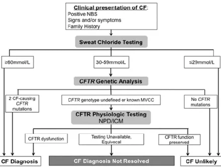
Initially a diagnosis of CF relied on the clinical phenotype, with recognition of the characteristic signs and symptoms of "classical" CF. There has been a surprising degree of difficulty encountered worldwide in establishing the diagnosis in a minority of cases and consequently, healthcare providers continue to face uncertain cases and challenging diagnostic dilemmas (Farrell et al., 2017c). However, the increased use of prenatal screening for maternal CF carrier status, prenatal ultrasound screening (that might reveal meconium ileus, meconium peritonitis, bowel obstruction, or echogenic bowel), and newborn screening (NBS) has resulted in the routine diagnosis of asymptomatic or minimally symptomatic infants and provided an opportunity to foster their normal growth and development (Farrell et al., 2017b). Nevertheless, the extensive NBS programmes implemented have identified increasing numbers of asymptomatic CF patients merely by identifying elevated serum concentrations of immunoreactive trypsinogen (IRT). This, poses new challenges to the CF diagnosis paradigm, especially when associated with borderline sweat Cl- (see below) and/or inconclusive CFTR genotypes (Farrell et al., 2017c). These inconclusive CF diagnosis through NBS, are designated as "CF screen positive with an inconclusive diagnosis" (CFSPID), also called "CFTR-related metabolic syndrome" (CRMS) (Farrell et al., 2017a). Diagnosis of CF in non-screened populations can be challenging because the age of onset and severity of symptoms can differ greatly as a result of highly variable degrees of CFTR dysfunction.
Figure 1.2 provides a simplified algorithm for the application of these consensus criteria to individuals with a suspicion of CF due to a positive NBS result, the appearance of signs or symptoms, or recognition of an immediate family member with history of CF (most often a sibling but may also include a parent or child). It should be noted that a positive NBS result does not mean the infant has CF; the probability of a CF diagnosis following a positive result varies greatly depending on the NBS method used (Farrell et al., 2017c).
6 Figure 1.2- Schematic representation of CF Diagnosis. Established guidelines to diagnose CF
according to clinical presentation and other types of evidence. Adapted from (Farrell et al., 2017c)
A confirmed CF diagnosis can only be established when an individual has both a clinical presentation of the disease and evidence of CFTR dysfunction. The tests conducted to establish CFTR dysfunction are done hierarchically, with sweat chloride being usually the first, followed by CFTR genetic analysis, and then CFTR functional tests like altered transepithelial nasal potential difference (NPD) or intestinal current of voltage measurements (ICM/IVM) (Sousa et al., 2012). For a positive diagnosis of CF two positive sweat tests are required and/or two CF-causing mutations identified. Only very rarely can an individual with a negative sweat chloride <30 mmol/L (see below) be considered to have CF and this diagnosis is only considered if all possible alternative diagnoses are excluded and if additional tests (genetic, functional tests) demonstrate dysfunctional CFTR. If only 1 CFTR gene variant is identified, further ("extended") CFTR gene analysis should be performed. CF is confirmed if both alleles possess CF-causing mutations. If two (or one) of these mutations is undefined, or if a mutation of varying clinical consequence (MVCC) is present CF is possible but not confirmed; CF is unlikely if the only mutations found have been described as non-CF-causing. If a CF diagnosis is not resolved, it can be considered as CRMS or CFSPID, if following NBS, or CFTR-RD (Farrell et al., 2017c).
However, depending on the ethnic background of the populations tested (Bobadilla et al., 2002), there are cases positively diagnosed with CF, from infancy to adulthood, that present "non-classical", or milder forms of the disease, representing 2% to 10% of all CF diagnoses. Most non-classical CF patients meet the same diagnostic criteria as classic CF patients, however, they may have a later presentation or have involvement of only one or two organ systems and usually with milder symptoms (Paranjape and Zeitlin, 2008). The majority of
7 patients with non-classical CF carry one severe and one mild or two mild CFTR mutations. A mild mutation is categorized as a gene alteration that still allows the resulting CFTR protein to partially function to a variable degree (Groman et al., 2005; Sousa et al., 2012).
Certain individuals show conflicting results from the diagnostic tools available, such as inconclusive genetic testing, borderline sweat tests with both non-CF and CF features. For such individuals with clinical phenotypes not fully meeting the CF diagnostic criteria it is also difficult to exclude CF and as such additional functional tests (NPD or ICM/IVM) should be carried out.
Sweat test
Although NBS is now widely implemented, to confirm a diagnosis of CF two positive sweat tests are required, with values ≥ 60 mmol/L indicating a diagnosis of CF and < 30 mmol/L indicating CF as very unlikely. For individuals with a clinical phenotype or positive NBS and sweat chloride levels 30-59 mmol/L further testing for CFTR mutations and function (such as NPD and ICM/IVM are indicated.
Genetic tests for CFTR
The discovery of the CF gene in 1989 (Riordan et al., 1989) provided a valuable tool for the genetic diagnosis of the disease. However, there are a large number of identified variants of the CFTR gene (currently more than 2000), most of which of unknown disease prognosis (Cystic Fibrosis Mutation Database, 2017). A more recent CFTR mutation database (CFTR2: https://cftr2.org) reports just the mutations of known disease prognosis.
All patients with clinical suspicion of CF and positive or borderline sweat test should be evaluated through genotype analysis. Traditionally, identification of CFTR mutations was performed by testing a panel of a given number of mutations (usually 5-150 mutations), usually customized for the most common mutations present in the local populations. In Europe, it has become clear which mutations should be analysed in an initial screening, since 90% of CF related mutations can be detected using specific methods for only 30 mutations (De Boeck, et al., 2011). However, there is a wide geographic variability and in the South of Europe the spectrum of mutations is much wider than in the North. So, other strategies can be used to identify mutations, with the direct DNA sequencing of the complete gene (or at least of all 27 exons and the respective flanking intronic regions) being the ultimate gold standard.
8 Active transport of ions across the nasal epithelium results in a potential difference (PD), which can be measured by a voltmeter between two electrodes: 1) an exploring electrode placed on the surface of the nasal epithelium and 2) a reference electrode (generally subcutaneous), which represents the internal surface (Keenan et al., 2015). NPD measurements should be performed in both nostrils and reported as an averaged result. This approach reduces variability between both nostrils and repeated measurements. This diagnosis method has become the standard reporting method in CF diagnostic and is widely used in research applications. Basal values for NPD are usually interpreted as normal (< -12 mV), intermediate (-12 to -7.7 mV) or abnormal (> -7.7 mV) (Ooi et al., 2012). CF patients usually present an increased magnitude of baseline PD (reflecting Na+ hyperabsorption), a dramatic response to amiloride, a transient or blunted response to low Cl- solution and isoproterenol (β-adrenergic agonist) and an exaggerated response to ATP (reflecting Cl -secretion through Calcium-activated chloride channels (CaCC)) (Taylor et al., 2009).
Intestinal Current/Voltage Measurements (ICM/IVM)
Bioelectrical measurements on rectal epithelia can be applied as a functional diagnostic tool to aid in establishing a diagnosis of CF, if sweat test results are equivocal and/or if CFTR-disease causing mutations are not readily identified by genetic testing. For Intestinal Current/Voltage Measurement (ICM/IVM), an intestinal biopsy (obtained by suction or forceps) and a special micro-Ussing chamber are needed. Through this method the ex vivo transepithelial short-circuit current (Isc) or transepithelial voltage (Vte) is measured and reflects the net ion fluxes across the tissue, which is calculated as equivalent current (Ieq-sc) by Ohm’s law in the case of voltage measurements. In CF, the intestinal CFTR-mediated Cl- secretion is impaired, while absorptive processes remain unchanged or may be enhanced. The values of Isc (or Ieq-sc) in response to forskolin (Fsk), an activator of CFTR, can lead to normal, absent or reduced currents. Under normal conditions, the Isc responds to Fsk alone and to Fsk with carbachol (CCH), which enhance the driving force for Cl- to exit the cell by stimulating basal K+ channels. This response consists of two components: a first lumen-negative current that is caused by the apical chloride efflux, followed by a larger lumen-negative current, caused by apical chloride secretion under the higher driving force (Sousa et al., 2012). In ICM/IVM of CF individuals, the apical Cl- efflux in response to Fsk and CCH does not occur, instead only a lumen-positive peak in response to CCH is observed corresponding to K+ efflux. A biphasic response may also be observed under CCH+Fsk (first lumen-positive corresponding to K+ channels efflux and then lumen-negative corresponding to Cl- efflux) due to residual CFTR-mediated Cl- efflux in milder forms of CF (Sousa et al., 2012).
9 Crossley and colleagues (Crossley et al., 1979) from Auckland, New Zealand, established the foundation for CF NBS in the 1970s by measuring immunoreactive trypsinogen (IRT) on a dried blood spot specimen, using a radioimmunoassay. In CF, the trypsinogen release into the circulation appears to be enhanced by abnormal pancreatic duct secretions. Thus, IRT levels are found to be elevated in CF and determination of IRT levels in newborns constitutes the assay used in most CF neonatal screening programmes (Rosenfeld et al., 2016). IRT values tend to remain raised for several months in newborns with CF, whereas in false positives they usually return to normal within the first weeks of life. Screening for CF can have different algorithms and use an increased IRT level from the dried blood spot specimen as the first stage. Most frequently, the second stage incorporates molecular testing for CFTR mutations on the first specimen (IRT/DNA). In some programmes, IRT concentrations are analysed on a second specimen collected between 1 and 2 weeks of life (IRT/IRT), whereas other programmes combine the two strategies, following 2 increased IRT results with CFTR mutation analysis performed on the second specimen (IRT/IRT/DNA) (Farrell et al., 2017b). An important outcome of recent studies is the discovery that Pancreatitis Associate Protein (PAP) may provide advantages for programmes that routinely collect samples after 36h of age and can be incorporated into these programmes in different ways (Farrell et al., 2017c). The careful selection of the mutations on the CFTR screening panel is critical in order to ensure adequate coverage for the racial and ethnic composition of the population being screened. The most important is that each programme adopts an algorithm that is adapted to the characteristics of the population being tested and also matches the health system conditions of each country. This approach has demonstrated long-term benefits such as early nutritional treatment, reduced hospitalizations (due to pulmonary exacerbations) and improved survival. In the 1950s, median life expectancy for patients with CF was a few months; the main causes of death were meconium ileus and malnutrition subsequent to pancreatic malabsorption. During the past six decades, median age of survival has increased progressively, and is now more than 40 years in developed countries (Farrell et al., 2017b). Replacement of pancreatic enzymes and intensive therapy guided by multidisciplinary teams have brought a major improvement in the treatment of CF and survival. Besides, the new guidelines for diagnosis of CF are very important to identify the patients as CF and begin health care at the earliest possible stage (Table 1.1).
10 Table 1.1 – Guidelines for CF diagnosis. Summary from the 2015 CF Foundation guidelines for
11
2. CFTR: from Gene to Protein and Function: CFTR Mutations &
Functional Classes
2.1 – CFTR: From Gene to Protein and Function
The CFTR gene is located on the long (q) arm of chromosome 7, bands 31-32 (7q31-7q32), and is one of the largest human genes, spanning ~190 kb. After transcription and splicing, the mRNA comprising 6.5 Kb is translated into the CFTR protein (or ABCC7) with 1480 amino acid (aa) residues (Riordan et al., 1989). The gene consists of 27 exons and 26 introns (Figure.1.3). The sizes of both exons and introns vary greatly, with the exons ranging from 38 bp in exon 16 to 724 bp in exon 14. Regarding intron length, the smallest intron (intron 25) comprises 600 bp, and the largest (intron 11) 28,085 bp (Ellsworth et al., 2000).
Figure.1.3 - Schematic representation of the CFTR gene, mRNA and protein. N– N-terminus;
TM– transmembrane domain; NBD – nucleotide-binding domain; R – regulatory domain; C – C terminus CFTR gene consist of 27 exons and introns. After transcription and mRNA splicing, a 1480 amino acid sequence is translated. Post-transcriptional modification of the protein glycosylation, occurs before translation to the cell membrane. The CFTR protein act at the apical membrane of epithelial cells (more details in section 2.2.1). [Image from MD Amaral, with permission].
As mentioned above, CFTR is a multidomain protein containing 1480 aa residues, that functions as a cAMP-activated and phosphorylation-regulated Cl- and bicarbonate channel at the apical membrane of epithelial cells. CFTR is a member of the ATP-binding cassette (ABC) transporter superfamily, also known as ABCC7 (Gadsby et al., 2006). Like other ABC transporters, it has two membrane spanning domains (MSDs) with six transmembrane segments each, portions of which form the pore through which anions pass. Additionally, it has two nucleotide binding domains (NBD1 and NBD2) and a regulatory domain (RD). Both NBDs bind and hydrolyse ATP, which drives channel opening and closure, respectively. The
12 RD, which is absent in all other ABC transporters, contains multiple consensus sites for phosphorylation by various kinases. Phosphorylation of the RD by protein kinase A (PKA) in response to cyclic AMP is regarded as the major determinant for opening of the channel (Riordan et al., 1989; Riordan, 2005).
Proteins belonging to the ABC transporter family are responsible for active transport of substrates across cell membranes, where ATP hydrolysis serves as the source of energy to drive the transport. However, CFTR is functionally distinct from other ABC transporters as it enables bidirectional permeation of anions, rather than vectorial transport of solutes (Riordan, 2005). Several hypotheses were raised regarding the function of CFTR protein. The model currently accepted for the mechanism of channel function and regulation proposes that (Figure 1.4) the first step is the phosphorylation of the RD by cAMP-dependent PKA (and by PKC) then ATP binds to the NBDs, which subsequently dimerize opening the channel (Sheppard and Welsh, 1999; Hwang and Sheppard, 2009) with the consequent alteration in the MSDs conformation, leading to the opening of the channel pore. It has also been shown that intramolecular interaction between the RD and both the N-terminus and NBD1 are required to regulate CFTR function (Baker et al., 2007). Even though the mechanism of CFTR channel gating is not fully understood, opening and closing of this Cl- channel is tightly regulated by the cellular balance of kinase and phosphatase activity and by ATP levels. Furthermore, the open probability (Po) of the channel is controlled by the extent of RD phosphorylation at its multiple sites (Hwang and Sheppard, 2009).
Figure 1.4 -Conformational changes of the CFTR Cl− channel during channel gating. The
simplified model shows a CFTR channel under quiescent (left) and activated (right) conditions. Communication between the NBDs and MSDs via the intracellular loops is either orthogonal (e.g. NBD1–MSD2) according to the most recent CFTR structural models based on Sav168834. P (phosphorylation of the RD); Pi (inorganic phosphate); PKA (cAMP-dependent protein kinase); PPase (protein phosphatase). In and Out denote the intra- and extracellular sides of the membrane, respectively. [Image reproduced from Hwang and Sheppard, 2009].
13 Besides its function as Cl- channel, CFTR has also been shown to regulate several other channels and transporters, thus being a general regulator of ion transport in epithelia. CFTR was found to be expressed in luminal membranes of both secretory and absorptive epithelia, playing a predominant role in both cAMP- and, as more recently shown (Faria et al., 2009; Lérias et al., 2018) in Ca2+-activated Cl- secretion.
2.2 – CFTR mutations & Functional Classification
As to date, 2026 mutations have been reported in the CFTR gene (Cystic Fibrosis Mutation Database, 2017), a number that comprises both disease causing mutations and polymorphisms that do not affect the CF phenotype. From these mutations, 39% are missense mutations, 16% frameshift mutations, 11% splicing mutations, 8% nonsense mutations, 2% In frame in/dels, 3% large in/dels, 0.8% mutations in the promoter, 13% sequence variation and 7% are of unknown consequences (Cystic Fibrosis Mutation Database, 2017). Currently these mutations are grouped into 7 functional classes defined according to the molecular changes caused by the different CFTR variants, with these evolving into theratypes, i.e., groups of mutations which can be corrected by the same therapeutic strategy (Figure 1.5).
Figure 1.5 – Functional classes of CFTR mutations/ theratypes. Wild-type CFTR protein at the
plasma membrane of epithelial cells properly functioning as a Cl- channel; (I) class I mutations: prevent translation; (II) class II: defective processing; (III) class III: defective regulation; (IV) class IV: defective conductance; (V) class V: reduced synthesis; (VI) class VI: decreased stability; (VII) class VII: no mRNA. Adapted from (De Boeck and Amaral, 2016)
14
Class I – Mutations that lead to no protein production
Class I mutations are those that can give origin to premature terminations codons (PTCs), such as nonsense mutations or some frameshift mutations, which are thus associated with more severe CF phenotypes (Elborn, 2016). Examples of class I mutations include modification of the codons that codify for the glycine in the position 542, the arginine in position 553, or the glutamine in position 637, into a stop codon (G542x, R553X and Q637X, respectively). G542X and R553X are two of the most common mutations in Europe following the F508del mutation (WHO, 2004). These mutations lead to significant degradation of the respective transcripts by nonsense mediated decay (NMD) so as to prevent production of truncated proteins that might be translated from these transcripts (De Boeck and Amaral, 2016). These mutations will be discussed in greater detail in section 3.
Class II – Mutants that affect intracellular traffic
Class II mutations are those that lead to incorrect folding and processing and thus prevent the protein from reaching the apical membrane of epithelial cells. F508del, the most common CF causing mutation, is a member of this class. By not being correctly folded, F508del CFTR protein is retained in the endoplasmic reticulum (ER) and targeted for degradation, thus not being fully-glycosylated in the Golgi nor transported to the apical membrane.
Class III - Mutations affecting channel gating
These mutations cause a defective response of CFTR to the phosphorylation of the RD by PKA after activation by cAMP. They are often located in the NBDs, affecting their interaction with ATP, and thus interfering with the correct gating of the channel. An example of a class III mutation is G551D (glycine to aspartate) which is quite frequent in Ireland (14%) and is thus also called the "Celtic mutation".
Class IV - Mutations that lead to defective chloride transport
Class IV mutations, such as R334W (mutation of an arginine to a histidine), lead to the synthesis of proteins which can be transported to the membrane and respond to stimulus, but have a reduced ion transport function due to defective conductance. Patients with these mutations have a milder CF phenotype.
Class V – Mutations that lead to reduced levels of protein
This class includes mutations that lead to reduced levels of protein, such as those affecting splicing of pre-mRNA such as 3272-26A>G or the TG12T5 sequence in intron 8 or missense mutations such as A455E (alanine to glutamate), which lead to reduced levels of transcripts
15 and thus low levels of functional protein. These mutations are also associated with milder CF phenotypes.
Class VI – Reduced stability or altered regulation of separate ion channels
In this group are included membrane-rescued mutations such as F508del, 120del23 (the deletion of the first codon), N287Y (asparagine to tyrosine). This class also includes variants with nonsense or frameshift mutations that originate PTCs in the last exon of the CFTR gene and lead to the production of carboxyl terminus (C-terminus)-truncated proteins. These proteins while being correctly processed, transported to the membrane and presenting normal function, are unstable and have an increased turnover at the cell surface. This is another class of mutations that leads to a milder CF phenotype.
Class VII – No mRNA, unrescuable mutations
Class VII mutations are the so-called "unrescuable mutations", because CFTR cannot be pharmacologically rescued per se, and include for example large deletions such as the del2,3(21kb) mutation.
16
3 Nonsense-mediated mRNA decay and CFTR nonsense mutations
The expression of protein-coding genes in eukaryotes involves the orchestration of transcriptional and posttranscriptional processes. To ensure the fidelity of these processes, the eukaryotic cell has evolved several quality control mechanisms. The best characterized mRNA quality control system in eukaryotes is the nonsense mediated decay (NMD) of mRNA. This surveillance mechanism detects and ensures the rapid degradation of faulty mRNAs harbouring premature translation termination codons (PTCs) that would otherwise have resulted in the synthesis of C-terminally truncated proteins. Hence, the quality control function of NMD relies on protecting the cell from the deleterious dominant-negative or gain-of-function effects of these truncated proteins (Kurosaki and Maquat, 2016; Nickless et al., 2017).
Nonsense-mediated mRNA decay
First discovered in yeast and then extensively studied in Caenorhabditis elegans,
Drosophila, mouse, human cells, and other model systems, NMD is a RNA surveillance
pathway that acts at the interface between transcription and translation (Nickless et al., 2017). Three key steps constitute the NMD pathway: 1) the identification of the PTC; 2) the assembly of the surveillance complex on the mRNA and 3) the degradation of the targeted RNA. The mechanism by which PTCs are recognized and discriminated from natural stop codons is translation-dependent and leads to the recruitment of NMD trans-acting factors, called upframeshift (UPF) proteins, such as UPF1, UPF2 and UPF3 (Lykke-Andersen and Heick Jensen, 2015).
During translation, stop codons are recognized by eukaryotic release factors (eRFs) eRF1 and eRF3, which are present in the translation apparatus, and in turn recruit UPF1, followed by the recruitment of the serine/threonine protein kinase SMG1. These proteins, together with the eRFs form the surveillance complex (SURF), and eventually lead to translation termination involving ribosomal dissociation (Figure 1.6). However, recognition of PTCs leads to the interaction of the SURF complex with UPF2 and 3 present at a downstream exon junction complex (EJC) leading to UPF1 phosphorylation that triggers transcript degradation (Hug et al., 2015; Nickless et al., 2017).
Within the nucleus, pre-mRNAs are synthesized and immediately undergo cis modifications binding various proteins during pre-mRNA processing, prior to mRNA formation. These include capping at the 5′ end, i.e., addition of a 7-methylguanosine residue (Cap) to protect the transcript from 5′-to-3′ exoribonucleases and provide a binding platform for the Cap-binding protein (CBP) complex (CBC), composed of CBP80–CBP20 (Da Costa et al., 2017).
17 In mammalian cells, and according to the classical EJC-dependent model, NMD depends on the interaction of the translation termination complex with a dynamic multiprotein assembly, the so-called EJC (Lykke-Andersen and Heick Jensen, 2015). NMD is tightly intertwined with mRNA biogenesis even at its earliest stages, since it is promoted by both the CBC and EJC. The latter consists of four core proteins- eukaryotic translation initiation factor 4A3 (eIF4A3), cancer susceptibility candidate 3 (CASC3), RNA-binding motif protein 8A (RBM8A or Y14), and mago-nashi homolog (MAGOH)- which are deposited ~20-24 nucleotides (nt) upstream of ~80% of all exon-exon junctions (Hir et al., 2016; Kurosaki and Maquat, 2016). This splicing-dependent "mark" serves to guide the NMD machinery once the mRNA is exported to the cytoplasm for translation-dependent inspection by NMD factors.
EJCs upstream of and within mRNA coding regions are removed by ribosomes during translation in the cytoplasm, however, because PTCs shorten the length of the coding region, any downstream EJCs that normally reside within the coding region would fail to be removed from what becomes the 3′UTR thus triggering NMD (Kurosaki and Maquat, 2016).
Initially, UPF1 associates with SMG1 and acts as a "clamp", interacting directly with eRF1 and eRF3 to form the SURF complex in the vicinity of the PTC (Hug et al., 2015; Da Costa et al., 2017) (Figure 1.6). UPF1 is the central NMD factor, it is an ATP-dependent RNA helicase containing an abundance of serine and threonine residues located within its N- and C- termini that function as phosphate-acceptor sites (Popp and Maquat, 2017). SMG1 is a phosphatidylinositol 3-kinase-related protein kinase that phosphorylates many of these residues, and this activity is initially held in check by the SMG8-SMG9 complex (Yamashita, 2013). Two subunits of the SMG1c complex, the SMG8 and SMG9 associate tightly with SMG1 and regulate its activity through the induction of conformational changes, with SMG8 binding to the preformed SMG9-SMG1 complex and maintaining the kinase in its inactive state (Yamashita et al., 2009; Arias-Palomo et al., 2011). Subsequently, the SURF complex interacts with UPF2, UPF3b and an EJC downstream of the PTC to form the decay-inducing complex (DECID), that triggers UPF1 phosphorylation and dissociation of eRF1 and eRF3 (Figure 1.6) (Da Costa et al., 2017). UPF1 phosphorylation has two consequences: first, phosphorylated UPF1 represses further translation initiation through an interaction with eIF3 that is critical for mRNA decay; second, phosphorylated UPF1 residues form platforms exposing new regions of UPF1 onto which RNA-degradative enzymes are either directly or indirectly recruited, resulting in targeted, rather than indiscriminate, mRNA destruction (Lykke-Andersen and Heick Jensen, 2015). As a consequence of the remodelling of NMD complexes, UPF1 adopts its active helicase conformation, due to the reorganization of its inhibitory domains through association with UPF2. The activated NMD complex (UPF1, UPF2 and UPF3b) is translocated from its position upstream of the EJC towards the 3’ of the EJC (Hug