1. Orthopaedic Surgeon/ Chief of Service, Professor of the Faculty of Medicine, University Clinic of Orthopaedics, Coimbra University Hospitals, Faculty of Medicine, University of Coimbra.
2. Orthopaedic Resident, University Clinic of Orthopaedics, Coimbra University Hospitals.
3. Orthopaedic Surgeon/ Graduated Assistant, University Clinic of Orthopaedics, Coimbra University Hospitals.
creased fluid pressure and implant motion may play a role, the final pathway seems to be related to the host response to the wear particles generated by the diffe rent friction systems of the prostheses: polyethylene-on--metal, metal-onpolyethylene-on--metal, polyethylene-on-ceramic, and ceramic-on-ceramic bearing surfaces1,2,3.
The cellular response to particulate debris, a foreign body reaction, can produce periprosthetic osteolysis that leads to bone loss and consequently to aseptic loose ning of the hip prostheses. Polyethylene particles seem to be the major culprit1,3.
Aseptic loosening remains a dominant cause for im-plant failure requiring revision. Revision hip surgery with extensive bone loss is never an easy prospect for the patient or surgeon. It is technically challenging, in-volves higher costs, and often lower success rates.
Particles of polyethylene and other debris are dis-pensed through the joint fluid. Fluid flows according to pressure gradients, and any area of bone accessed by joint fluid is a potential site for deposition of wear de-bris. Schmalzried, Jasty and Harris described “the ef-fective joint space” where debris and fluids migrate around the components of the prostheses and cause bone lysis out of the joint space. This concept explains the development of osteolysis at the tip of a well-fixed femoral component, or over the dome of an acetabular component with holes in the metal backing2,3.
Pelvic and femoral osteolysis are generally progres-sive processes of particle-induced bone loss. The pat-tern of lysis depends on the implant design. Many pa-tients with osteolytic lesions remain asymptomatic un-til catastrophic failure occurs from dislocation or periprosthetic fracture. Once the osteolysis is detected, more frequent follow-up is advisable. Radiographs should be made at 3- to 6-month intervals rather than yearly4,5,6.
The purpose of this paper is to report an unusual case of a patient with a stable total revision hip
pros-Rapid progression of a severe femoral bone
loss in a stable revision hip prosthesis:
causes and management
Fernando Judas1, Alexandre Marques2, Luís Maximino3, Francisco Lucas3
AbstrAct
We report a case of a severe bone loss of the proximal fe mur with a rapid progression, in a 72-year-old male patient with a stable total revision hip prosthesis. The pa tient had a persistent mechanical thigh pain. The blood laboratory values were normal. Infection disease and osteolytic bone tumor were excluded.
A surgical procedure was performed. The native bone of the proximal femur was resorbed and replaced by a dense fibrous tissue with some sclerotic bone frag-ments. A large amount of a brownish fluid and a red--brown and friable tissue were found in the pseudo-joint cavity. The proximal femur was reconstructed using a large amount of cryopreserved cancellous bone allograft, with retention of the femoral prosthesis.
The mechanism of the bone lesion can be related not only to the host response to the wear particles, but mainly to the fluid pressure in the effective joint space. Femoral progressive osteolysis in a stable hip pros-thesis is an indication for surgery in useful time, before adverse bone remodeling can begin and lead to major bone loss.
Keywords: Severe femoral bone loss; Osteolysis;
Revi-sion hip prosthesis.
IntroductIon
Bone loss has been reported in association with either loose or well-fixed total hip prostheses. Although
thesis who had a rapid and severe bone resorption of the proximal femur, like an osteolytic bone tumor. We suggest a possible explanation for the development of the impressive bone loss and we describe the surgical technique used for femur reconstruction.
cAsE rEport
A 72-year-old man was submitted in January 2006 to a total revision hip arthroplasty in the right side due to aseptic loosening of a primary cementless prosthesis CLS®, with 11-years follow-up. The aetiology of the
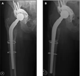
primary prosthesis was a degenerative arthritis of the hip. In the revision surgery, an acetabular metallic support ring with a cemented polyethylene cup and a revision conical stem were implanted. The pelvic and femoral bone loss were reconstructed with morselized cancellous bone allograft (Fig. 1a). No complications were reported in the first three years of the postopera -tive course.
A dislocation of the revision prosthesis occurred in May 2009 by indirect traumatism. The dislocation was reduced under general anesthesia and signs of osteo -lysis in the proximal femur were detected. The patient
returned to his normal activity after 15 days.
In August 2010 the radiographic studies showed an aggravation of the femoral osteolysis in the stage II, ac-cording to the HUC Classification System6.
Neverthe-less the femoral stem was stable by the excellent fixa-tion of the distal part and the acetabular component re-mained well-fixed (Fig.1b). The patient had a thigh pain, that was aggravated by weight-bearing and re-lieved by analgesics (metamizole) and resting. The pa-tient walked with support aids, two forearm crutches, and was medicated with warfarin to control atrial fi -brillation.
Due to the constant mechanical pain and weakness, he visited our Department five months later. Plain ra-diographs revealed a severe expansive osteolytic lesion of the proximal femur. The bone was almost entirely resorbed (Fig. 2) and a surgical intervention was per-formed. The blood laboratory values were normal.
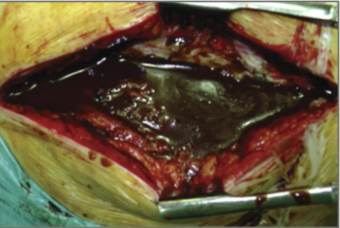
The hip was exposed through a standard posterior approach. The proximal femur bone was replaced by a pseudojoint cavity composed by a dense fibrous tis-sue with some sclerotic bone fragments. A capsular distension was detected. When the pseudojoint cavi-ty was incised, a large amount of a brownish fluid (400 cc) escaped under great pressure (Fig. 3).
The inner surfaces of the pseudojoint cavity and the
FIGurE 1.Periprosthetic osteolysis. a) Radiograph of a revision hip prosthesis at 1-year follow-up. The prosthesis was stable with absence of periprosthetic osteolysis. b) At 4-years follow-up the radiograph revealed moderate proximal femoral osteolysis, stage II according to the HUC Classification System
A b
FIGurE 2.Five months later, painful and severe expansive osteolytic lesion of the proximal femur. The bone was almost entirely resorbed. The acetabular and femoral components remained mechanically stable
joint space were cleaned of a red-brown, uniform and friable tissue. At macroscopic examination it seemed like a chronic connective granulation tissue possibly derived from hematoma resorption, with no evidence of acute inflammation. The histopathologic examina-tion of the specimens revealed fibrous and necrotic material, including some segments of granulation tis-sue with an exuberant angiomatous component.
The proximal femur was reconstructed using a large amount of cryopreserved cancellous bone allograft (300 cc) from HUC Tissue Bank, with retention of the femoral prosthesis. The morselized bone was im-pacted to fill the space of the pseudojoint cavity around the femoral implant (Figs. 4a and 4b).
Final culture results of fluid and soft-tissue speci-mens obtained at the time of surgery were negative for infection.
No complications were reported in the postopera-tive course. Immediately after surgery, the patient was mobilized with protected weight-bearing. Three weeks after surgery, the mechanical tight pain disappeared. At 7-months follow-up, the patient was clinically able to walk without external support; had a Trendelenburg sign and reported no groin or thigh pain. He was very pleased with the surgery result.
dIscussIon
The current clinical case illustrates a rare example of an extensive and aggressive osteolysis of the proximal femur. Investigations have indicated that the cellular response to wear particles is central to the
development of osteolysis in total hip arthroplasties. Mechani -cal factors were also responsible for osteolysis. Whether the initiating factor is biologic or mechanical is a subject of debate2,3.
In the current case, four years after the revision hip surgery plain radiographs showed a moderate osteoly -sis of the proximal femur. The primary factor in this ins tance seems to have been the biological reaction to the wear particles of polyethylene, cement and metal, either alone or in concert.
However, an unusual progression of the lytic bone loss was detected during a short period of five months that led to the femoral bone resorption. In this situa-tion, it is vital to exclude the presence of infection or bone tumor, because periprosthetic osteolysis is usual -ly a progressive and slow process that also leads to a progressive bone stock loss.
Infection must be suspected whenever resorption of bone is considerable, the endosteal surface of the bone is scalloped, periosteal elevation is present, and the erythrocyte sedimentation rate or C-reactive pro-tein level are high. The clinical and laboratory studies eliminated an infection process in this clinical case, and the final culture results of fluid and soft-tissue specimens obtained at the time of surgery were nega-tive for infection.
Although the incidence of malignant tumors in pa-tients undergoing total hip arthroplasties is known to be lower than in the general population, there are seve -ral reports on the development of malignant tumors at
FIGurE 4.Management of the expansive bone loss. a) The proximal femur was reconstructed using cryopreserved cancellous bone allograft (HUC Tissue Bank), the prosthesis was not replaced b) Radiograph at 7 months of the postoperative period showing the process of the allograft bone incorporation
A
FIGurE 3.Intraoperative photograph showing a large amount of a brownish fluid under great pressure (400 cc) in the pseudojoint cavity and in the joint space
the site of total hip arthroplasties. Malignant fibrous histiocytoma at the site of an endoprosthesis of the hip constitutes a distinct rarity, and has been attributed to the implants or to their alloy constituents7. The
histopathologic examination of the specimen from the surgical operation revealed fibrous and necrotic mate-rial and the absence of malignant lesions.
A mechanical instability of the femoral component may originate expansive bone loss8, nevertheless we
can also eliminate this mechanism because in the cur-rent case the stem was stable.
We believe that the continued expansion of the os-teolysis during a short time of five months can be main-ly secondary to an intense fluid pressure, supported by a failure of venous and lymphatic drainage. In fact, a large volume of articular fluid under a high pressure was found in the pseudojoint cavity and in the joint space at the time of surgery. No granulomatous mem-branes suggestive of a biological reaction to wear particles were detected in periprosthetic tissues. Osteo -lysis can also occur in the absence of wear particles by fluid pressure in the effective joint space9.
Van der Vis and Aspenberg showed that fluctuating pressure applied to bone can result in the death of os-teocytes near the implant and subsequent bone re-sorption. Thus, there is evidence that fluid pressure and ensuing flow could be a main cause of not only pain, but also osteolysis10,11. It is also true that bone
erosion by aneurisms can be caused by constant mecha -nical pressure, analogous to what happens between periprosthetic osteolytics lesions and arthrosis cysts.
The exact pathogenesis of the granulation tissue with exuberant angiomatous component present in the pseudojoint cavity and in the joint space is unclear. One hypothesis is the possibility that a hemorrhagic process may have contributed to its origin and also to the fluid pressure. This may be related to the fact that the patient was under oral anticoagulant therapy (warfarin) to control atrial fibrillation, and he had also des -cribed a constant and mechanical thigh pain during the preoperative time.
Severe bone loss of the proximal femur secondary to major osteolysis is a difficult situation to deal with, of-fering no ideal treatment option. The possible options include the implantation of a composite allograft/stem prosthesis, a modular type megaprosthesis, a custom-made megaprosthesis, the use of massive or cortical strut bone allografts, and impaction cancellous bone allografting12, 13, 14, 15, 16.
In the current case the replacement of the femoral
re-vision prosthesis by a new femoral implant was not an acceptable option, because the stem was mechanically stable.
The proximal femur reconstruction with bone allo-grafting seemed preferable because it is an attractive technique since it restores the bone stock around the implant17, 18, and it can be done without surgical
dislo-cation of the stem. We used cancellous bone allografts. The morselized bone was impacted and filled the space between the femoral stem and the inner surfaces of the pseudojoint cavity.
Cryopreserved cancellous bone allografts showed clinical efficacy for the reconstruction of bone defects19.
The validity of the impaction allografting technique has been demonstrated previously by histological and me-chanical criteria. Histologically, grafted bone chips were shown to be replaced by host bone in human retrieval studies and animal experiments21.
Femoral progressive osteolysis in a stable hip pros-thesis even in the absence of symptoms is clear indica-tion for surgery, before adverse bone remodeling may begin and lead to major bone loss. If allowed to progress, femoral reconstruction becomes more com-plex or impossible.
corrEpondEncE to Fernando Judas
University Clinic of Orthopaedics, Coimbra University Hospitals, Praceta Prof. Mota Pinto, Bloco de Celas 3000 Coimbra, Portugal
E-mail: fernandojudas@gmail.com rEFErEncEs
1. Canales V, Panisello JJ, Herrera A, Sola A, Mateo JJ, Caballero MJ. Extensive osteolysis caused by polyethylene particle migration in an anatomical hydroxyapatite-coated hip prosthesis: 10 years’ follow-up. Arthroplasty 2010; 25(7):1115-1124.
2. Harris WH. The problem is osteolysis. Clin Orthop 1995; 311:46.
3. Archibeck MJ, Jacobs JJ, Roebuck KA, Glant TT. The basic scien-ce of periprosthetic osteolysis. Instr Course Lect 2001;50:185--195.
4. Harris WH, McCarthy Jr JC, O’Neill DA. Femoral component loosening using contemporary techniques of femoral cement fi-xation. J Bone Joint Surg 1982; 64A:1063.
5. Maloney WJ, Woolson ST. Increasing incidence of femoral os-teolysis in association with uncemented Harris-Galante total hip arthroplasty: a follow-up report. J Arthroplasty 1996;11: 130.
6. Proença A, Judas F, Cabral R, Canha N. Revision surgery of hip prosthesis. Osteolysis reconstruction with bone allografts. 1996 Orthopaedics Department of Coimbra University Hospitals. 7. Schuh A, Zeiler G, Holzwarth U, Aigner T. Malignant fibrous
2004; 425:218-222.
8. Kalhor M, Notzli HP, Stover MD, Ganz G. Extreme ectasia of the femoral diaphysis secondary to loosening of a long Wagner stem: a case report. J Bone Joint Surg Am 2004; Volume 86--A(3):590-594.
9. Manley MT, D’Antonio JA, Capello WN, Edidin AA. Osteolysis: a disease of access to fixation interfaces. Clin Orthop 2002;405:129-137.
10. Aspenberg P, Van der Vis H. Migration, particles, and fluid pressure. A discussion of causes of prosthetic loosening. Clin Or -thop. 1998;352:75-80.
11. Van der Vis HM, Aspenberg P, Marti RK, Tigchelaar W, Van Noorden CJ. Fluid pressure causes bone resorption in a rabbit model of prosthetic loosening. Clin Orthop. 1998;350:201--208.
12. Bertani A, Helix M, Louis ML, Rochwerger A, Curvale G. Total hip arthroplasty in severe segmental femoral bone loss situa-tions: use of a reconstruction modular stem design. (JVC IX). Retrospective study of 23 cases. Orthop Traumatol Surg Res 2009; 95(7):491-497.
13. Head WC, Malinin TI, Mallory TH, Emerson RH Jr. Onlay cor-tical allografting for the femur. Orthop Clin North Am 1998;29:307-312.
14. Gross AE, Hutchison CR, Alexeeff M, Mahomed N, Leitch K, Morsi E. Proximal femoral allografts for reconstruction of bone stock in revision arthroplasty of the hip. Clin Orthop 1995;319:151-158.
15. Zmolek JC, Dorr LD. Revision total hip arthroplasty. The use of solid allograft. J Arthroplasty 1993;8:361-370.
16. Argenson JN, Gravier R. Femoral revision with impacted mas-sive or morselized graft. Rev Chir Orthop Reparatrice Appar Mot 2000;86:46-47.
17. Slooff TJJH, Schreurs BW, Gardeniers JWM: Revision total hip arthroplasty with impacted cancellous allografts and cement. 2001; In Pipino F (ed): Bone Cement and Cemented Fixation of Implants 40 Years of Clinical Practice and Prospective for the future; 2001; pp 163.
18. Gie GA, Ling RSM: Contained, morsellised, impacted allograft in revision total hip arthroplasty, the femoral side. 1998 Ort-hopaedics Today; 1:18.
19. Judas F. Contribution to the study of Morselized Bone Allografts and Biomaterials. PhD Thesis. 2002, Coimbra University. 20. Judas F, Figueiredo MH, Cabrita AM, Proença A: Incorporation
of impacted morselized bone allografts in rabbits. Transplant Proc 2005, 37(6):2802-2804.
21. Williams Amy, Szabo RM. Bone transplantation. Orthopaedics 2004; Vol 27, Nº 5:488-495.